"V体育官网" Skip to main page content
U.S. flag

An official website of the United States government

Dot gov

The . gov means it’s official. Federal government websites often end in . gov or . mil. Before sharing sensitive information, make sure you’re on a federal government site. VSports app下载.

Https

The site is secure V体育官网. The https:// ensures that you are connecting to the official website and that any information you provide is encrypted and transmitted securely. .

. 1998 Oct 19;143(2):297-307.
doi: 10.1083/jcb.143.2.297.

Serine phosphorylation of SR proteins is required for their recruitment to sites of transcription in vivo

Affiliations

Serine phosphorylation of SR proteins is required for their recruitment to sites of transcription in vivo (VSports注册入口)

T Misteli et al. J Cell Biol. .

Abstract

Expression of most RNA polymerase II transcripts requires the coordinated execution of transcription, splicing, and 3' processing. We have previously shown that upon transcriptional activation of a gene in vivo, pre-mRNA splicing factors are recruited from nuclear speckles, in which they are concentrated, to sites of transcription (Misteli, T. , J. F. Cáceres, and D. L VSports手机版. Spector. 1997. Nature. 387:523-527). This recruitment process appears to spatially coordinate transcription and pre-mRNA splicing within the cell nucleus. Here we have investigated the molecular basis for recruitment by analyzing the recruitment properties of mutant splicing factors. We show that multiple protein domains are required for efficient recruitment of SR proteins from nuclear speckles to nascent RNA. The two types of modular domains found in the splicing factor SF2/ ASF exert distinct functions in this process. In living cells, the RS domain functions in the dissociation of the protein from speckles, and phosphorylation of serine residues in the RS domain is a prerequisite for this event. The RNA binding domains play a role in the association of splicing factors with the target RNA. These observations identify a novel in vivo role for the RS domain of SR proteins and suggest a model in which protein phosphorylation is instrumental for the recruitment of these proteins to active sites of transcription in vivo. .


Figures
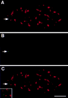
Figure 1
Figure 1
Localization of the transcriptionally inactive pem gene in HeLa cells. HeLa cells carrying the pem gene were grown for 24 h in the presence of 1 μg/ml tetracycline and fixed. The RNA was digested with RNAse, cells were denatured, and the pem gene was localized by in situ hybridization (B) and splicing factor SC35 localized by indirect immunofluorescence (A). In more than 95% of cells, splicing factor SC35 was found not to be associated with the transcriptionally inactive pem locus (C). The position of the pem locus is indicated by an arrow. Bar, 5 μm.
Figure 2
Figure 2
(A) Recruitment of SF2/ASF mutants to transcription sites. Endogenous SF2/ASF (a–c), full-length SF2/ASF (d–f), or SF2/ASF mutants lacking RRM1 (g–i), RRM2 (j–l), or the RS domain (m–o) were transfected into pem-HeLa cells in the presence of tetracycline to inhibit pem expression. 14 h after transfection, tetracycline was removed to induce pem transcription for 4 h. Cells were fixed, and the SR protein was detected using an anti-SF2/ASF antibody (a, red) or an anti–T7 epitope antibody (d, g, j, and m, red). Pem RNA was detected by in situ hybridization (b, e, h, k, and n, green). Endogenous SF2/ASF and full-length SF2/ ASF were recruited to and accumulate at the site of pem transcription. Deletion of any single domain prevented recruitment. Arrows and arrowheads indicate the sites of pem transcription. (B) A test line was drawn so as to include a splicing factor compartment and the site of pem RNA. The relative fluorescence intensity along the line for both the splicing factor signal (red) and the pem RNA signal (green) was measured in arbitrary units. The fluorescence intensity peaks for endogenous and wild-type SF2/ASF protein coincided with the peak for the pem RNA, indicating recruitment of splicing factor to the site of pem RNA. For mutants of SF2/ASF, the two peaks were separated, indicating the absence of the splicing factor mutants from the site of pem RNA. Bar, 5 μm.
Figure 2
Figure 2
(A) Recruitment of SF2/ASF mutants to transcription sites. Endogenous SF2/ASF (a–c), full-length SF2/ASF (d–f), or SF2/ASF mutants lacking RRM1 (g–i), RRM2 (j–l), or the RS domain (m–o) were transfected into pem-HeLa cells in the presence of tetracycline to inhibit pem expression. 14 h after transfection, tetracycline was removed to induce pem transcription for 4 h. Cells were fixed, and the SR protein was detected using an anti-SF2/ASF antibody (a, red) or an anti–T7 epitope antibody (d, g, j, and m, red). Pem RNA was detected by in situ hybridization (b, e, h, k, and n, green). Endogenous SF2/ASF and full-length SF2/ ASF were recruited to and accumulate at the site of pem transcription. Deletion of any single domain prevented recruitment. Arrows and arrowheads indicate the sites of pem transcription. (B) A test line was drawn so as to include a splicing factor compartment and the site of pem RNA. The relative fluorescence intensity along the line for both the splicing factor signal (red) and the pem RNA signal (green) was measured in arbitrary units. The fluorescence intensity peaks for endogenous and wild-type SF2/ASF protein coincided with the peak for the pem RNA, indicating recruitment of splicing factor to the site of pem RNA. For mutants of SF2/ASF, the two peaks were separated, indicating the absence of the splicing factor mutants from the site of pem RNA. Bar, 5 μm.
Figure 3
Figure 3
Quantitative analysis of recruitment. Cells were scored for accumulation of the wild-type or mutant SR proteins at the site of pem transcription, as detected by in situ hybridization and indirect immunofluorescence. The percentage of cells (n = 200 from 3 experiments) showing accumulation of splicing factor at the site of transcription is shown ±SD. Endogenous and wild-type SR proteins were efficiently recruited to the site of transcription in more than 90% of cells, whereas deletion mutants of SF2/ASF, SRp40, SC35, or SRp20 were typically found at the site of pem RNA in ∼30% of cells (see Results).
Figure 4
Figure 4
Distinct roles in the recruitment process for RRMs and the RS domain. HeLa cells were cotransfected with a β-TM minigene and the indicated SF2/ASF construct. 14 h after transfection, SF2/ASF was detected by immunofluorescence using an anti–T7 epitope antibody (red), and β-TM RNA was visualized by in situ hybridization (green). (A) Full-length SF2/ASF was efficiently recruited to sites of β-TM transcription. (B) Deletion of the first RRM prevented recruitment and resulted in the diffuse distribution of SF2/ASF-ΔRRM1. Identical results were obtained for SF2/ASF lacking RRM2 (data not shown). (C) Deletion of the RS domain resulted in the retention of SF2/ASF-ΔRS in native speckles at sites distinct from β-TM transcription sites. Bar, 5 μm.
Figure 5
Figure 5
The RS domain of SF2/ ASF is required for recruitment in living cells. Transcription of BK virus was triggered by addition of 50 μg/ml cAMP to the growth medium, and splicing factor distribution was imaged at the indicated times after induction of transcription. After acquisition of the final image, the cell was fixed, and the position of the induced RNA was detected by fluorescence in situ hybridization. (Top) Full-length SF2/ASF formed a peripheral extension from an existing speckle (red), and SF2/ASF accumulated at the site of BK early gene transcription (arrow). (Bottom) Deletion of the RS domain prevented dissociation of the splicing factor and its accumulation at the site of transcription. Pseudocolored images are shown. Speckles appear in red against a yellow/green background representing the nucleoplasmic pool of the splicing factor. The spectrum of pseudocolors represents the intensities of the fluorescence signal measured for each pixel (blue lowest, red brightest). Arrow, position of the RNA signal. The time after induction of gene expression is indicated in minutes. Bar, 1.5 μm.
Figure 6
Figure 6
Phosphorylation of serine residues in the RS domain is required for recruitment. (A) Amino acid sequence of the RS domain of SF2/ASF. The RS domain of SF2/ASF was either deleted or the RS-dipeptides substituted with either RG- or GS-dipeptides. (B) Immunoblot analysis of the phosphorylation state of SF2/ASF in vivo. The indicated T7-tagged constructs were transfected into BHK cells, and total cell lysates were probed by Western blotting with (+) or without (−) alkaline phosphatase digestion. Full-length SF2/ASF and SF2/ASF-GS were phosphorylated in vivo, and SF2/ASF-ΔRS and SF2/ASF-RG were not phosphorylated in vivo.
Figure 6
Figure 6
Phosphorylation of serine residues in the RS domain is required for recruitment. (A) Amino acid sequence of the RS domain of SF2/ASF. The RS domain of SF2/ASF was either deleted or the RS-dipeptides substituted with either RG- or GS-dipeptides. (B) Immunoblot analysis of the phosphorylation state of SF2/ASF in vivo. The indicated T7-tagged constructs were transfected into BHK cells, and total cell lysates were probed by Western blotting with (+) or without (−) alkaline phosphatase digestion. Full-length SF2/ASF and SF2/ASF-GS were phosphorylated in vivo, and SF2/ASF-ΔRS and SF2/ASF-RG were not phosphorylated in vivo.
Figure 7
Figure 7
Phosphorylation of serine residues in the RS domain is required for recruitment in living cells. The recruitment assay was performed as described in Fig. 5. (Top) The phosphorylated SF2/ASF-GS mutant was recruited to the newly formed site of transcription (arrow). (Bottom) The unphosphorylated SF2/ASF-RG mutant was retained in nuclear speckles and did not accumulate at the transcription site (arrow). Time after induction of gene expression is indicated in minutes. Images are pseudocolored; speckles appear in red against a green/ yellow background representing the nucleoplasmic pool of the splicing factor. Arrow, position of the RNA signal. Bar, 1.5 μm.

References

    1. Báuren G, Wieslander L. Splicing of Balbiani ring 1 gene pre-mRNA occurs simultaneously with transcription. Cell. 1994;76:183–192. - "VSports注册入口" PubMed
    1. Beyer AL, Osheim YN. Splice site selection, rate of splicing, and alternative splicing on nascent transcripts. Genes Dev. 1988;2:754–765. - PubMed
    1. Cáceres JF, Krainer AR. Functional analysis of pre-mRNA splicing factor SF2/ASF structural domains. EMBO (Eur Mol Biol Organ) J. 1993;12:4715–4726. - PMC - PubMed
    1. Cáceres JF, Misteli T, Screaton G, Spector DL, Krainer AR. Role of the modular domains of SR-proteins in subnuclear localization and alternative splicing specificity. J Cell Biol. 1997;138:225–238. - PMC - PubMed
    1. Cao W, Jamison SF, Garcia-Blanco MA. Both phosphorylation and dephosphorylation of ASF/SF2 are required for pre-mRNA splicing in vitro. RNA. 1997;3:1456–1467. - PMC - PubMed

V体育安卓版 - Publication types

MeSH terms