"V体育2025版" Skip to main page content
U.S. flag

An official website of the United States government

Dot gov

The . gov means it’s official. Federal government websites often end in . gov or . mil VSports app下载. Before sharing sensitive information, make sure you’re on a federal government site. .

Https

The site is secure. The https:// ensures that you are connecting to the official website and that any information you provide is encrypted and transmitted securely V体育官网. .

. 1998 Jun 15;141(6):1423-32.
doi: 10.1083/jcb.141.6.1423.

"VSports最新版本" The regulation of reactive oxygen species production during programmed cell death

Affiliations

"V体育2025版" The regulation of reactive oxygen species production during programmed cell death

S Tan et al. J Cell Biol. .

Abstract

Reactive oxygen species (ROS) are thought to be involved in many forms of programmed cell death. The role of ROS in cell death caused by oxidative glutamate toxicity was studied in an immortalized mouse hippocampal cell line (HT22). The causal relationship between ROS production and glutathione (GSH) levels, gene expression, caspase activity, and cytosolic Ca2+ concentration was examined. An initial 5-10-fold increase in ROS after glutamate addition is temporally correlated with GSH depletion. This early increase is followed by an explosive burst of ROS production to 200-400-fold above control values. The source of this burst is the mitochondrial electron transport chain, while only 5-10% of the maximum ROS production is caused by GSH depletion. Macromolecular synthesis inhibitors as well as Ac-YVAD-cmk, an interleukin 1beta-converting enzyme protease inhibitor, block the late burst of ROS production and protect HT22 cells from glutamate toxicity when added early in the death program VSports手机版. Inhibition of intracellular Ca2+ cycling and the influx of extracellular Ca2+ also blocks maximum ROS production and protects the cells. The conclusion is that GSH depletion is not sufficient to cause the maximal mitochondrial ROS production, and that there is an early requirement for protease activation, changes in gene expression, and a late requirement for Ca2+ mobilization. .


Figures
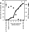
Figure 2
Figure 2
Inhibitors of macromolecular synthesis and ICE proteases prevent ROS production. Inhibitors were added to HT22 cells at 2-h intervals during a 10-h exposure to 5 mM glutamate, and DCF fluorescence and cell survival were measured. ROS production is presented as the ratiometric increase in median DCF fluorescence versus the control value (black circles). Cell survival was measured by trypan blue exclusion and is presented as the percent of the control value (black squares). Each set of data represents the mean of two trials that were repeated four times, plus or minus the standard error. (A) Effect of actinomycin D on ROS production and cell survival. 0.1 μg/ml actinomycin D was added to HT22 cells at 2-h intervals during a 10-h exposure to 5 mM glutamate. (B) Effect of cycloheximide on ROS production and cell survival. 300 nM cycloheximide was added to HT22 cells during a 10-h exposure to 5 mM glutamate. (C) Effect of Ac-YVAD-cmk on ROS production and cell survival. 50 μM Ac-YVAD-cmk was added to HT22 cells as in parts A and B.
Figure 2
Figure 2
Inhibitors of macromolecular synthesis and ICE proteases prevent ROS production. Inhibitors were added to HT22 cells at 2-h intervals during a 10-h exposure to 5 mM glutamate, and DCF fluorescence and cell survival were measured. ROS production is presented as the ratiometric increase in median DCF fluorescence versus the control value (black circles). Cell survival was measured by trypan blue exclusion and is presented as the percent of the control value (black squares). Each set of data represents the mean of two trials that were repeated four times, plus or minus the standard error. (A) Effect of actinomycin D on ROS production and cell survival. 0.1 μg/ml actinomycin D was added to HT22 cells at 2-h intervals during a 10-h exposure to 5 mM glutamate. (B) Effect of cycloheximide on ROS production and cell survival. 300 nM cycloheximide was added to HT22 cells during a 10-h exposure to 5 mM glutamate. (C) Effect of Ac-YVAD-cmk on ROS production and cell survival. 50 μM Ac-YVAD-cmk was added to HT22 cells as in parts A and B.
Figure 1
Figure 1
GSH depletion and ROS production after exposure to glutamate or BSO. (A) Glutamate (5 mM) or BSO (50 μM) were added to cells, and GSH and ROS were measured as a function of time. The amount of GSH was calculated as nmoles GSH per mg protein and is presented as a percentage of the control value. ROS are presented as the ratiometric increase in median DCF fluorescence versus the control sample. Both GSH and ROS were quantified at 2-h intervals up to 10 h after the addition of glutamate. GSH data are the average of five trials. GSH depletion by BSO is an average of four trials. ROS data are averages of eight trials. (black squares) GSH, glutamate; (black circles) ROS, glutamate; (white squares) GSH, BSO; (white circles) ROS, BSO. (B) The glutamate data from the above and similar experiments were plotted in terms of GSH concentration (% maximum) vs. log ROS (% maximum).
Figure 3
Figure 3
Effect of uncoupling mitochondria on ROS production. (A) 5 μM of the protonophore FCCP was added to the cells at 2-h intervals after glutamate exposure to see how late the inhibitor could be added and still protect cells. ROS levels are expressed as the ratiometric increase in median DCF fluorescence versus the control value (black circles). Cell survival is the percent of the control (black squares). (B) In vitro production of H2O2 by mitochondria from HT22 cells was determined by fluorescence spectrometry using the fluorescent substrate, p-HPAA. Basal H2O2 production was initiated by providing mitochondria with 6 mM succinate. Once H2O2 production was established, 0.2 μM FCCP was added, resulting in inhibition of H2O2 production. 0.1 μM H2O2 was added to indicate the increase in relative fluorescence with a known concentration of H2O2. (C) In vitro production of H2O2 by mitochondria from HT22 cells was established as in B. Basal H2O2 production was initiated by providing the mitochondria with 6 μM succinate. 100 μM pargyline had no effect on H2O2 production, while 100 μM clorgyline decreased H2O2 production to less than 10% of the basal level. 0.1 μM H2O2 was added to indicate the increase in relative fluorescence with a known concentration of H2O2.
Figure 3
Figure 3
Effect of uncoupling mitochondria on ROS production. (A) 5 μM of the protonophore FCCP was added to the cells at 2-h intervals after glutamate exposure to see how late the inhibitor could be added and still protect cells. ROS levels are expressed as the ratiometric increase in median DCF fluorescence versus the control value (black circles). Cell survival is the percent of the control (black squares). (B) In vitro production of H2O2 by mitochondria from HT22 cells was determined by fluorescence spectrometry using the fluorescent substrate, p-HPAA. Basal H2O2 production was initiated by providing mitochondria with 6 mM succinate. Once H2O2 production was established, 0.2 μM FCCP was added, resulting in inhibition of H2O2 production. 0.1 μM H2O2 was added to indicate the increase in relative fluorescence with a known concentration of H2O2. (C) In vitro production of H2O2 by mitochondria from HT22 cells was established as in B. Basal H2O2 production was initiated by providing the mitochondria with 6 μM succinate. 100 μM pargyline had no effect on H2O2 production, while 100 μM clorgyline decreased H2O2 production to less than 10% of the basal level. 0.1 μM H2O2 was added to indicate the increase in relative fluorescence with a known concentration of H2O2.
Figure 4
Figure 4
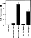
ROS production in cystine-free medium. Cells were exposed to either cystine-free medium supplemented with 10% dialyzed fetal bovine serum or 5 mM glutamate. The ratiometric increase in median DCF fluorescence versus the control value was determined after 4 and 8 h exposure for each sample.
Figure 5
Figure 5
ROS production and intracellular Ca2+ changes. ROS were monitored (DCF fluorescence, horizontal axis) at the same time as intracellular Ca2+ levels (Indo-1, vertical axis) during a 10-h exposure to 5 mM glutamate. Data from 10,000 live cells were collected for each graph.
Figure 6
Figure 6
Ca2+ changes and the mitochondrial electron transport chain. Ca2+ levels were monitored at 2-h intervals in cells treated with 5 μM FCCP + glutamate (black squares) or with glutamate alone (black circles) during a 10-h time course. Ca2+ increase is expressed as the ratiometric increase in the number of cells with high fluorescence (Fig. 5, top) with respect to the control. Cell survival is expressed as a percent of the control (dotted lines) and is normalized to the number of cells surviving after the exposure to 5 μM FCCP in the absence of glutamate. Survival in FCCP alone at 10 h was 40%.
Figure 7
Figure 7
Cytosolic Ca2+ changes that are due to the influx of Ca2+. Glutamate was added to all samples for 10 h, while 20 μM CoCl2 was added at 2-h intervals after glutamate to determine how late into the glutamate exposure CoCl2 could be added and still protect cells from death. Cell viability was determined after 24 h. Ca2+ changes were detected as an increase in Indo-1 fluorescence over a wavelength designated as high (Fig. 5) and presented as a ratiometric increase with respect to the control (black circles). ROS production is measured using DCF as in previous figures (black squares). Cell survival is shown as a percentage of the control (bars). The * symbol indicates that P < 0.05 between the 6- and the 8-h time points.
Figure 8
Figure 8
Intracellular Ca2+ cycling during glutamate exposure. Glutamate was added to all samples for 10 h, and 150 μM ruthenium red was added to the cells at 2-h intervals after the addition of glutamate. Ca2+ changes were detected as an increase in Indo-1 fluorescence over a wavelength designated as high, and data are presented as a ratiometric increase with respect to the control (black circles). ROS were detected using DCF and are expressed as the ratio of the median DCF fluorescence with respect to the control (black squares). Cell survival at 10 h is expressed as a percentage of the control (bars).

References

    1. Abe J, Takahashi M, Ishida M, Lee J-D, Berk BC. c-Src is required for oxidative stress-mediated activation of big mitogen-activated protein kinase 1 (BMK1) J Biol Chem. 1997;272:20389–20394. - PubMed
    1. Ames BN, Shigenaga MK, Hagen TM. Oxidants, antioxidants, and the degenerative diseases of aging. Proc Natl Acad Sci USA. 1993;90:7915–7922. - PMC - PubMed
    1. Bass DA, Parce JW, Dechatelet LR, Szejda P, Seeds MC, Thomas M. Flow cytometric studies of oxidative product formation by neutrophils: a graded response to membrane stimulation. J Immunol. 1983;130:1910–1917. - PubMed (VSports注册入口)
    1. Boveris A, Chance B. The mitochondrial generation of hydrogen peroxide. General properties and effect of hyperbaric oxygen. Biochem J. 1973;134:707–716. - "VSports在线直播" PMC - PubMed
    1. Boveris A, Oshino N, Chance B. The cellular production of hydrogen peroxide. Biochem J. 1972;128:617–630. - PMC (V体育官网) - PubMed

Publication types

MeSH terms