Skip to main page content
U.S. flag

An official website of the United States government

Dot gov

The . gov means it’s official. Federal government websites often end in VSports app下载. gov or . mil. Before sharing sensitive information, make sure you’re on a federal government site. .

Https

The site is secure. The https:// ensures that you are connecting to the official website and that any information you provide is encrypted and transmitted securely. V体育官网.

. 1998 Apr 1;12(7):1046-57.
doi: 10.1101/gad.12.7.1046.

Defects in limb, craniofacial, and thymic development in Jagged2 mutant mice

Affiliations

Defects in limb, craniofacial, and thymic development in Jagged2 mutant mice

R Jiang et al. Genes Dev. .

Abstract

The Notch signaling pathway is a conserved intercellular signaling mechanism that is essential for proper embryonic development in numerous metazoan organisms VSports手机版. We have examined the in vivo role of the Jagged2 (Jag2) gene, which encodes a ligand for the Notch family of transmembrane receptors, by making a targeted mutation that removes a domain of the Jagged2 protein required for receptor interaction. Mice homozygous for this deletion die perinatally because of defects in craniofacial morphogenesis. The mutant homozygotes exhibit cleft palate and fusion of the tongue with the palatal shelves. The mutant mice also exhibit syndactyly (digit fusions) of the fore- and hindlimbs. The apical ectodermal ridge (AER) of the limb buds of the mutant homozygotes is hyperplastic, and we observe an expanded domain of Fgf8 expression in the AER. In the foot plates of the mutant homozygotes, both Bmp2 and Bmp7 expression and apoptotic interdigital cell death are reduced. Mutant homozygotes also display defects in thymic development, exhibiting altered thymic morphology and impaired differentiation of gamma delta lineage T cells. These results demonstrate that Notch signaling mediated by Jag2 plays an essential role during limb, craniofacial, and thymic development in mice. .


Figures (V体育平台登录)

Figure 1
Figure 1
Targeted disruption of the mouse Jag2 gene. (A) Targeting scheme. The top line shows the genomic organization of a portion of the Jag2 gene. Exons are indicated by black boxes. Additional uncharacterized exons are present 3′ of the exons indicated. The middle line represents the structure of the targeting vector. A 5.0-kb deletion was created that removes exons encoding the DSL domain and half of the first EGF repeat. Shown at the bottom is the predicted structure of the Jag2 locus following homologous recombination of the targeting vector. Probes used for Southern blot analysis are indicated. (E) EcoRI; (N) NotI; (S) SacI; (Sp) SpeI. (B) DNA isolated from embryos of the intercross of Jag2+/ΔDSL heterozygous mice was digested with EcoRI, blotted, and hybridized with the indicated probe. Genotypes of progeny are indicated at the top of the lane. Lack of hybridization of the DSL domain probe to DNA of the Jag2ΔDSL homozygotes confirms that the fragment containing the exon encoding the DSL domain is deleted in the transmitted Jag2ΔDSL allele.
Figure 2
Figure 2
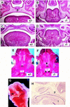
Cleft palate in Jag2ΔDSL homozygotes. (A–D) Frontal sections of E18 embryos. Sections A and B are more ventral than sections C and D. Genotypes are indicated in each panel. Note in the Jag2ΔDSL homozygote fusion of the tongue with the unelevated palatal shelves (white arrows in D). (E,F) Ventral view of stained skeletal preparations of neonatal skulls. The dorsal extent of the palatine shelves is indicated with white arrows in the heterozygote (E). In the Jag2ΔDSL homozygote (F), the palatine shelves have not grown toward and fused in the dorsal midline. The maxillary shelves are also more lateral in the Jag2ΔDSL homozygote. (G,H) Jag2 RNA expression in wild-type mouse embryos. In the head of a E10.5 mouse embryo (G), Jag2 is expressed in the epithelial cell layer of the branchial arches and the area surrounding the nasal pits. (H) Sagittal section of an E12.5 mouse embryo revealed high levels of Jag2 expression in the epithelium and muscles of the tongue, and epithelium of the palate and nasal pharynx. (m) Maxillary shelf; (p) palate; (pl) palatine shelf; (t) tongue.
Figure 3
Figure 3
Expression of Jag2 and Notch1 in limb buds of wild-type embryos. Whole mount in situ hybridization of mouse embryos at E9.5 and E10.5. (A) Prior to formation of the AER at E9.5, Jag2 is expressed throughout the limb ectoderm. (B) After AER formation at E10.5, Jag2 expression becomes restricted to the AER (arrows). (C) Low levels of Notch1 expression are also detected in the AER at E10.5 (arrows).
Figure 4
Figure 4
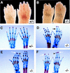
Limb defects in Jag2ΔDSL homozygotes. (A,B) Feet of neonatal mice. A forefoot (left) and hindfoot (right) is shown in each panel. Jag2ΔDSL homozygous neonates (B) exhibit syndactyly of both fore- and hindlimbs, although the hindlimbs are more severely affected. (C–F) Stained skeletal preparations of neonatal limbs of a heterozygous (C) and three Jag2ΔDSL homozygous mice (D–F). Both a fore- and a hindfoot are shown in each panel. The forefoot is on the right in C and E, and on the left in D and F. The homozygotes display several defects affecting distal phalanges, including secondary osseous fusions of both fore- and hindfeet (white arrows in D–F), primary chondrogenic fusions (black arrow in D), and terminal splitting of a distal phalanx (arrowheads in E,F).
Figure 5
Figure 5
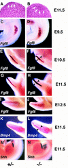
Jag2ΔDSL homozygotes have a hyperplastic AER and exhibit altered marker gene expression. (A,B) Histological analysis of forelimb buds of Jag2ΔDSL homozygous mutant embryo (B) and littermate control (A) at E11.5. The extent of the AER is indicated by the white arrows. (C–J) Time course of Fgf8 expression in the limb bud. (C,D) At E9.5, Fgf8 is expressed in a broad band in the forelimb bud ectoderm of both the Jag2ΔDSL homozygote and the littermate control. (D,E) At E10.5, the Fgf8 expression domain in the AER of the forelimb is broader in the some of the Jag2ΔDSL homozygotes than in wild-type or heterozygous littermates. (G,H) At E11.5, the Fgf8 expression domain is expanded in the AER of both fore- and hindlimbs of all Jag2ΔDSL homozygotes. (I,J) At E12.5, the Fgf8 expression remains broader in the mutants, and is also more irregular along the anterior margin of the limb bud. (K,L) Bmp4 expression at E11.5. (M,N) Shh expression at E11.5.
Figure 6
Figure 6
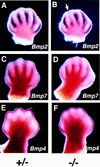
Expression of Bmp2 and Bmp7 is reduced in limbs of Jag2ΔDSL homozygotes. (A,B) Bmp2 expression. Bmp2 expression is reduced in Jag2ΔDSL homozygous mutants between the developing digits 2 and 3 (white arrow). (C,D) Bmp7 expression. Bmp7 expression is reduced in distal regions of the interdigital mesenchyme in the Jag2ΔDSL mutants. (E,F) Bmp4 expression. Bmp4 expression in distal mesenchyme is unaffected in the Jag2ΔDSL mutants. Note the altered morphology of the foot plate in the Jag2ΔDSL homozygous mutants (B,D,F). All embryos shown are at E13.5.
Figure 7
Figure 7
Visualization of interdigital cell death by Nile Blue Sulphate vital staining. Limb buds were isolated at E13.5. Plantar (ventral) apects of hindfeet are shown. Apoptotic cells that stain blue with Nile Blue Sulphate are observed between all forming digits of wild-type and heterozygous littermates, as well as in the anterior (a) and posterior (p) necrotic zones (A) In Jag2ΔDSL homozygotes (B,C), the numbers of staining cells are greatly reduced in the regions where digits 2, 3, and 4 would normally form. There is no apparent reduction of staining cells in the region between digits 1 and 2, and in the anterior and posterior necrotic zones.
Figure 8
Figure 8
Jag2ΔDSL homozygotes exhibit impaired differentiation of γδ T cells and altered thymic morphology. (A,B) FACS profiles of fetal thymocytes from a representative control littermate (A) and a Jag2ΔDSL homozygous mutant (B) assessed for CD4 vs. CD8 expression, proportion of TCRαβ+ cells in the CD4+CD8+ population, and TCRγδ+ cells in the CD4CD8 population. Broken lines denote the background staining levels that were used to establish gates for respectively assessing labeling of CD4+CD8+ and CD4CD8 cells by phycoerythrin-conjugated TCR αβ- or γδ-specific monoclonal antibodies. Annotated numbers denote the percent positive cells of the indicated phenotype. (C–E) Altered morphology of the fetal thymus in Jag2ΔDSL homozygotes at E18. (C) Thymus section from heterozygous littermate. (D,E) Thymus sections from two Jag2ΔDSL homozygotes. In the heterozygote, both darker-stained cortical regions and multiple lighter-stained medullary regions (arrows) can be observed. In the Jag2ΔDSL homozygotes, both the number and the size of the developing medullary regions are reduced.

References

    1. Artavanis-Tsakonas S, Matsuno K, Fortini ME. Notch signaling. Science. 1995;268:225–232. - PubMed
    1. Bettenhausen B, Hrabe de Angelis M, Simon D, Guenet J-L, Gossler A. Transient and restricted expression during mouse embryogenesis of Dll1, a murine gene closely related to Drosophila Delta. Development. 1995;121:2407–2418. - PubMed (V体育2025版)
    1. Blaumueller CM, Qi H, Zagouras P, Artavanis-Tsakonas S. Intracellular cleavage of Notch leads to a heterodimeric receptor on the plasma membrane. Cell. 1997;90:281–291. - "V体育平台登录" PubMed
    1. Conlon RA, Reaume AG, Rossant J. Notch1 is required for the coordinate segmentation of somites. Development. 1995;121:1533–1545. - PubMed (VSports app下载)
    1. Crossley PH, Monowada G, MacArthur CA, Martin GR. Roles for FGF8 in the induction, initiation, and maintenance of chick limb development. Cell. 1996;84:127–136. - PubMed

Publication types (V体育官网入口)

Associated data