Skip to main page content (V体育平台登录)
U.S. flag

An official website of the United States government

Dot gov

The . gov means it’s official. Federal government websites often end in . gov or . mil. Before sharing sensitive information, make sure you’re on a federal government site VSports app下载. .

Https

The site is secure. The https:// ensures that you are connecting to the official website and that any information you provide is encrypted and transmitted securely V体育官网. .

. 1997 Dec 15;11(24):3482-96.
doi: 10.1101/gad.11.24.3482.

Requirement for NF-kappaB in osteoclast and B-cell development

Affiliations

Requirement for NF-kappaB in osteoclast and B-cell development

V体育ios版 - G Franzoso et al. Genes Dev. .

"VSports注册入口" Abstract

NF-kappaB is a family of related, dimeric transcription factors that are readily activated in cells by signals associated with stress or pathogens. These factors are critical to host defense, as demonstrated previously with mice deficient in individual subunits of NF-kappaB. We have generated mice deficient in both the p50 and p52 subunits of NF-kappaB to reveal critical functions that may be shared by these two highly homologous proteins. We now demonstrate that unlike the respective single knockout mice, the p50/p52 double knockout mice fail to generate mature osteoclasts and B cells, apparently because of defects that track with these lineages in adoptive transfer experiments. Furthermore, these mice present markedly impaired thymic and splenic architectures and impaired macrophage functions. The blocks in osteoclast and B-cell maturation were unexpected VSports手机版. Lack of mature osteoclasts caused severe osteopetrosis, a family of diseases characterized by impaired osteoclastic bone resorption. These findings now establish critical roles for NF-kappaB in development and expand its repertoire of roles in the physiology of differentiated hematopoietic cells. .


Figures
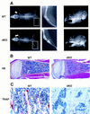
Figure 1
Figure 1
Severe osteopetrosis in p50 and p52 double knockout mice. (A) Radio-opaque long bone shafts and failure of incisor teeth to erupt in double knockout mice. Shown are whole body anterior–posterior (left panels) and skull lateral (right panels) radiographs of a 4-week-old double knockout mouse (dKO, bottom panels) and a wild-type littermate control (WT, top panels). (Middle panels) Enlargement of the boxed areas in the left panels to document the absence of a normal marrow cavity in the femur of a mutant mouse. (B) Normal marrow cavity of an 8-day-old control heterozygote mouse (WT) (p50(+/−), p52(+/−), which was indistinguishable from a true wild-type mouse) and marrow cavity from an 8-day-old double knockout (dKO) mouse, which is largely filled with unremodeled osteocartilaginous matrix. Shown are hematoxylin and eosin (HE) (B) and TRAP (C) staining of longitudinal sections through proximal tibiae. (C) Absence of TRAP+ cells in double knockout mice. A different pair of 8-day-old mutant mouse (dKO) and control littermate (WT) was used in C [WT control in C was p50(+/+), p52(+/−)], which was indistinguishable from a true wild-type animal). Osteopetrosis and lack of osteoclasts were confirmed histologically in six double knockout mice, aged 3–4 weeks. Mean values (±s.d.) are for cancellous bone volumes in sections of humeri from five dKO mice 42 ± 7% vs. 3.4 ± 1% in seven p50(+/+), p52(+/−) mice. Original magnifications were 4× (HE) and 40× (TRAP).
Figure 2
Figure 2
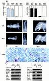
The osteopetrotic phenotype is due to a defect intrinsic to the osteoclast cell lineage. (A) Impaired development of double knockout osteoclasts in vitro. Primary osteoblasts (OBl) isolated from the calvariae of newborn wild-type (WT) or double knockout (dKO) mice were cocultured with spleen cells (Spl) isolated from either wild-type or mutant mice for 20 days on dentine slices in the presence of 10−8 m 1,25 dihydroxyvitamin D3. TRAP+ osteoclasts (left) and resorption pits (right) were counted after the culture period. Means and standard errors were calculated from six dentine slices per group. Data shown are representive of three separate experiments. (B) Osteopetrosis is cured by wild-type fetal liver transfer. Formation of radiologically clear marrow cavities within the shafts of long bones (left and middle panels) and normal tooth eruption (right panels) in adoptively transferred double knockout (dKO, bottom panels) and littermate control (WT, p50(+/−), p52(+/−), top panels) mice. Four-day-old double knockout mice and littermate controls were lethally irradiated and injected intraperitoneally (i.p.) with wild-type fetal liver cells (day 14). Whole body (left and middle panels) and skull lateral (right panels) radiographs were taken 4.5 weeks after injection. Middle panels represent an enlargement of the inset in the left panels. (C) Presence of numerous TRAP+ osteoclasts in proximal tibia sections of adoptively transferred double knockout mice. The control littermate that was adoptively transferred was a p50(+/+), p52(+/−) mouse. Mice were analyzed 3.5 weeks after fetal lever cell transfer, when their cancellous bone volumes were 7.8% and 4.7% in two rescued double knockout mice and 5.5% and 6.2% in two rescued wild-type mice. (D) Altered gene expression in double knockout mice. RT–PCR was performed on RNA extracted from thioglycollate-elicited macrophages obtained from two pairs of 3-week-old littermates (WT, p50(+/+), p52(+/−), and dKO). Cells were isolated from the peritoneal cavity, allowed to adhere to plastic, and then treated with LPS for 2 hr or left untreated, as indicated. PCR primers and animal genotypes are as indicated. Identical results were obtained in two independent experiments, and data from a representative pair of mice are shown. Data shown in the left and right panels are from two independent RT reactions (GAPDH controls at the bottom of each set).
Figure 3
Figure 3
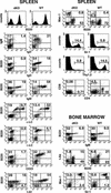
Defects of B and T lymphocytes in double knockout mice. Three-color FCM analysis of spleen and BM cell suspensions from 13- to 15-day-old double knockout mice (dKO, left panels) and p50(+/+), p52(+/−) littermate controls (WT, right panels). Two-color profiles or single-color histograms are displayed. The data are taken from analyses of three representative sets of mice, but the results were confirmed with two additional pairs. The following antibodies (listed top to bottom and left to right) were used. Spleen: B220–biotin, IgD–FITC vs. IgM–biotin; IgD-FITC vs. B220–PE; IgM–biotin vs. B220–PE; κ-light chain (LC)–FITC vs. B220–PE; λ-LC–FITC vs. B220–PE; CD23–FITC vs. B220–biotin; I-Ab-biotin vs. B220–FITC; B220–FITC vs. Mac-1–PE; Gr-1–FITC; Thy1.2–biotin; and CD4–biotin vs. CD8a–PE. Bone marrow: IgM–biotin vs. B220–PE; B220–FITC vs. class II (I-Ab)–biotin; and Gr-1–FITC vs. Mac-1–PE. Numbers reflect the percentage of positively stained cells. The percentage of B220+ BM cells was modestly increased in mutant mice, whereas class II expression was more intense in wild-type BM, even though fully mature cells were nearly absent, as judged by lack of expression of IgD (not shown).
Figure 4
Figure 4
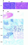
Histological abnormalities in spleens of double knockout mice. (A) Spleens of mutant mice lack white pulp. Bouin’s-fixed paraffin-embedded sections were obtained from spleens of a 10-day-old double knockout mouse (dKO, right panels) and a wild-type littermate (WT, left panels) and stained with HE, as indicated. (B) Absence of B- and T-cell areas in spleens from 2- to 4-week-old double knock-out mice (dKO) and littermate controls (WT) (right and left panels, respectively). Control littermates in A and B were p50(+/+), p52(+/−), except for macrophage marker panels, in which the control was p50(+/−), p52(+/+). Acetone-fixed, frozen sections were processed with anti-B220, anti-CD3, anti-macrophage (Mac-1 and F4/80) or anti-dendritic cell (M342) antibodies, as indicated. HRP-conjugated secondary antibodies were used, except for the B220 staining where the secondary antibody was AP conjugated. Stained cryosections from representative pairs of littermates are shown.
Figure 5
Figure 5
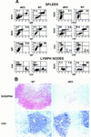
Developmental arrest of B cells is due to defects that track with to the B-cell lineage. Lethally irradiated RAG-1-deficient mice were injected with BM cells isolated from 22-day-old double knockout mice (dKO) or p50(+/+), p52(+/−) or p50(+/−), p52(+/+) (WT; these mice are indistinguishable from wild type) littermate control donor mice, as indicated. Fifteen weeks later mice were challenged i.p. with TNP–KLH (100 μg) adsorbed to alum. Mice were analyzed 9-days after challenge. (A) Three-color FCM analysis of spleen and lymph nodes of adoptively transferred RAG-1-deficient mice. Two-color profiles or single-color histograms are displayed in black and white, and genotypes of donor animals are as indicated. The following antibodies (listed top to bottom and left to right) were used. Spleen: IgM–biotin vs. B220–PE; IgD–FITC vs. B220–PE; IgD–FITC vs. IgM–biotin; CD23–PE vs. B220–biotin; I-Ab–biotin vs. B220–PE, and CD4–biotin vs. CD8a–PE. Lymph nodes: CD4–biotin vs. CD8a–PE and IgM–biotin vs. B220–PE. Numbers in the quadrants reflect the percentage of total spleen and lymph node cells in that quadrant. FCM data are representative of three paired sets of adoptively transferred RAG-1 mice. (B) Stained cryosections from representative double knockout (dKO, right panels) and p50(+/−), p52(+/+) (WT, left panels) control littermate marrow-transferred RAG-1-deficient animals are shown. Splenic cryosections were stained with PNA (brown) and anti-B220 antibodies (red) (top panels) or anti-CD-3 antibodies (blue; bottom panels), as indicated.
Figure 5
Figure 5
Developmental arrest of B cells is due to defects that track with to the B-cell lineage. Lethally irradiated RAG-1-deficient mice were injected with BM cells isolated from 22-day-old double knockout mice (dKO) or p50(+/+), p52(+/−) or p50(+/−), p52(+/+) (WT; these mice are indistinguishable from wild type) littermate control donor mice, as indicated. Fifteen weeks later mice were challenged i.p. with TNP–KLH (100 μg) adsorbed to alum. Mice were analyzed 9-days after challenge. (A) Three-color FCM analysis of spleen and lymph nodes of adoptively transferred RAG-1-deficient mice. Two-color profiles or single-color histograms are displayed in black and white, and genotypes of donor animals are as indicated. The following antibodies (listed top to bottom and left to right) were used. Spleen: IgM–biotin vs. B220–PE; IgD–FITC vs. B220–PE; IgD–FITC vs. IgM–biotin; CD23–PE vs. B220–biotin; I-Ab–biotin vs. B220–PE, and CD4–biotin vs. CD8a–PE. Lymph nodes: CD4–biotin vs. CD8a–PE and IgM–biotin vs. B220–PE. Numbers in the quadrants reflect the percentage of total spleen and lymph node cells in that quadrant. FCM data are representative of three paired sets of adoptively transferred RAG-1 mice. (B) Stained cryosections from representative double knockout (dKO, right panels) and p50(+/−), p52(+/+) (WT, left panels) control littermate marrow-transferred RAG-1-deficient animals are shown. Splenic cryosections were stained with PNA (brown) and anti-B220 antibodies (red) (top panels) or anti-CD-3 antibodies (blue; bottom panels), as indicated.
Figure 6
Figure 6
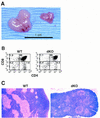
Impaired thymus glands in double knockout mice. (A) Macroscopic appearance of thymic glands obtained from a pair of 17-day-old littermates: (right) double knockout; (left) p50(+/+), p52(+/−). Original magnification was 1.3×. The dKO thymus shown is one of the larger ones seen in mutant mice. (B) FCM analysis of a thymic cell suspension from a 5-day-old double knockout mouse (dKO, right panel) and a littermate control (p50(+/+), p52(+/−); WT, left panel). CD4–biotin vs. CD8a–PE two-color profiles are displayed. Results were confirmed with two additional pairs of mice. Numbers in the quadrants reflect the percentage of total cells in that quadrant. (C) Altered microarchitecture in thymic glands of double knockout mice. Representative, Bouin’s-fixed paraffin-embedded sections were obtained from thymic glands of a 10-day-old double knockout mouse (dKO, right) and a wild-type littermate control (WT, left) and stained with HE, as indicated.
Figure 7
Figure 7
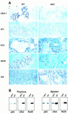
Histologic abnormalities in thymus glands of double knockout mice. (A) Thymic glands were obtained from 15- to 17-day-old double knockout mice (dKO, right) and littermate controls [WT, left; all controls were p50(+/+), p52(+/−), except for the I-Ab staining where the genotype of the control mouse was p50(+/−), p52(+/+)]. Acetone-fixed, frozen sections were processed with UEA-1 (staining MECs) or anti-MEC (4F1), anti-cortical ephitelial cell (6C3), anti-dendritic cell (M342), and anti-class II (I-Ab) antibodies, as indicated. Stained cryosections from representative pairs of littermates are shown. (B) Reduced expression of RelB and c-Rel in spleens and thymus glands of double knockout mice. Western blot analyses of thymus glands (left panels) and spleens (right panels) from 13-day-old double knockout (dKO) and control littermates [WT, p50(+/+), p52(+/−)] mice are shown. Antibodies to various NF-κB family members were used as indicated.
Figure 8
Figure 8
Model for osteoclast development indicating stage at which NF-κB appears to be required (see text).

References

    1. Agger R, Witmer-Pack M, Romani N, Stossel H, Swiggard WJ, Metlay JP, Storozynsky E, Freimuth P, Steinman RM. Two populations of splenic dendritic cells detected with M342, a new monoclonal antibody to an intracellular antigen of interdigitating dendritic cells and some B lymphocytes. J Leukocyte Biol. 1992;52:34–42. - PubMed
    1. Anderson G, Moore NC, Owen JJ, Jenkinson EJ. Cellular interactions in thymocyte development. Annu Rev Immunol. 1996;14:73–99. - PubMed
    1. Baeuerle PA, Henkel T. Function and activation of NF-κB in the immune system. Annu Rev Immunol. 1994;12:141–179. - V体育安卓版 - PubMed
    1. Baldwin AS. The NF-κB and IκB proteins: New discoveries and insights. Annu Rev Immunol. 1996;14:649–681. - PubMed
    1. Beg AA, Baltimore D. An esssential role for NF-κB in preventing TNF-α-induced cell death. Science. 1996;274:782–784. - PubMed

Publication types (VSports app下载)

Substances