Skip to main page content
U.S. flag

An official website of the United States government

Dot gov

The . gov means it’s official. Federal government websites often end in . gov or . mil VSports app下载. Before sharing sensitive information, make sure you’re on a federal government site. .

Https

The site is secure. The https:// ensures that you are connecting to the official website and that any information you provide is encrypted and transmitted securely. V体育官网.

. 1997 Nov;8(11):2119-32.
doi: 10.1091/mbc.8.11.2119.

"V体育官网" Nuclear pore complex number and distribution throughout the Saccharomyces cerevisiae cell cycle by three-dimensional reconstruction from electron micrographs of nuclear envelopes

Affiliations
Free PMC article

Nuclear pore complex number and distribution throughout the Saccharomyces cerevisiae cell cycle by three-dimensional reconstruction from electron micrographs of nuclear envelopes

M Winey et al. Mol Biol Cell. 1997 Nov.
Free PMC article

Abstract

The number of nuclear pore complexes (NPCs) in individual nuclei of the yeast Saccharomyces cerevisiae was determined by computer-aided reconstruction of entire nuclei from electron micrographs of serially sectioned cells. Nuclei of 32 haploid cells at various points in the cell cycle were modeled and found to contain between 65 and 182 NPCs. Morphological markers, such as cell shape and nuclear shape, were used to determine the cell cycle stage of the cell being examined VSports手机版. NPC number was correlated with cell cycle stage to reveal that the number of NPCs increases steadily, beginning in G1-phase, suggesting that NPC assembly occurs continuously throughout the cell cycle. However, accumulation of nuclear envelope observed during the cell cycle, indicated by nuclear surface area, is not continuous at the same rate, such that the density of NPCs per unit area of nuclear envelope peaks in apparent S-phase cells. Analysis of the nuclear envelope reconstructions also revealed no preferred NPC-to-NPC distance. However, NPCs were found in large clusters over regions of the nuclear envelope. Interestingly, clusters of NPCs were most pronounced in early mitotic nuclei and were found to be associated with the spindle pole bodies, but the functional significance of this association is unknown. .


V体育安卓版 - Figures
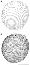
Figure 2
Figure 2
Surface of the nuclear envelope models was defined by a mesh of triangles (see MATERIALS AND METHODS). The contours (A, front only) of the S-phase nucleus shown in Figure 1C are connected by a triangular mesh (B) covering the surface defined by the contours. Bar, 0.5 μm.
Figure 5
Figure 5
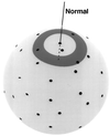
Schematic representation of the SDA protocol. An individual NPC (black dots) is chosen as the point of reference and localized by a line (normal) through the center of the NPC perpendicular to the surface of the nuclear envelope (light gray). Density is measured in a series of concentric rings (medium gray, only one is shown) of defined inner and outer radii around the normal. The area within each ring is calculated and the number of NPCs within the ring is determined by measuring distances along the surface from the normal (see MATERIALS AND METHODS). With the area and NPC numbers determined, the NPC density in each ring can be derived. This analysis is reiterated for a given nuclear envelope model by using each NPC as the center with fixed ring sizes, so that the mean NPC density in a given ring can be determined and plotted as shown in Figure 6.
Figure 6
Figure 6
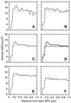
NPC SDA (see MATERIALS AND METHODS) of nuclei showing NPC density (Y-axis) as a function of distance from an NPC, averaged over all NPCs in the model or sets of models. (A) SDA for the mitotic nucleus 23 (Table 1). (B) SDA for mitotic nucleus 26 (Table 1). (C) SDA for all the mitotic nuclei (Table 1, models 19–26). (D) SDA in C (thick lines) compared with results from 40 control SDAs. A control SDA was computed by randomizing NPC positions in each of the eight mitotic nuclei and summing the resulting SDAs. The three graphs in light lines are the mean and the mean plus and minus two standard deviations. (E) The cumulative SDAs for all the G1- and S-phase nuclei (Table 1, models 1–10 and 11–18, respectively). (F) Cumulative SDA for the late anaphase nuclei (Table 1, models 27–32). The dashed lines show the mean density from 0.60 to 0.92 μm.
Figure 1
Figure 1
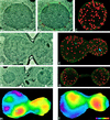
Model building of nuclear envelopes is carried out by using digitized electron micrographs of serially sectioned nuclei (A) on which the outer nuclear envelope and the position of the NPCs are manually indicated (B, outer nuclear envelope is green and NPCs are red). After modeling on images of every section of a given nucleus, the 3D model can be displayed (C). Perspective and a gradient of lighting with depth have been used to differentiate NPCs on the front and back surfaces of the model. The magenta-colored contour near the bottom of the model (C) represents the section shown in A and B. Various statistics from this model can be extracted using the IMOD program (MATERIALS AND METHODS, see Table 1). The spherical nucleus shown in A–C is from an S-phase cell (Table 1, cell 15) and is similar to the nuclei seen in all the G1- and S-phase cells. Representative digitized electron micrographs of a mitotic (D; Table 1, cell 26; arrows indicate SPBs) and a late anaphase (F; Table 1, cell 32; arrow indicates a piece of the nucleus in the neck region) are shown, as well as the models of the nuclei in these cells (E and G, respectively; blue objects in E are SPBs). Two projections (H and I) of a smoothed surface density map of the mitotic nucleus in D and E, generated as described in MATERIALS AND METHODS. The projections show the front (I) and back (H) surfaces of the model with respect to the orientation shown in E. Positions of the SPBs are indicated by gray objects (one each in H and I). The color bar (I inset) shows the colors corresponding to densities from zero (magenta) to 26.7 NPCs/μm2 (red). Scale bars, 0.5 μm.
Figure 3
Figure 3
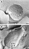
Freeze–fracture images of the nuclear envelope (see MATERIALS AND METHODS) from two different (A and B) cells, where B appears to be a mitotic nucleus. The position of a few NPCs is indicated by arrows. Bars, 0.2 μm.
Figure 4
Figure 4
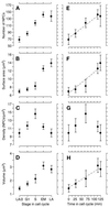
Graphs of mean NPC number (A), nuclear surface area (B), NPC density (C), and nuclear volume (D) versus cell cycle stage. The stages G1, S, early mitotic (EM), and late anaphase (LA) are defined in the text. The category LA/2 is defined by dividing the value for the late anaphase cells in two to facilitate comparison between the late anaphase and the G1 nuclei and is not significantly different from analyzing each lobe of the LA nuclei individually. The bars show 74% confidence limits, so that nonoverlapping bars indicate that the means are significantly different (p ≤ 0.05, one-tailed t test). These five cell cycle stages are also plotted (E–H) by their relative position in the cell cycle as defined by Yeh et al. (1995; see also RESULTS). For ease of plotting, the LA/2 numbers were put at time 0, and the points are then G1, S, EM, and LA, from left to right. The error bars are 95% confidence intervals to facilitate comparison between the means and the straight lines.
Figure 7
Figure 7
SDA of NPCs relative to SPBs as the center. The axes are as in Figure 6, but the scale on the Y-axis is different. The SDAs presented are cumulative for the G1- and S-phase nuclei (A, n = 4 SPBs), the early mitotic nuclei (B, n = 8 SPBs), or the late anaphase nuclei (C, n = 7 SPBs) in which SPBs could be unambiguously placed. The dashed lines show the mean density from 1.0 to 1.5 μm.

References

    1. Bucci M, Wente SR. In vivo dynamics of nuclear pore complexes in yeast. J Cell Biol. 1997;136:1185–1199. - PMC - PubMed
    1. Byers B. Cytology of the yeast life cycle. In: Strathern JN, Jones EW, Broach JR, editors. Molecular Biology of the Yeast Saccharomyces. I. Life Cycle and Inheritance. Cold Spring Harbor, NY: Cold Spring Harbor Laboratory; 1981. pp. 59–96.
    1. Byers B, Goetsch L. Behavior of spindles and spindle plaques in the cell cycle and conjugation of Saccharomyces cerevisiae. J Bacteriol. 1975;124:511–523. - PMC - PubMed
    1. Davis LI. The nuclear pore complex. Annu Rev Biochem. 1995;64:865–896. - PubMed
    1. Ding R, McDonald KL, McIntosh JR. Three-dimensional reconstruction and analysis of mitotic spindles from the yeast, Schizosaccharomyces pombe. J Cell Biol. 1993;120:141–151. - PMC - PubMed

Publication types

VSports注册入口 - MeSH terms

LinkOut - more resources