<small lang="vpeQX"></small> Skip to main page content
U.S. flag

An official website of the United States government

Dot gov

The VSports app下载. gov means it’s official. Federal government websites often end in . gov or . mil. Before sharing sensitive information, make sure you’re on a federal government site. .

Https

The site is secure. The https:// ensures that you are connecting to the official website and that any information you provide is encrypted and transmitted securely. V体育官网.

. 2022 May 24;14(11):2583.
doi: 10.3390/cancers14112583.

"VSports最新版本" Model Cell Lines and Tissues of Different HGSOC Subtypes Differ in Local Estrogen Biosynthesis

Affiliations

Model Cell Lines and Tissues of Different HGSOC Subtypes Differ in Local Estrogen Biosynthesis

Renata Pavlič et al. Cancers (Basel). .

Abstract (V体育2025版)

Ovarian cancer (OC) is highly lethal and heterogeneous. Several hormones are involved in OC etiology including estrogens; however, their role in OC is not completely understood. Here, we performed targeted transcriptomics and estrogen metabolism analyses in high-grade serous OC (HGSOC), OVSAHO, Kuramochi, COV632, and immortalized normal ovarian epithelial HIO-80 cells. We compared these data with public transcriptome and proteome data for the HGSOC tissues. In all model systems, high steroid sulfatase expression and weak/undetected aromatase (CYP19A1) expression indicated the formation of estrogens from the precursor estrone-sulfate (E1-S). In OC cells, the metabolism of E1-S to estradiol was the highest in OVSAHO, followed by Kuramochi and COV362 cells, and decreased with increasing chemoresistance. In addition, higher HSD17B14 and CYP1A2 expressions were observed in highly chemoresistant COV362 cells and platinum-resistant tissues compared to those in HIO-80 cells and platinum-sensitive tissues. The HGSOC cell models differed in HSD17B10, CYP1B1, and NQO1 expression. Proteomic data also showed different levels of HSD17B10, CYP1B1, NQO1, and SULT1E1 between the four HGSOC subtypes. These results suggest that different HGSOC subtypes form different levels of estrogens and their metabolites and that the estrogen-biosynthesis-associated targets should be further studied for the development of personalized treatment VSports手机版. .

Keywords: COV362; HIO-80; Kuramochi; OVSAHO; differentiated; high-grade serous ovarian carcinoma; immunoreactive; mesenchymal subtype; ovarian cancer; proliferative. V体育安卓版.


Conflict of interest statement

The authors declare no conflict of interest.

Figures (VSports最新版本)
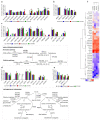
Figure 1
Figure 1
Gene expression of the (a) uptake transporters, (b) efflux transporters, (c) estrogen biosynthetic enzymes (with a schematic representation of local estrogen biosynthesis), (d) estrogen metabolic enzymes (with a schematic representation of estrogen metabolism), and (e) estrogen receptors in the HIO-80, OVSAHO, Kuramochi, and COV362 cell lines. (f) A heatmap with a dendrogram of the evaluated genes (excluding the weakly/not expressed genes CYP1A2, CYP3A5, CYP3A7, HSD3B1, HSD3B2, and SULT2A1) clustered based on Euclidean distance and Ward’s linkage. The expression of the genes of interest was evaluated in three individual experiments. Kruskal–Wallis with Dunn’s multiple comparison tests; *, p < 0.05. Data are presented as means ± SD. Normalized mRNA values for individual genes are shown in Supplementary Table S2.
Figure 2
Figure 2
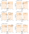
Pairwise comparison of the gene expression in the HIO-80, OVSAHO, Kuramochi, and COV362 cells, presented as volcano plots. FC, fold change; horizontal dashed line, the cutoff for experimental significance (dark orange; −log (1.3); p < 0.05); vertical dashed lines, the cutoff for genes similarly expressed in both cell lines (FC, ±2.0); vertical grey line (x = 0), genes not expressed in either cell line; red dots, differentially expressed genes; black dots, non-differentially expressed genes. Fold regulation and p values (Mann–Whitney U tests) of gene expression for individual cell pairs are presented in Supplementary Table S4.
Figure 3
Figure 3
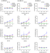
E1-S metabolism in HIO-80, OVSAHO, Kuramochi, and COV362 cells. Time courses for the estrogen metabolites E1-S, E1, E2, and E2-S following the addition of 2.3 nM E1-S (left), 8.5 nM E1-S (middle), or 85 nM E1-S (right) to the cells. Data are presented as means ± SD of two individual experiments. Several statistically significant differences (Mann–Whitney U test) in the gene expression are presented in Supplementary Table S5. Values on the graphs are presented in Supplementary Table S6.
Figure 4
Figure 4
The expression of genes for (a) uptake transporters, (b) efflux transporters, (c) estrogen biosynthetic enzymes, (d) estrogen metabolic enzymes, and (e) estrogen receptors in the HGSOC tissues. (f) A heatmap with a dendrogram of all evaluated genes clustered based on the Euclidean distance and Ward’s linkage. The data from the Ovarian Serous Cystadenocarcinoma (TCGA, PanCancer Atlas) study were downloaded from cBioPortal on 10 January 2022. Data are presented as means ± SD (n = 300). Statistically significant differences (One-way ANOVA with Bonferroni correction) are shown in Supplementary Table S7.
Figure 5
Figure 5
The expression of (a) HSD17B14 and (b) CYP1A2 in platinum-sensitive and platinum-resistant HGSOC tissues. Data from the Ovarian Serous Cystadenocarcinoma (TCGA, PanCancer Atlas) study were downloaded from cBioPortal on 10 January 2022. Data are presented as means ± SD (n (sensitive) = 105, n (resistant) = 43). Mann–Whitney U test; *, p < 0.05.
Figure 6
Figure 6
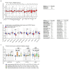
The normalized protein levels in (a) HGSOC (study IDs PDC000114, PDC000113) [33], (left) and significant differences between individual proteins (right), (b) normal fallopian tube and HGSOC tissues (study ID PDC000110 [34], left) and significant differences between individual proteins in the HGSOC tissues (right), (c) different subtypes of HGSOC (study IDs PDC000114, PDC000113) [33]). All the data were downloaded from the NCI, PDC server on 12 January 2022 and are shown as mean ± SD. One-way ANOVA with Bonferroni correction (a,b) and Tukey’s tests (c); *, p < 0.05; **, p < 0.01; ***, p < 0.001; FC, fold change; bold, differences that are more important for interpretation; »>« denotes »levels are higher than«.

References

    1. Sung H., Ferlay J., Siegel R.L., Laversanne M., Soerjomataram I., Jemal A., Bray F. Global Cancer Statistics 2020: GLOBOCAN Estimates of Incidence and Mortality Worldwide for 36 Cancers in 185 Countries. CA A Cancer J. Clin. 2021;71:209–249. doi: 10.3322/caac.21660. - "VSports" DOI - PubMed
    1. Stewart C., Ralyea C., Lockwood S. Ovarian Cancer: An Integrated Review. Semin. Oncol. Nurs. 2019;35:151–156. doi: 10.1016/j.soncn.2019.02.001. - DOI - PubMed
    1. Desai A., Xu J., Aysola K., Qin Y., Okoli C., Hariprasad R., Chinemerem U., Gates C., Reddy A., Danner O., et al. Epithelial ovarian cancer: An overview. World J. Transl. Med. 2014;3:1–8. doi: 10.5528/wjtm.v3.i1.1. - DOI (VSports最新版本) - PMC - PubMed
    1. Kossaï M., Leary A., Scoazec J.Y., Genestie C. Ovarian Cancer: A Heterogeneous Disease. Pathobiol. J. Immunopathol. Mol. Cell. Biol. 2018;85:41–49. doi: 10.1159/000479006. - DOI - PubMed
    1. Vaughan S., Coward J.I., Bast R.C., Jr., Berchuck A., Berek J.S., Brenton J.D., Coukos G., Crum C.C., Drapkin R., Etemadmoghadam D., et al. Rethinking ovarian cancer: Recommendations for improving outcomes. Nat. Rev. Cancer. 2011;11:719–725. doi: 10.1038/nrc3144. - DOI - PMC - PubMed