Skip to main page content
U.S. flag

An official website of the United States government

Dot gov

The . gov means it’s official. Federal government websites often end in . gov or . mil VSports app下载. Before sharing sensitive information, make sure you’re on a federal government site. .

Https

The site is secure. The https:// ensures that you are connecting to the official website and that any information you provide is encrypted and transmitted securely V体育官网. .

. 2022 May;19(5):547-553.
doi: 10.1038/s41592-022-01461-y. Epub 2022 May 2.

"VSports最新版本" NEAT-seq: simultaneous profiling of intra-nuclear proteins, chromatin accessibility and gene expression in single cells

Affiliations

NEAT-seq: simultaneous profiling of intra-nuclear proteins, chromatin accessibility and gene expression in single cells

Amy F Chen et al. Nat Methods. 2022 May.

Abstract

In this work, we describe NEAT-seq (sequencing of nuclear protein epitope abundance, chromatin accessibility and the transcriptome in single cells), enabling interrogation of regulatory mechanisms spanning the central dogma VSports手机版. We apply this technique to profile CD4 memory T cells using a panel of master transcription factors (TFs) that drive T cell subsets and identify examples of TFs with regulatory activity gated by transcription, translation and regulation of chromatin binding. We also link a noncoding genome-wide association study single-nucleotide polymorphism (SNP) within a GATA motif to a putative target gene, using NEAT-seq data to internally validate SNP impact on GATA3 regulation. .


Conflict of interest statement

Competing Interests

The remaining authors declare no competing interests.

"V体育官网入口" Figures
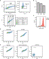
Extended Data Fig. 1
Extended Data Fig. 1. Staining of nuclear and cytosolic proteins using oligo-antibodies blocked with EcoSSB.
a) Sorting of cells expressing low, mid, or high levels of nuclear GFP that have been stained with an anti-GFP oligo-conjugated antibody. b) Quantitative PCR for the conjugated oligo from equal cell numbers of sorted populations in (a) for n = 2 technical replicates. c) Staining of K562 cells and mouse ESCs for endogenous GATA1 protein using an anti-GATA1 antibody linked to an 80bp oligo with 3’-Cy5. d) Flow cytometry plot of HEK293T cells expressing cytosolic GFP and stained with an anti-GFP antibody linked to a 100 bp single stranded DNA oligo with 3’-Cy5 modification. e) Flow cytometry plot of nuclear GFP-expressing HEK293T cells with a GFP antibody linked to a Cy5-modified ssDNA oligo using the conditions indicated. “NEAT-seq”: NEAT-seq fixation, permeabilization, and staining conditions using oligo-antibodies pre-incubated with EcoSSB. “Dextran sulfate block”: NEAT-seq fixation and permeabilization conditions with inCITE-seq staining conditions (i.e with dextran sulfate blocking agent). “inCITE-seq”: inCITE-seq fixation, permeabilization, and staining conditions. Spearman correlation is shown.
Extended Data Fig. 2
Extended Data Fig. 2. NEAT-seq species mixing experiment data quality.
a-b) Cutoffs for annotating a cell as human, mouse, or mixed based on percentage of reads in a cell mapping to the human genome for ATAC-seq (a) and RNA-seq (b) data. c) Scatterplot of number of ATAC-seq reads mapping to the human vs mouse genome in each cell prior to removing HTO doublets, with each cell colored by its classification as a human cell, mouse cell, mixed species doublet, or an HTO doublet. d) Same as (c) but for RNA-seq reads. e) Fragment length distribution of ATAC-seq data generated using NEAT-seq. f) Average Tn5 insertions across transcriptional start sites normalized to the flanking region +/− 2kb from the start site (i.e TSS enrichment) from scATAC-seq data generated using NEAT-seq. g) Log-transformed Tn5 insertions in ATAC-seq peaks for NEAT-seq data vs bulk ATAC-seq data in K562 cells. Pearson correlation is shown. h) A comparison of RNA-seq counts from bulk data vs NEAT-seq in K562 cells (left) or standard 10X Multiome data in GM12878 cells (right). Values are log-transformed TPM (for single cell data) or FPKM (for bulk data). Pearson correlation is shown. i) Western blot of mESC and K562 cell lysate using oligo-conjugated SOX2 antibody pre-incubated with EcoSSB and detected with IR800 secondary antibody (Licor). GAPDH was also probed as a loading control with IR700 secondary antibody. Imaging was performed on a Licor Odyssey imaging system. Images shown are representative results of 2 independent experiments.
Extended Data Fig. 3
Extended Data Fig. 3. CD4 memory T cell subset annotations.
a) scRNA-seq UMAP of CD4 memory T cells colored with scATAC-seq cluster IDs. TCM = central memory, Act. = recently activated cells, Uncom. = uncommitted memory cells. b) RNA expression of master TF drivers and canonical cell surface markers of CD4 memory cell subsets in each scATAC-seq cluster. TBX21 = Tbet transcript, RORC = RORγT transcript, IKZF2 = Helios transcript. c) Gene accessibility for cytokines induced in different CD4 T cell subsets overlayed on the scATAC-seq UMAP. d) RNA levels for the cytokines in (c). e) chromVAR deviation scores for the naïve and CM T cell TFs, LEF1 and TCF7, overlayed on scATAC-seq UMAP. f) RNA expression of the CM marker, CCR7, overlayed on the scATAC-seq UMAP.
Extended Data Fig. 4
Extended Data Fig. 4. Enrichment of TF ADTs in the T cell subtype driven by the TF.
a) Unsmoothed ADT counts for each TF overlayed on the scATAC-seq UMAP after the indicated normalization method (raw counts, NPC normalization, and total ADT counts normalization). b) Log2-transformed, NPC-normalized ADT counts for each TF separated by scATAC-seq cluster for cells stained with antibody concentration 1 (see methods). c) Same as (b) but for antibody concentration 2. d) Scatterplots with marginal histograms of log2-transformed read-normalized RNA vs log2-transformed NPC-normalized ADT counts for each TF. Colored data points represent cells belonging to the scATAC-seq cluster most enriched in expression of the indicated TF. e) Unsmoothed, normalized RNA counts of the indicated TFs overlayed on the scATAC-seq UMAP.
Extended Data Fig. 5
Extended Data Fig. 5. Correlation of ADT levels with gene locus accessibility, RNA, and motif accessibility for each TF.
a) Correlation across all cells for each measurement. Values were first smoothed across neighboring cells using MAGIC imputation to account for dropouts. Pearson correlations are shown. b) The data in (a) but averaged across cells within each scATAC-seq cluster.
Extended Data Fig. 6
Extended Data Fig. 6. Post-transcriptional regulation of GATA3.
a) Flow cytometry plots of GATA3-transfected and WT HEK293T cells stained with an EcoSSB-bound oligo-conjugated GATA3 antibody, using a fluorescent secondary antibody for detection. b) Log2-transformed RNA levels of the indicated gene in cells expressing high RNA and low protein (“low”) vs high RNA and high protein for GATA3 (“high”). The mean RNA expression for each group is shown above the violin plot. N = 140 cells examined over one independent experiment for both “high” and “low” populations. Boxplots show median with bounds of the box representing the 25th and 75th percentiles and the whiskers extending to the value closest to but not exceeding 1.5 times the interquartile range. Data extending beyond the whiskers are plotted individually as outliers.
Extended Data Fig. 7
Extended Data Fig. 7. Identification of DNA regulatory elements and genes correlated with master TF protein expression.
Spearman correlations between ATAC-seq peak accessibility and NPC-normalized TF ADT counts across single cells. Cutoffs for significant correlations are indicated by dashed lines (see Methods). Points in red indicate peaks containing a binding motif for the TF. TF motif enrichment in significantly correlated peaks was calculated using a hypergeometric test. b) Spearman correlations between read-normalized RNA counts and NPC-normalized TF ADT counts across single cells. Cutoffs for significant correlations are indicated by dashed lines (see Methods). Significantly correlated genes known to be enriched or play a functional role in the relevant T cell subset are labeled. c) CCR4 ATAC-seq tracks in CD4 memory cells separated into quintiles by GATA3 ADT levels, along with significantly correlated peak-gene linkages (adj. p < 0.05). Spearman correlations are shown. Peaks containing a GATA3 motif are indicated. d-e) CCR4 RNA expression (d) and accessibility at the highlighted GATA3 motif-containing peak (e) as a function of GATA3 ADT levels. Mean is shown with standard error of the mean of n = 768 +/− 1 cells per group.
Extended Data Fig. 8
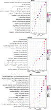
Extended Data Fig. 8. GO term enrichment for candidate target genes in TF-driven peak-gene linkages.
Enriched GO terms in the target gene list were identified by hypergeometric test using enrichGO in the clusterProfiler R package, using all genes with at least one RNA count in the dataset as a background gene list. Adjusted p-values were calculated using the Benjamini-Hochberg procedure.
Extended Data Fig. 9
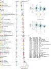
Extended Data Fig. 9. Single-tissue eQTL analysis for the rs62088464-TSEN54 variant-gene pair from GTEx portal.
a) Normalized effect size (NES) across various tissues for the protective (G) vs risk (A) allele on TSEN54 expression. The risk allele preserves the GATA3 motif. A negative NES value indicates a gene with expression that is associated with the risk allele. Error bars indicate 95% confidence intervals. b) Normalized TSEN54 expression grouped by rs62088464 genotype for lung and esophagus mucosa. c) Genes ordered by NES across tissues for rs62088464. P-values for (b) and (c) are outputs from the GTEx portal (release v8) and are calculated from a two-sided t-test comparing the observed NES in a tissue to a null NES of 0.
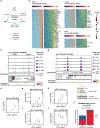
Fig. 1:
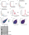
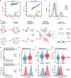
Fig. 1:. An intra-nuclear staining protocol using oligo-antibodies enables simultaneous profiling of nuclear protein, chromatin accessibility and RNA transcripts in single cells.
a) Flow cytometry plot of HEK293T cells expressing nuclear-localized GFP and stained with an anti-GFP antibody linked to an 80 bp single stranded DNA oligo with 3’-Cy5 modification. Spearman correlation is shown. b) Schematic of NEAT-seq workflow. c) Distribution of centered log ratio (CLR) transformed counts of HTOs from anti-NPC antibodies (HTO5–9). d) Scatterplot of number of reads mapping to the human vs mouse genome in each cell after removing HTO doublets, with each cell colored by its classification as a human cell, mouse cell, or mixed species doublet. e-g) Distribution of RNA UMIs (e), genes (f), and ATAC-seq fragments (g) per cell. Boxplots show median with bounds of the box representing the 25th and 75th percentiles and the whiskers extending to the value closest to but not exceeding 1.5 times the interquartile range. Outliers extending beyond the whiskers are not shown. h) CLR-transformed counts of ADTs corresponding to GATA1, OCT4, and SOX2 in cells classified as human or mouse cells based on mapping of ATAC-seq and RNA-seq reads to each genome.
Fig. 2:
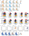
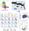
Fig. 2:. Profiling of CD4 memory T cells using NEAT-seq reveals modes of regulation across the central dogma.
a) scATAC-seq UMAP of CD4 memory T cells with cell type classifications. Annotations with cell numbers in parentheses: Th1 (1562), Th2 (939), Th17 (1855), Treg (583), TCM = central memory (2512), Act. = recently activated cells (116), Uncom. = uncommitted memory cells (905). b) Top enriched motifs in peaks that are more accessible in each cluster. c) Plots on scATAC UMAP of TF chromVAR deviations (motif accessibility), accessibility surrounding the TF gene locus (gene accessibility), RNA levels, and protein levels as measured by ADTs for the indicated TFs. n = 8472 cells in each plot except for ADTs, where n = 3841 cells. d) Scatterplot of log2-transformed, normalized RNA vs ADT counts for GATA3 with cutoffs shown for high RNA, high protein, and low protein indicated. e) Differentially expressed genes between cells with high RNA and high protein vs high RNA and low protein for GATA3 based on a two-sided Wilcoxon rank sum test. Adjusted p-values indicate Benjamini-Hochberg corrected values. Points in red represent genes with adjusted p-value < 0.05 and log2 fold change > 0.5.
Fig. 3:
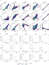
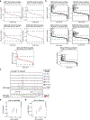
Fig. 3:. Identification of peak-gene linkages associated with master TF protein expression.
a) Diagram showing criteria used to identify candidate TF-peak-gene linkages for each TF. b) Heatmaps of peak-gene linkages correlated with abundance of the indicated TF identified based on criteria in (a). Genes that are in the top 200 genes significantly enriched in the T cell subset driven by the corresponding TF based on data by Schmiedel et al. are labeled. c) CCR6 ATAC-seq tracks in CD4 memory cells separated into quintiles by RORgT ADT levels, along with significantly correlated peak-gene linkages (adj. p < 0.05). Spearman correlations are shown. Peaks containing a RORgT motif are indicated. d-f) CCR6 RNA expression (d) and accessibility at the highlighted RORgT motif-containing peaks (e-f) as a function of RORgT ADT levels. Mean is shown with standard error of the mean of n = 768 +/− 1 cells per group. g) TSEN54 ATAC-seq tracks as in (c), but for GATA3 ADT, with the indicated GWAS SNP location highlighted. h-i) Accessibility at the highlighted SNP-containing peak (h) and TSEN54 RNA expression (i) as a function of GATA3 ADT levels. Mean is shown with standard error of the mean, n = approximately 768 cells per group. j) Read-normalized Tn5 insertions per cell per million reads mapping to the risk vs protective allele in cells above the 90th percentile vs cells below the 50th percentile of GATA3 ADT levels. P-value is calculated using a one-sided binomial test on T:C ratio of insertion counts.

VSports最新版本 - References

    1. Ma S. et al. Chromatin Potential Identified by Shared Single-Cell Profiling of RNA and Chromatin. Cell 183, 1103–1116.e20 (2020). - PMC - PubMed
    1. Stoeckius M. et al. Simultaneous epitope and transcriptome measurement in single cells. Nat. Methods 14, 865–868 (2017). - PMC (V体育官网入口) - PubMed
    1. Swanson E. et al. Simultaneous trimodal single-cell measurement of transcripts, epitopes, and chromatin accessibility using TEA-seq. Elife 10, (2021). - PMC (V体育官网入口) - PubMed
    1. Mimitou EP et al. Scalable, multimodal profiling of chromatin accessibility, gene expression and protein levels in single cells. Nat. Biotechnol (2021) doi:10.1038/s41587-021-00927-2. - DOI - PMC - PubMed
    1. Gerlach JP et al. Combined quantification of intracellular (phospho-)proteins and transcriptomics from fixed single cells. Sci. Rep 9, 1469 (2019). - PMC - PubMed

Methods References

    1. Bujalowski W. & Lohman TM Escherichia coli single-strand binding protein forms multiple, distinct complexes with single-stranded DNA. Biochemistry 25, 7799–7802 (1986). - PubMed
    1. Lohman TM & Overman LB Two binding modes in Escherichia coli single strand binding protein-single stranded DNA complexes. Modulation by NaCl concentration. J. Biol. Chem 260, 3594–3603 (1985). - V体育官网入口 - PubMed
    1. Reddy MS, Guhan N. & Muniyappa K. Characterization of single-stranded DNA-binding proteins from Mycobacteria. The carboxyl-terminal of domain of SSB is essential for stable association with its cognate RecA protein. J. Biol. Chem 276, 45959–45968 (2001). - PubMed (V体育官网入口)
    1. Parks Benjamin. GreenleafLab/matcha. "V体育2025版" https://github.com/GreenleafLab/matcha.
    1. Stuart T. et al. Comprehensive Integration of Single-Cell Data. Cell 177, 1888–1902.e21 (2019). - PMC - PubMed

Publication types