"V体育官网入口" Skip to main page content
U.S. flag

An official website of the United States government

Dot gov

The VSports app下载. gov means it’s official. Federal government websites often end in . gov or . mil. Before sharing sensitive information, make sure you’re on a federal government site. .

Https

The site is secure. The https:// ensures that you are connecting to the official website and that any information you provide is encrypted and transmitted securely V体育官网. .

. 2021 Jun 23;13(13):3145.
doi: 10.3390/cancers13133145.

Telomerase and Pluripotency Factors Jointly Regulate Stemness in Pancreatic Cancer Stem Cells

Affiliations

Telomerase and Pluripotency Factors Jointly Regulate Stemness in Pancreatic Cancer Stem Cells (V体育2025版)

Karolin Walter et al. Cancers (Basel). .

Abstract

To assess the role of telomerase activity and telomere length in pancreatic CSCs we used different CSC enrichment methods (CD133, ALDH, sphere formation) in primary patient-derived pancreatic cancer cells. We show that CSCs have higher telomerase activity and longer telomeres than bulk tumor cells. Inhibition of telomerase activity, using genetic knockdown or pharmacological inhibitor (BIBR1532), resulted in CSC marker depletion, abrogation of sphere formation in vitro and reduced tumorigenicity in vivo. Furthermore, we identify a positive feedback loop between stemness factors (NANOG, OCT3/4, SOX2, KLF4) and telomerase, which is essential for the self-renewal of CSCs. Disruption of the balance between telomerase activity and stemness factors eliminates CSCs via induction of DNA damage and apoptosis in primary patient-derived pancreatic cancer samples, opening future perspectives to avoid CSC-driven tumor relapse. In the present study, we demonstrate that telomerase regulation is critical for the "stemness" maintenance in pancreatic CSCs and examine the effects of telomerase inhibition as a potential treatment option of pancreatic cancer. This may significantly promote our understanding of PDAC tumor biology and may result in improved treatment for pancreatic cancer patients VSports手机版. .

Keywords: cancer stem cells; pancreatic cancer; self-renewal; stemness; telomerase; telomere length. V体育安卓版.


Conflict of interest statement

The authors declare no conflict of interest.

Figures
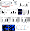
Figure 1
Figure 1
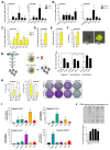
Telomerase activity stabilizes telomere length in pancreatic CSCs: (A) telomere length in primary PDAC cells (red dot) with respect to patient age measured by FlowFISH; (B) schematic illustration showing the CSC enrichment methods for CD133 and ALDEFLUOR via FACS or in sphere culture; (CE) RT-qPCR analysis of TERT and TERF1 mRNA levels in primary pancreatic cancer stem cells enriched and selected by FACS for CD133 (C), or ALDEFLUOR (D), and cultured as spheres (E) (CSCs) vs. corresponding control (non-CSCs) (n = at least 3 independent experiments). (F) Immunofluorescence staining and quantification for TERT (red) in CD133− and CD133+ FACSorted cells (n = 5 independent experiments). Cells were counterstained with DAPI (nuclear marker, blue), 4× magnification is shown. (GI) Telomerase activity measurement in CD133 (G) or ALDEFLUOR (H) negative vs. positive cells and in adherent vs. sphere cell-cultures (I) (n = 3 independent FACSortings and sphere culture experiments). (J) Representative pictures of Q-FISH with telomeres (green) and DAPI (blue) and violin plot showing telomere length analysis in CD133 negative (non-CSCs) and positive cells (CSCs) 40× magnification is shown. The mean is depicted in numbers and as black line. n ≥ 3 from independent FACSortings with each >150 measurements per group. In (CJ), data are presented as mean ± SEM. * p ≤ 0.05 (Mann–Whitney-U test).
Figure 2
Figure 2
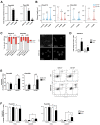
Interplay between pluripotency/stemness factors and telomerase activity in pancreatic CSCs: (A,B) RT-qPCR analysis of pluripotency/stemness-associated genes in primary pancreatic CSCs enriched by CD133 expression (A) or ALDEFLUOR activity (B) vs. CD133− and ALDEFLUOR- non-CSCs (n = at least 3 independent FACSortings). (CF) Quantitative RT-PCR (C), telomerase activity (D), flow cytometry analysis for CD133 (E) and sphere formation capability (F), compared in NANOG negative cells (white) vs. NANOG positive cells (yellow) using a NANOG-YNL reporter. (G) Schematic illustration and quantification on the production of virions in parental pseudorabies viruses (PRV-NIA3 and vBecker) compared to telomerase activity-dependent virus production (PRV-TER) in adherent HPDE and Panc185 cells as compared to Panc185 spheres. (H) Clonogenic Assay in NANOG-YNL positive and negative cells after withdrawing BIBR1532 (n = ≥3 independent experiments. ns = not statistically significant) (I) Gene expression levels of stemness/pluripotency genes and TERT in HEK293T cells after inducing expression of SOX2, OCT3/4, KLF4 and NANOG, compared to the empty vector control (n = 4 independent experiments). In (AI), data are represented as mean ± SEM. * p ≤ 0.05 (Mann–Whitney-U test). (J) Telomerase activity after inducing expression of SOX2, OCT3/4, KLF4 and NANOG, compared to the empty vector control. Representative TRAP assays and quantification are depicted.
Figure 3
Figure 3
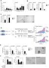
Targeting telomerase activity with a small molecule inhibitor (BIBR1532): (A,B) The effects of BIBR1532 treatment on telomerase activity (A), and telomere length (B) in CD133− cells (blue) and in the CD133+ CSC population (red) were quantified. For illustrative purposes, the telomere length measurements of Figure 1H were re-used here (indicated by transparent color). The mean is depicted in numbers and as black line, >150 measurements per group. (C) Quantification of gH2AX foci (>50 cells per group) in CD133− and CD133+ cells after 7 days vehicle or BIBR1532 treatment. Quantification and representative pictures of immunofluorescence staining of gH2AX foci are provided. (D) Senescent cells quantified by flow cytometry staining for SA-beta-Gal in CD133− (non-CSCs) and CD133+ (CSCs) after BIBR1532 or solvent treatment (7 days). (E,F) Apoptosis quantified by flow cytometry using double staining for CD133 and AnnexinV or (E) Caspase3/7 (F). In (AF), data are represented as mean ± SEM. n = 3 independent experiments. * p ≤ 0.05 (Mann–Whitney-U test), ns = not statistically significant.
Figure 4
Figure 4
Telomerase inhibition as treatment strategy for pancreatic cancer (stem) cells: (A) Effects of the small molecule telomerase inhibitor BIBR1532 on the expression of TERT and pluripotency-associated genes as measured by RT-qPCR (n = 4 independent experiments). (B) Flow cytometry analyses for CD133 or ALDEFLUOR after BIBR1532 or solvent treatment (n = 3 independent experiments). (C) Quantification and representative pictures of spheres after BIBR1532 treatment (n = 3 independent experiments). (D) Schematic overview of BIBR1532 pre-treatment and in vivo experiment, number of tumorigenic cells within the whole population shown as cancer stem cell (CSC) frequencies as determined by extreme limiting dilution assays (ELDA) in nude mice, and tumor volume measured after injection of 100,000 BIBR1532 or solvent treated Panc215 and Panc354 cells (n ≥ 4 mice for each group). (E) Viability of PDX-derived organoids treated with gemcitabine and BIBR1532 at the indicated concentrations. (F) Quantification and representative pictures of sphere formation assays after single agent or combination treatment with gemcitabine, Olaparib, and BIBR1532 (n = 5 independent experiments). In (AC,E,F) data are represented as mean ± SEM. * p ≤ 0.05 (Mann–Whitney-U test). In (D) data are represented as mean ± SEM. * p ≤ 0.05 (Area under the curve).
Figure 5
Figure 5
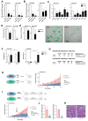
Knockdown of TERT diminishes pancreatic cancer stem cells: (A,B) Gene expression of TERT (A) and telomerase activity (B) in Panc215 and Panc354 cells transduced with shRNA-340159 and shRNA-340160 compared to scrambled (shSCR) control. (C) Expression levels of stemness/pluripotency-associated genes using shRNA-340160 (shTERT) for TERT mediated knockdown in Panc215 and Panc354 cells. (D,E) Flow cytometry analyses for CD133 (D) and sphere formation (E) upon TERT knock-down (KD) compared to scrambled (shSCR) control. (F) Quantification of apoptosis in shSCR and shTERT transduced cells (Panc215 and Panc354) using flow cytometry analysis for AnnexinV (n = at least 3 independent experiments). (G) In vivo tumor-initiating potential with CSC frequencies (number of tumorigenic cells within the whole population) as determined by ELDA in nude mice after TERT-KD compared to scrambled control (n = 8 animals); (H) Schematic illustration of in vivo experiment with cells carrying an inducible TERT-KD (Dox shTERT) or scrambled (Dox SCR) control construct. Cells were treated with doxycycline (DOX) 7 days before s.c. xenografting in nude mice. Graph shows time-dependent growth of subcutaneously engrafted tumors (n = 4 mice per group). (I) Schematic illustration of in vivo experiment and visual representation of time-dependent tumor growth of subcutaneously (s.c.) engrafted tumors arising from TERT-KD and SCR cells, over the course of doxycycline treatment 28 days after s.c. xenografting in nude mice (n = 6 mice per group). Tumor growth is depicted until the first control mice had to be sacrificed. 4× magnification is shown (J) Gene expression of TERT and telomerase activity compared in TERT-KD and SCR tumors (induced with DOX at day 28) after sacrificing mice 43 days after xenografting. (K) representative H&E staining of at day 43 explanted TERT-KD and scrambled tumors (induced with DOX at day 28). In (AG,J) data are represented as mean ± SEM. * p ≤ 0.05 (Mann–Whitney-U test). In (H,I) data are represented as mean ± SEM. * p ≤ 0.05 (Area under the curve).

References (V体育安卓版)

    1. Rahib L., Smith B.D., Aizenberg R., Rosenzweig A.B., Fleshman J.M., Matrisian L.M. Projecting cancer incidence and deaths to 2030: The unexpected burden of thyroid, liver, and pancreas cancers in the United States. Cancer Res. 2014;74:2913–2921. doi: 10.1158/0008-5472.CAN-14-0155. - DOI - PubMed
    1. Siegel R.L., Miller K.D., Jemal A. Cancer statistics, 2015. CA Cancer J. Clin. 2015;65:5–29. doi: 10.3322/caac.21254. - DOI - PubMed
    1. Von Hoff D.D., Ervin T., Arena F.P., Chiorean E.G., Infante J., Moore M., Seay T., Tjulandin S.A., Ma W.W., Saleh M.N., et al. Increased survival in pancreatic cancer with nab-paclitaxel plus gemcitabine. N. Engl. J. Med. 2013;369:1691–1703. doi: 10.1056/NEJMoa1304369. - DOI - PMC - PubMed
    1. Conroy T., Desseigne F., Ychou M., Bouche O., Guimbaud R., Becouarn Y., Adenis A., Raoul J.L., Gourgou-Bourgade S., de la Fouchardiere C., et al. FOLFIRINOX versus gemcitabine for metastatic pancreatic cancer. N. Engl. J. Med. 2011;364:1817–1825. doi: 10.1056/NEJMoa1011923. - DOI - PubMed
    1. Hermann P.C., Huber S.L., Herrler T., Aicher A., Ellwart J.W., Guba M., Bruns C.J., Heeschen C. Distinct populations of cancer stem cells determine tumor growth and metastatic activity in human pancreatic cancer. Cell Stem Cell. 2007;1:313–323. doi: 10.1016/j.stem.2007.06.002. - DOI - PubMed