Skip to main page content
U.S. flag

An official website of the United States government

Dot gov

The . gov means it’s official. Federal government websites often end in . gov or VSports app下载. mil. Before sharing sensitive information, make sure you’re on a federal government site. .

Https

The site is secure. The https:// ensures that you are connecting to the official website and that any information you provide is encrypted and transmitted securely. V体育官网.

. 2021 Jan 15;81(2):384-399.
doi: 10.1158/0008-5472.CAN-20-1488. Epub 2020 Nov 10.

VSports手机版 - Frizzled-7 Identifies Platinum-Tolerant Ovarian Cancer Cells Susceptible to Ferroptosis

Affiliations

Frizzled-7 Identifies Platinum-Tolerant Ovarian Cancer Cells Susceptible to Ferroptosis (V体育官网)

Yinu Wang et al. Cancer Res. .

Abstract

Defining traits of platinum-tolerant cancer cells could expose new treatment vulnerabilities VSports手机版. Here, new markers associated with platinum-tolerant cells and tumors were identified using in vitro and in vivo ovarian cancer models treated repetitively with carboplatin and validated in human specimens. Platinum-tolerant cells and tumors were enriched in ALDH+ cells, formed more spheroids, and expressed increased levels of stemness-related transcription factors compared with parental cells. Additionally, platinum-tolerant cells and tumors exhibited expression of the Wnt receptor Frizzled-7 (FZD7). Knockdown of FZD7 improved sensitivity to platinum, decreased spheroid formation, and delayed tumor initiation. The molecular signature distinguishing FZD7+ from FZD7- cells included epithelial-to-mesenchymal (EMT), stemness, and oxidative phosphorylation-enriched gene sets. Overexpression of FZD7 activated the oncogenic factor Tp63, driving upregulation of glutathione metabolism pathways, including glutathione peroxidase 4 (GPX4), which protected cells from chemotherapy-induced oxidative stress. FZD7+ platinum-tolerant ovarian cancer cells were more sensitive and underwent ferroptosis after treatment with GPX4 inhibitors. FZD7, Tp63, and glutathione metabolism gene sets were strongly correlated in the ovarian cancer Tumor Cancer Genome Atlas (TCGA) database and in residual human ovarian cancer specimens after chemotherapy. These results support the existence of a platinum-tolerant cell population with partial cancer stem cell features, characterized by FZD7 expression and dependent on the FZD7-β-catenin-Tp63-GPX4 pathway for survival. The findings reveal a novel therapeutic vulnerability of platinum-tolerant cancer cells and provide new insight into a potential "persister cancer cell" phenotype. SIGNIFICANCE: Frizzled-7 marks platinum-tolerant cancer cells harboring stemness features and altered glutathione metabolism that depend on GPX4 for survival and are highly susceptible to ferroptosis. .


Conflict of interest statement

Conflicts of Interest: The authors have no conflicts of interest to declare.

Figures

Figure 1.
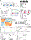
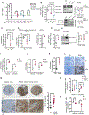
Figure 1.. Stemness and ferroptosis signatures are enriched in Pt-T OC cells.
(A) Representative FACS side scatter analysis of the ALDH(+) population (left), and percentage (mean ± SD, n=5) of ALDH(+) cells (right) in parental (WT), cisplatin tolerant (CDDP), and carboplatin tolerant (Carbo) SKOV3 cells. (B) Representative images (top), and numbers (mean ± SD, n=4) of spheroids (bottom) formed by 1000 parental (WT), CDDP, and carboplatin tolerant (Carbo) SKOV3 cells after 7 days of culture under non-attachment conditions. (C) mRNA levels (fold-change ± SD, n=3) of stemness-related TFs (Sox2, Oct4 and Nanog) measured by real-time RT-PCR in CCDP and Carbo tolerant vs. parental SKOV3 cells (WT). (D) GSEA shows upregulated stemness pathway (Stem Cell_UP) in SKOV3 CDDP-tolerant vs. parental cells (FDR=3.22E-04). (E, F) FACS side scatter analysis of percentage of ALDH(+) cells (E), and fold-change (mean ± SD, n=3) ALDH1A1, Nanog, and Oct4 mRNA expression levels (F) in OVCAR3 xenografts treated with PBS (Ctrl) or carboplatin (Carbo). (G) Hierarchical clustering heatmap for DEG (FDR<0.05) ferroptosis-related genes in SKOV3_CDDP vs. control cells (n=3 replicates/group). (H) Representative pictures of a colony formation assay (left), and numbers (mean ± SD, n=3) of colonies (right) developed from 4000 cells isolated from Pt-R human tumors treated with DMSO (Ctrl), ML210 (500nM), DFOA (800nM), or ML210 plus DFOA for 24 hours. (I) Fluorescence histograms (top), and mean (± SD, n=3) fluorescence (bottom) of BODIPY 581/591-C11 staining show lipid peroxidation in SKOV3 and OVCAR5 parental (WT) cells and cisplatin tolerant (CDDP) cells treated with DMSO or ML210 (1 uM) for 20 hours. (J) Survival curves for WT, cisplatin tolerant (CDDP) and carboplatin tolerant (Carbo) SKOV3 cells in response to cisplatin (left) or GPX4 inhibitor ML210 (right). Cisplatin and ML210 IC50 values are shown. (K-L) Survival curves of cells from primary HGSOC tumors treated with cisplatin (left), or GPX4 inhibitor ML210 (right). IC50 for cisplatin and ML210 are shown. For all comparisons: *P<0.05, **P<0.01, ***P<0.001.
Figure 2.
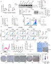
Figure 2.. Frizzled 7 (FZD7) is upregulated in Pt-T OCs.
(A) Fold-change (mean ± SD, n=3) of FZD7 mRNA expression levels measured by real-time RT-PCR in cisplatin tolerant (CDDP) compared with parental OVCAR3, OVCAR5, SKOV3 cells. (B) Western blotting for FZD7 in parental and Pt-T SKOV3, OVCAR5 and OVCAR3 cells and Pt-T (-CDDP). Quantification shows fold change of FZD7 expression (n=3 experiments). (C-D) FZD7 mRNA expression levels (mean fold-change ± SD, n = 3) (C), and FZD7 protein levels measured by western blotting (D; including quantification in 3 experiments) in Pt-R PEO4 vs. PEO1 cells. (E) Fold-change (mean ± SD) of FZD7 mRNA levels in carboplatin-treated (Carbo) and control (Ctrl) SKOV3 (E) and OVCAR3 (F) xenografts (n = 3 per group). (G) FACS side scatter analysis (left) and average (±SD, n = 3) of FZD7(+) cells dissociated from HGSOC tumors. (H) FACS side scatter analysis of FZD7(+) cells (left), and percentage (mean ± SD, n=3) of FZD7(+) cells (right) in WT and Pt-T SKOV3, OVCAR5 and COV362 cells. (I) NSG mice carrying PDX received carboplatin (15mg/kg weekly) to induce platinum tolerance. Arrows indicate carboplatin treatment. Mean volumes (± SD) are shown (n=5). (J) Mean fold change (± SD, n=5) for Sox2, Nanog, Oct4, ALDH1A, and ALDH1A2 mRNA expression levels in Pt-T vs. control PDXs. (K, L) Mean fold-change of FZD7 mRNA levels (K), and representative images of FZD7 IHC staining (L) in Pt-T vs. control PDXs (Ctrl). (M) FZD7 IHC staining and H-scores (mean ± SD) (right) in sections of fallopian tube (n=6), chemo-naïve OC tumors (n=117) and Pt-T tumors (n=23) included in two tissue microarrays. For all comparisons: *P< 0.05, **P<0.01, and ***P<0.001.
Figure 3.
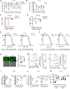
Figure 3.. Functional role of FZD7 in OC cells.
(A) FZD7 mRNA levels (mean fold-change ± SD, n=3–4) in FZD7 (+) and FZD7 (−) cells FACS selected from SKOV3, OVCAR5, and COV362 cells and HGSOC tumors. (B-D) Cell viability (mean fold-change ± SD, n=4) of FZD7(+) and FZD7(−) cells from OVCAR5 (n=3) (B), SKOV3 (n=3) (C) or COV362 (n=4) (D) cells, plated, treated with the indicated doses of CDDP for 24 hours, and cultured for additional three days. (E, F) Representative pictures and numbers (mean ± SD, n = 4–5) of spheroids formed after 7 days of culture by FZD7(+) and FZD7(−) cells FACS sorted from SKOV3 (E) and OVCAR5 (F) OC cells. Spheroids were counted or cell numbers were estimated by CellTiter-Glo 3D cell viability assay. (G-I) mRNA levels (fold-change ± SD) of Nanog in FZD7(+) compared with FZD7(−) cells from SKOV3 (G) (n=3), OVCAR5 (H) (n=8) and COV362 (I)(n=3) cells. For all comparisons: *P<0.05, **P<0.01, ***P<0.001.
Figure 4.
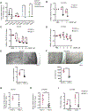
Figure 4.. FZD7 regulates stemness characteristics.
(A) (Left) FZD7 mRNA levels (mean fold-change ± SD, n=3) in SKOV3 cells transduced with shRNAs targeting FZD7 (shFZD7) vs. control shRNAs (shctrl). (Right) Numbers of spheroids (mean ± SD, n=4) formed by 2,000 shFZD7 or shctrl SKOV3 cells cultured for 14 days and counted under a microscope. (B) FZD7 mRNA expression levels (mean fold-change ± SD, n=4; left) and numbers of spheroids (n=5; right) in OVCAR5 cells transduced with shRNAs directed at FZD7 (shFZD7) vs. control shRNAs (shctrl). Cell viability assessed numbers of cells growing as spheroids using the CellTiter-Glo kit. (C) Sox2 and Nanog mRNA levels (mean fold-change ± SD, n=3–4) in OVCAR5 cells transduced with shFZD7 vs. shctrl. (D) FZD7 mRNA levels (mean fold-change ± SD, n=3) in Pt-R primary HGSOC cells transduced with shRNAs targeting FZD7 (shFZD7) vs. control shRNA (shctrl). (E) ALDH1A1 mRNA levels (mean fold-change ± SD, n = 3) in primary tumor cells transduced with shFZD7 cells vs. shctrl. (F) Average fold-change (± SD, n = 3) of FZD7 mRNA in SKOV3 (left) and OVCAR5 (right) cells transfected with FZD7-pcDNA3.1 vs. empty vector (ctrl). (G) Spheroid formation estimated with a CellTiter-Glo viability kit (bottom) from 1,000 ctrl and FZD7 expressing SKOV3 or OVCAR5 cells (described in F) and cultured for 7 days (n =4–5 per group). (H-J) Effects of CDDP on cell survival measured by CCK8 assays in SKOV3_shctrl and SKOV3_shFZD7 (H), SKOV3 cisplatin tolerant cells (SKOV3_CDDP) transduced with shRNAs targeting FZD7 (shFZD7_1, _2) or control shRNA (I), and SKOV3 cells transfected with FZD7-pcDNA3.1 (FZD7) or empty vector (Ctrl) (J). Cells were treated with cisplatin for 24 hours and cultured for additional 3 days (n=3–4). Cisplatin IC50 value is shown. (K) Days to tumor initiation (mean ± SD, n = 10) of sq xenografts induced by 2×10⁶ shctrl and shFZD7 transduced OVCAR5 cells. (L) FACS side scatter analysis of ALDH(+) cells (top), and percentage (mean ± SD, n=3) of ALDH(+) cells (bottom) from cell suspensions generated from OVCAR5_shctrl and OVCAR5_shFZD7 xenografts. (M) Numbers (mean ± SD, n=5) of spheroids (bottom) formed during 14 days by 1000 cells derived from cell suspensions from OVCAR5_shctrl or OVCAR5_FZD7 xenografts. (N-O). In vivo limited dilution assay used serially diluted numbers (2,500, 5,000, and 10,000) of OVCAR5_shctrl and shFZD7 cells injected sq into nude mice (n=4 replicates per group). (N) Average tumor weights (± SD) are shown (for the 10,000 cells group). (O) Stem cell frequencies were calculated by using the Extreme Limiting Dilution Analysis (http://bioinf.wehi.edu.au/software/elda/; p = 0.000254). (P) A Venn diagram shows the number overlapping and unique DEGs between OVCAR5-derived OCSCs (ALDH+CD133+) versus non-OCSCs (ALDH-CD133-), OVCAR5 cisplatin tolerant (CDDP-R) vs. parental (WT), and FZD7(+) versus FZD7(−) OVCAR5 cells (FDR < 0.05). ALDH+CD133/ALDH−CD133- and FZD7+/FZD7− cells were sorted by FACS. For all comparisons: *P<0.05, **P<0.01, ***P<0.001.
Figure 5.
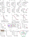
Figure 5.. FZD7 regulates GPX4 and intracellular redox states.
(A) Fold-change (mean ± SD, n=3–4) of GPX4 mRNA levels in FZD7(+) vs. FZD7(−) cells FACS sorted from SKOV3, OVCAR5, COV362 cells and cell suspensions from HGSOC tumors. (B) Fold-change (mean ± SD, n=4) of GPX4 mRNA expression levels in OVCAR5 cells transduced with shRNAs targeting FZD7 (shFZD7) vs. control shRNAs (shctrl). (C) Western blotting for FZD7, GPX4 and GAPDH in SKOV3 and OVCAR5 cells stably transduced with shctrl and shFZD7. Quantification shows fold change of FZD7 and GPX4 expression across 3 experiments. (D-E) GPX4 mRNA expression levels (fold-change ± SD, n = 3) in SKOV3 Pt-T cells (CDDP) (D), and Pt-R primary human HGSOC cells (E) transduced with shRNAs directed at FZD7 (shFZD7) vs. control shRNAs (shctrl). (F, G) Average fold-change (± SD, n=3) of GPX4 mRNA levels in SKOV3 (F) and OVCAR5 (G) cells transfected with FZD7-pcDNA3.1 vs. empty vector (ctrl). (H) Western blotting for FZD7, GPX4 and GAPDH in OVCAR5 cells transfected with FZD7-pcDNA3.1 vs. empty vector (ctrl). Quantification shows fold change of FZD7 and GPX4 expression across 3 experiments. (I-K) GPX4 mRNA expression (mean fold-change ± SD) in SKOV3 and OVCAR5 Pt-T (CDDP) vs. parental cells (WT) (I; n = 3/group). (J) SKOV3 and OVCAR5 xenografts (n=3 per group; J) and PDX tumors (K; n=5 per group) treated with PBS (Ctrl) or carboplatin. (L) Representative images of GPX4 IHC staining in sections of control (Ctrl) and Pt-T PDXs. (M) Representative pictures of GPX4 IHC (left) and H-scores (mean ± SD) (right) in sections of fallopian tube (n = 6) and Pt-R HGSOC tumors (n = 23). (N) Lipid peroxidation in SKOV3 and OVCAR5 cells transduced with scrambled shRNA (shctrl) or shRNAs targeting FZD7 (shFZD7) and expressed as average luciferase units/3×106 cells (± SD, n=4) (O) Intracellular lipid peroxidation measured in SKOV3 and OVCAR5 cells transfected with FZD7 or control vector (Ctrl) and expressed as average luciferase units/3×106 cells ± SD (n=4). For all comparisons: *P<0.5, **P<0.01, ***P<0.001.
Figure 6.
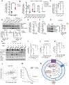
Figure 6.. FZD7 marks a cell population susceptible to GPX4 inhibitors.
(A) Average fold-change (±SD, n = 3) in mRNA expression levels of selected glutathione metabolism genes in OVCAR5 cells transduced with shRNAs targeting FZD7(shFZD7) vs. control shRNA (A), and in OVCAR5 cells transfected with FZD7-pcDNA3.1 (FZD7) vs. control vector (Ctrl) (B) (C-D) Viability of FZD7(+) and FZD7(−) cells sorted from OVCAR5 and COV362 (D) cells and treated with DMSO or ML210 (OVCAR5, 2μM; COV362, 1μM) for 72 hours. Data are presented as average fold-change (± SD, n = 4) of absorbance values relative to control. (E) Survival curves of OVCAR5 (left) and SKOV3 (right) cells transduced with control shRNAs (shctrl) or shRNAs targeting FZD7 (shFZD7) and treated with ML210 for 3 days (n=3–4). ML210 IC50 values are shown below. (F) Survival curves of OVCAR5 (left) and SKOV3 (right) cells transfected with FZD7-pcDNA3.1 or control vector and treated with ML210 for 3 days. ML210 IC50 values are shown below (n=3–4). (G) Images (left) and quantification of intracellular ROS levels (right) in OVCAR5 cells transfected with control shRNA (shctrl) or shRNA targeting FZD7 (shFZD7). Data are presented as means (± SD) of DCF fluorescence intensity per cell area (n = 15). (H) Histograms of fluorescence intensity (left) and mean (± SD, n = 3) (right) of BODIPY 581/591-C11 in OVCAR5 (left) and SKOV3 (right) cells transfected with vector (ctrl), FZD7-pCDNA3.1 (FZD7), control shRNA (shctrl), or shRNAs targeting FZD7 (shFZD7). (I, J) Mean (± SD, n=3) fluorescence intensity of BODIPY 581/591-C11 show effects of ML210 (1 μM for 20 hours) on lipid peroxidation levels in SKOV3 (I) and OVCAR5 (J) cells transfected with empty vector (ctrl), FZD7-pcDNA3.1 (FZD7), control shRNA (shctrl), or shRNA against FZD7 (shFZD7). (K) Intracellular ROS levels in OVCAR5 cells transfected with shctrl and shFZD7 treated with DMSO, ML210 (2μM, 24 hours) and ML210 + DFOA (800nM, 24 hours) measured by assessing DCFHDA oxidation. Average intensity per cell area (± SD) is shown (n=15). For all comparisons: *P<0.05, **P<0.01, ***P<0.001.
Figure 7.
Figure 7.. FZD7 regulates GPX4 expression and gluthatione metabolism by activating the canonical β catenin/p63 pathway.
(A) P63 mRNA levels (fold-change ± SD, n=4) in Pt-T PDXs (carbo) vs. controls (Ctrl). (B) P63 mRNA levels (fold-change ± SD, n=3–6) in FZD7(+) versus FZD7(−)cells sorted from SKOV3, OVCAR5, and COV362 cell lines, and cell suspensions from human tumors. (C) P63 mRNA expression levels (fold-change ± SD, n = 4) in OVCAR5 cells transduced with shRNAs targeting FZD7 (shFZD7) vs. control shRNA (shctrl). (D) Western blot for β-catenin, P63, and GAPDH in SKOV3 and OVCAR5 cells transduced with shRNAs targeting FZD7 (shFZD7) or control shRNA (shctrl). Quantification shows fold change of β-catenin and P63 expression across 3 experiments. (E, F) P63 mRNA levels (fold-change ± SD, n=3) in OVCAR5 (E) and SKOV3 (F) cells transfected with FZD7 vs. control vector (Ctrl). (G) Western blot for β-catenin, P63, and GAPDH in OVCAR5 cells transfected with control (ctrl) or FZD7-pcDNA3.1 (FZD7). Quantification shows fold change of β-catenin and P63 expression across 3 experiments. (H) Western blot for FZD7, β-catenin, P63, GPX4 and GAPDH in OVCAR5 cells transfected with shctrl, shFZD7 (J) treated with WNT3a (150ng/ul) and/or IWR-1-endo (1 μM) for 24 hours (n = 2). (I-K) FZD7(I), P63 (J), and GPX4 (K) mRNA expression levels (fold-change ± SD, n=3–4) in OVCAR5 cells transduced with control shRNAs (Ctrl_shctrl, Ctrl_shP63, FZD7_shctrl) or transfected with FZD7 expression vector and subsequently transduced with shRNA targeting P63 (FZD7_shP63). For all comparisons: *P<0.05, **P<0.01, ***P<0.001. (L) Scatter plot shows the correlation between P63 and FZD7 mRNA expression levels in HGSOC tumors (n=419) profiled in the TCGA database. Pearson correlation coefficients and P-values are shown. (M) Kaplan-Meier survival curves for HGSOC patients profiled in the TCGA having high (n=61) or low (n=318) P63 (TP63-012) mRNA expression levels. High or low levels were defined based on statistically determined cutoff point that maximizes absolute value of the standardized two-sample linear rank statistic. (N) Model demonstrates the proposed mechanism by which FZD7 engages the anti-oxidant pathway governed by GPX4.

References

    1. Berek JS, Bertelsen K, du Bois A, Brady MF, Carmichael J, Eisenhauer EA, et al. Advanced epithelial ovarian cancer: 1998 consensus statements. Ann Oncol. 1999;10 Suppl 1:87–92. - PubMed
    1. Tummala MK, and McGuire WP. Recurrent ovarian cancer. Clin Adv Hematol Oncol. 2005;3(9):723–36. - PubMed
    1. Hangauer MJ, Viswanathan VS, Ryan MJ, Bole D, Eaton JK, Matov A, et al. Drug-tolerant persister cancer cells are vulnerable to GPX4 inhibition. Nature. 2017;551(7679):247–50. - VSports最新版本 - PMC - PubMed
    1. Sharma SV, Lee DY, Li B, Quinlan MP, Takahashi F, Maheswaran S, et al. A chromatin-mediated reversible drug-tolerant state in cancer cell subpopulations. Cell. 2010;141(1):69–80. - PMC - PubMed
    1. Liau BB, Sievers C, Donohue LK, Gillespie SM, Flavahan WA, Miller TE, et al. Adaptive Chromatin Remodeling Drives Glioblastoma Stem Cell Plasticity and Drug Tolerance. Cell Stem Cell. 2017;20(2):233–46 e7. - PMC - PubMed

Publication types

MeSH terms