<u dropzone="GpWxw"></u> V体育安卓版 - Skip to main page content
U.S. flag

An official website of the United States government

Dot gov

The . gov means it’s official. Federal government websites often end in . gov or . mil. Before sharing sensitive information, make sure you’re on a federal government site VSports app下载. .

Https

The site is secure V体育官网. The https:// ensures that you are connecting to the official website and that any information you provide is encrypted and transmitted securely. .

. 2020 Jun;19(6):1058-1069.
doi: 10.1074/mcp.TIR119.001720. Epub 2020 Mar 10.

MaxQuant Software for Ion Mobility Enhanced Shotgun Proteomics

Affiliations

"VSports" MaxQuant Software for Ion Mobility Enhanced Shotgun Proteomics

Nikita Prianichnikov et al. Mol Cell Proteomics. 2020 Jun.

Abstract (VSports在线直播)

Ion mobility can add a dimension to LC-MS based shotgun proteomics which has the potential to boost proteome coverage, quantification accuracy and dynamic range VSports手机版. Required for this is suitable software that extracts the information contained in the four-dimensional (4D) data space spanned by m/z, retention time, ion mobility and signal intensity. Here we describe the ion mobility enhanced MaxQuant software, which utilizes the added data dimension. It offers an end to end computational workflow for the identification and quantification of peptides and proteins in LC-IMS-MS/MS shotgun proteomics data. We apply it to trapped ion mobility spectrometry (TIMS) coupled to a quadrupole time-of-flight (QTOF) analyzer. A highly parallelizable 4D feature detection algorithm extracts peaks which are assembled to isotope patterns. Masses are recalibrated with a non-linear m/z, retention time, ion mobility and signal intensity dependent model, based on peptides from the sample. A new matching between runs (MBR) algorithm that utilizes collisional cross section (CCS) values of MS1 features in the matching process significantly gains specificity from the extra dimension. Prerequisite for using CCS values in MBR is a relative alignment of the ion mobility values between the runs. The missing value problem in protein quantification over many samples is greatly reduced by CCS aware MBR. MS1 level label-free quantification is also implemented which proves to be highly precise and accurate on a benchmark dataset with known ground truth. MaxQuant for LC-IMS-MS/MS is part of the basic MaxQuant release and can be downloaded from http://maxquant. org. .

Keywords: Bioinformatics; bioinformatics software; label-free quantification; mass spectrometry; quantification. V体育安卓版.


Conflict of interest statement

* The authors have declared a conflict of interest. The authors state that they have potential conflicts of interest regarding this work: S. K. , H V体育ios版. K. , M. L. , and S. B. are employees of Bruker.

Figures

None
Graphical abstract
Fig. 1.
Fig. 1.
Elements of feature detection. A, Raw data is mapped to a common grid. Although the ion mobility scans already form a regular grid, the m/z values of data centroids are irregular. All intensity values within an ion mobility window and within a mass range are mapped to a common mass grid with a spacing that is monotone increasing with m/z. To obtain the processed intensities on the common grid, raw intensities are averaged using a Gaussian kernel with a locally adapted width according to the effective resolution. B, The raw data cube is sliced along the ion mobility axis to obtain planes with signal intensity as a function of m/z and retention time. In these planes, features can be detected with the algorithms used for feature detection in the conventional MaxQuant workflow for LS-MS data without ion mobility. C, The result of the conventional LC-MS MaxQuant algorithm are feature boundaries in each m/z-retention time plane. These boundaries are closed curves each surrounding the base area of a peak. D, The base areas of peaks found in C, are clustered between consecutive planes to obtain closed surfaces surrounding the three-dimensional base volumes of the 4D features.
Fig. 2.
Fig. 2.
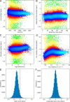
Mass recalibration. A–D, Residual mass errors after the dependence of all but one variable have been recalibrated, showing the dependence of the residual mass error on m/z (A), retention time (B), logarithm of the peak intensity (C) and ion mobility (D). Colors reflect the density of data points. E, Mass error distribution before recalibration. F, Mass error distribution after recalibration has been applied.
Fig. 3.
Fig. 3.
Retention time and ion mobility alignment. A, Difference in retention time between matched feature pairs plotted against the retention time of the feature in one of the runs. The point density is color-coded in plots A–D. B, Same as in A, but after retention time alignment has been applied. C, Like A, but now the difference in 1/K0 within the feature pair is plotted against 1/K0 in one of the runs, indicating differences between runs in terms of ion mobility. D, Same as in C, but after ion mobility alignment has been applied.
Fig. 4.
Fig. 4.
Accuracy of matching between runs. A, Retention time match difference distribution before alignment. B, Retention time match difference distribution after alignment. C, 1/K0 match difference distribution before alignment. D, 1/K0 match difference distribution after alignment.
Fig. 5.
Fig. 5.
Specificity of ion mobility enhanced matching between runs. A, Matches were performed without retention time restriction and then divided into “true” (Δt < 42s) and “false” (Δt > 42s) matches. The percentage of these matches is shown as a function of the window size in 1/K0 that was applied to the matching. B, The gain in specificity by using ion mobility as a function of the window size in 1/K0.
Fig. 6.
Fig. 6.
Protein quantification coverage. A, Number of protein groups quantified in 10 replicates without and with matching between runs. B, Number of proteins groups quantified in N out of 10 replicates without and with matching between runs. C, Number of protein groups quantified in 208 short human plasma runs. D, Gain of quantified protein groups in C, by matching between runs.
Fig. 7.
Fig. 7.
LFQ on a benchmark dataset. A, LFQ intensity plotted against fold change between replicate groups. (Both logarithmic.) Vertical lines correspond to fold changes expected by the mixing of species-derived samples. B, Same as A, but with matching between runs. C–D, Histograms of data in A–B, projected on the horizontal axes.

References

    1. Kanu A. B., Dwivedi P., Tam M., Matz L., and Hill H. H. (2008) Ion mobility-mass spectrometry. J. Mass Spectrom. 43, 1–22 - PubMed
    1. Cumeras R., Figueras E., Davis C. E., Baumbach J. I., and Gràcia I. (2015) Review on Ion Mobility Spectrometry. Part 2: hyphenated methods and effects of experimental parameters. Analyst 140, 1391–1410 - PMC - PubMed
    1. May J. C., and McLean J. A. (2015) Ion mobility-mass spectrometry: Time-dispersive instrumentation. Anal. Chem. 87, 1422–1436 - PMC - PubMed
    1. Valentine S. J., Plasencia M. D., Liu X., Krishnan M., Naylor S., Udseth H. R., Smith R. D., and Clemmer D. E. (2006) Toward plasma proteome profiling with ion mobility-mass spectrometry. J. Proteome Res. 5, 2977–84 - PubMed
    1. Baker E. S., Livesay E. A., Orton D. J., Moore R. J., Danielson W. F., Prior D. C., Ibrahim Y. M., LaMarche B. L., Mayampurath A. M., Schepmoes A. A., Hopkins D. F., Tang K., Smith R. D., and Belov M. E. (2010) An LC-IMS-MS platform providing increased dynamic range for high-throughput proteomic studies. J. Proteome Res. 9, 997–1006 - V体育平台登录 - PMC - PubMed

"V体育平台登录" MeSH terms

Substances (V体育2025版)

VSports在线直播 - LinkOut - more resources