Skip to main page content
U.S. flag

An official website of the United States government

Dot gov

The . gov means it’s official. Federal government websites often end in . gov or . mil. Before sharing sensitive information, make sure you’re on a federal government site. VSports app下载.

Https

The site is secure. The https:// ensures that you are connecting to the official website and that any information you provide is encrypted and transmitted securely V体育官网. .

. 2020 Jan 17;19(1):10.
doi: 10.1186/s12943-019-1112-1.

The PI3K subunits, P110α and P110β are potential targets for overcoming P-gp and BCRP-mediated MDR in cancer

Affiliations

The PI3K subunits, P110α and P110β are potential targets for overcoming P-gp and BCRP-mediated MDR in cancer

Lei Zhang et al. Mol Cancer. .

Abstract

Background: PI3K/AKT is a vital signaling pathway in humans VSports手机版. Recently, several PI3K/AKT inhibitors were reported to have the ability to reverse cancer multidrug resistance (MDR); however, specific targets in the PI3K/AKT pathways and the mechanisms associated with MDR have not been found because many of the inhibitors have multiple targets within a large candidate protein pool. AKT activation is one presumed mechanism by which MDR develops during cancer treatment. .

Methods: The effects of inhibiting PI3K 110α and 110β by BAY-1082439 treatment and CRISPR/Cas9 knockout were examined to determine the possible functions of BAY-1082439 and the roles of PI3K 110α and 110β in the reversal of MDR that is mediated by the downregulation of P-gp and BCRP. Inhibition of AKT with GSK-2110183 showed that the downregulation of P-gp and BCRP is independent of generalized AKT inactivation V体育安卓版. Immunofluorescence, immunoprecipitation, MTT, flow cytometry and JC-1 staining analyses were conducted to study the reversal of MDR that is mediated by P-gp and BCRP in cancer cells. An ATPase assay and a structural analysis were also used to analyze the potential mechanisms by which BAY-1082439 specifically targets PI3K 110α and 110β and nonspecifically influences P-gp and BCRP. .

Results: By inhibiting the activation of the PI3K 110α and 110β catalytic subunits through both the administration of BAY-1082439 and the CRISPR/Cas9 deletion of Pik3ca and Pik3cb, the ATP-binding cassette transporters P-gp/ABCB1 and BCRP/ABCG2 were downregulated, thereby reestablishing the drug sensitivity of human epidermoid carcinoma and non-small cell lung cancer (NSCLC) MDR cells. Inhibition of AKT did not reverse the MDR mediated by P-gp or BCRP. The ABC family proteins and AKT may play MDR-enhancing roles independently. V体育ios版.

Conclusions: The reversal of the dual functions of ABC-transporter-mediated and AKT-activation-enhanced MDR through the inhibition or knockout of PI3K 110α or 110β promises to improve current strategies based on combined drug treatments to overcome MDR challenges VSports最新版本. .

Keywords: Breast cancer resistance protein (BCRP/ABCG2/ABCP/MXR); Cancer; Multidrug resistance (MDR); P-glycoprotein (P-gp/ABCB1/MDR1); P110α/PIK3CA; P110β/PIK3CB; PI3K; Reversal of MDR. V体育平台登录.


Conflict of interest statement

The authors declare that they have no competing interests.

V体育ios版 - Figures

Fig. 1
Fig. 1
BAY-1082439 reversed MDR of cancers via down-regulating P-gp and BCRP transporters. a MTT assay showing the ability of BAY-1082439 to reverse MDR mediated by P-gp over-expressed in KB-C2 cells and BCRP over-expressed in H460/MX20 cells. Viability of cells without treatment with BAY-1082439 and anti-cancer drugs (colchicine for KB-C2 and mitoxantrone for H460/MX20) was set as standard control (denominator) for comparing the combined effect of BAY-1082439. The negative control, parental drug-sensitive cells KB-3-1 and H460 were treated through the same procedure. IC50 are indicated by arrows. b Down-regulation of P-gp and BCRP by BAY-1082439 targeting PI3K 110α and 110β. The cells were cultured with drugs and BAY-1082439 (BAY, 10 μM), and analyzed by Western blot. Relative quantification was carried out with ImageQuant TL based on the intensity of the bands. Colchicine at 0.7 and 0.01 μM were applied to drug-resistant KB-C2 and drug-sensitive KB-3-1 cells, respectively. Mitoxantrone at 3 and 0.3 μM were applied to drug-resistant H460/MX20 and drug-sensitive H460 cells, respectively. c Localization and level of P-gp and BCRP in the MDR cancer cells determined by immuno-fluorescence (IF). Positive signals on cell membrane and nuclear envelope are depicted by orange and purple arrows, respectively. Inhibition of P-gp and BCRP by BAY-1082439 is depicted by arrows in light blue. KB-C2 and H460/MX80 cells were cultured with 1 μM of colchicine and mitoxantrone, respectively, followed by co-culture with 10 μM of BAY-1082439. By 24 h and 48 h of co-culture, respectively, KB-C2 and H460/MX80 cells showing inhibition of P-gp or BCRP expression were studied by IF analysis
Fig. 2
Fig. 2
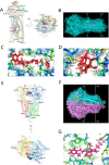
Model structure showing the interactions stabilizing BAY-1082439/P-gp and BAY-1082439/BCRP complexes. a Domains and positions of P-gp binding with BAY-1082439. b Structures showing the surface of BAY-1082439/P-gp complex. c Polar interaction between BAY-1082439 and P-gp. d Spatial structure of the cavity labeled with residues groups for P-gp to dock with BAY-1082439. e Binding domains and positions of BCRP docking with BAY-1082439. f Structures showing surface of BAY-1082439/BCRP complex. g Polar interaction between BAY-1082439 and BCRP
Fig. 3
Fig. 3
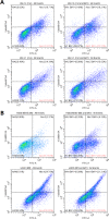
Flow cytometry analysis of BAY-1082439 in attenuating the anti-apoptosis ability of the MDR cell lines KB-C2 (a) and H460/MX80 (b) with the presence of colchicine (Col) and mitoxantrone (Mit) during 48 h of culture. The rate of cell necrosis (Nec) and apoptosis (Apo) with and without BAY-1082439 (BAY) was compared. The drug-sensitive cells KB-3-1 and H460 showing no apparent change of necrosis and apoptosis after treatment with BAY-1082439, were set as control for KB-C2 and H460/MX80, respectively. The dosage of BAY-1082439 was 10 μM. Colchicine was applied at 2 μM to KB-C2 and 0.02 μM to KB-3-1. Mitoxantrone was applied at 10 μM to H460/MX80 and 0.1 μM to H460. BAY-1082439 was applied at 10 μM to all the tested cells. Annexin V-FITC binding to phosphatidylserine (PS) that translocates to the external leaflet of the apoptotic cells was determined via FITC-A detection mode (Ex: 488 nm, Em: 525 nm). Propidium iodide (PI) that binds nucleotides of late-apoptotic or necrotic cells was determined via violet 610-A detection mode (Ex: 450 nm; Em: 610 nm) to avoid excitation of FITC. The four cell lines in healthy status were labelled with H1-H4
Fig. 4
Fig. 4
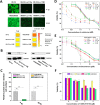
Reversal of MDR ability via knockout of target P110 subunits, including P110α (PIK3CA) and P110β (PIK3CB), from MDR cancer cell populations of KB-C2 and H460/MX80 with pCRISPR-CG01 all-in-one plasmid. a Map of pCRISPR-CG01. b Analysis with Western blot confirming the knockout of PIK3CA and PIK3CB that is of low abundance in KB-C2 and H460/MX80, as compared with GAPDH. To correct possible result deviation caused by exposure saturation of partial bands, less exposure of GAPDH bands was used to indicate relative cell counts. The absence or intensity-reduction of the target bands are depicted by red stars. Truncated proteins are depicted by head-down arrows. Relative quantification was carried out with ImageQuant TL based on the intensity of the bands. Statistical calculation was made based on three independent repeats. c PCR as adjuvant method to characterize target P110 subunit deficiency in KB-C2 and H460/MX80 cells. Missing bands or reduced copies of the target PCR products are depicted by red stars. New bands generated by chromosomal recombination are depicted by diamonds. Truncated fragments are depicted by head-down arrows. d-f MTT assay showing changes in MDR level of the KB-C2 and H460/MX80 cells with target PI3K 110α or 110β subunits knocked out. Colchicine and paclitaxel, the substrates of P-gp, were used for evaluation of reversal of KB-C2 over-expressing P-gp. The BCRP substrate mitoxantrone was used for analysis of reversal of MDR of H460/MX80 with BCRP over-expressed. The experiments were performed at least three times
Fig. 5
Fig. 5
Analysis of the mechanisms for reversal of P-gp or BCRP mediated MDR ability in the MDR cancer cells with P110α or P110β knocked out. a Immunofluorescence microscopic characterization of down-regulation of P-gp or BCRP expression in KB-C2 and H460/MX80 cells with target P110α and P110β subunits knocked out. PI3K 110α and 110 β subunit-k.o. KB-C2 (namely KB-C2-k.o.110α and KB-C2-k.o.110β, respectively) cells expressing low level of P-gp or BCRP were depicted by arrows in orange, as most cells of these two populations were hardly be detected with positive signals, as confirmed by IF-Cytell statistical analysis according to description in the methods. The cells were seeded to 96-well plate at a density of 5 × 103 cells per well. This experiment was independently repeated at least three times. b Western blot for the analysis of P-gp and BCRP expression in KB-C2 and H460/MX80 cells with target P110 subunits knocked out. c Relative protein expression level was calculated according to the band showing protein density and volume. d and e MTT assay showing poor effect of GSK-2110183 to reverse P-gp- or BCRP-mediated drug-resistance in KB-C2 or H460/MX20. f GSK-2110183 reduces cell viability via lower their ability to proliferate or anti-apoptosis. KB-3-1, H460, KB-C2 and H460/MX20 cells were cultured with gradient concentrations of GSK-2110183
Fig. 6
Fig. 6
Immunoprecipitation analysis showing the effect of inhibition of P110α and P110β on the expression level of AKT and some pivotal proteins regulated by AKT. Western blot for the analysis of Pan-AKT (a) and series of its downstream factors (b) was performed after the KB-C2 and H460/MX80 MDR cells and the KB-3-1 and H460 drug sensitive parental cells were co-cultured with 10 μM of BAY-1082439 for 48 h. In addition to BAY-1082439, KB-3-1 and KB-C2 cells were also co-cultured with 0.1 and 1 μM of colchicine, and H460 and H460/MX80 cells were also co-cultured with 1 and 10 μM of mitoxantrone, respectively. Molecular for each protein: AKT: 60 kDa; GSK3β: 47 kDa; P53: 53 kDa; Phospho-FOXO3a (Ser253): 82–97 kDa; caspase-9: 46 kDa; Bcl-xL: 23.7 kDa
Fig. 7
Fig. 7
Mechanism for ABC transporters regulated by the level of PI3K 110α or 110β subunit. The level of P110 subunits may regulate P-gp and BCRP directly, altering drug-sensitivity of the cells and yielding changed cell proliferation and survival ability. Knockout of P110 subunits induced down-regulation of P-gp and BCRP, and suggested the potentiality of P110α and P110β to be applied as targets for reversal of MDR of cancers. This route can be separate from AKT activation that had been regarded as a downstream effect in PI3K/AKT signaling pathway, playing an important role in the activation of many factors through phosphorylation. Italic characters indicate the major function of CRISPR/Cas9 deletion of P110α or P110β from the MDR cancer cells

References

    1. Cerniglia GJ, Dey S, Gallagher-Colombo SM, Daurio NA, Tuttle S, Busch TM, Lin A, Sun R, Esipova TV, Vinogradov SA, et al. The PI3K/Akt pathway regulates oxygen metabolism via pyruvate dehydrogenase (PDH)-E1alpha phosphorylation. Mol Cancer Ther. 2015;14(8):1928–1938. doi: 10.1158/1535-7163.MCT-14-0888. - DOI - PMC - PubMed
    1. Courtnay R, Ngo DC, Malik N, Ververis K, Tortorella SM, Karagiannis TC. Cancer metabolism and the Warburg effect: the role of HIF-1 and PI3K. Mol Biol Rep. 2015;42(4):841–851. doi: 10.1007/s11033-015-3858-x. - DOI - PubMed
    1. Qian P, He XC, Paulson A, Li Z, Tao F, Perry JM, Guo F, Zhao M, Zhi L, Venkatraman A, et al. The Dlk1-Gtl2 locus preserves LT-HSC function by inhibiting the PI3K-mTOR pathway to restrict mitochondrial metabolism. Cell Stem Cell. 2016;18(2):214–228. doi: 10.1016/j.stem.2015.11.001. - DOI - PMC - PubMed
    1. Liu X. Overstimulation can create health problems due to increases in PI3K/Akt/GSK3 insensitivity and GSK3 activity. Springerplus. 2014;3:356. doi: 10.1186/2193-1801-3-356. - "VSports" DOI - PMC - PubMed
    1. McKnight NC, Zhenyu Y. Beclin 1, an essential component and master regulator of PI3K-III in health and disease. Curr Pathobiol Rep. 2013;1(4):231–238. doi: 10.1007/s40139-013-0028-5. - DOI - PMC - PubMed

Publication types

V体育平台登录 - MeSH terms

Substances