Skip to main page content
U.S. flag

An official website of the United States government

Dot gov

The . gov means it’s official. Federal government websites often end in . gov or . mil VSports app下载. Before sharing sensitive information, make sure you’re on a federal government site. .

Https

The site is secure. The https:// ensures that you are connecting to the official website and that any information you provide is encrypted and transmitted securely V体育官网. .

. 2019 Jan 18;10(6):673-683.
doi: 10.18632/oncotarget.26586.

"VSports最新版本" Downregulation of miR-194-5p induces paclitaxel resistance in ovarian cancer cells by altering MDM2 expression

Affiliations

Downregulation of miR-194-5p induces paclitaxel resistance in ovarian cancer cells by altering MDM2 expression

"VSports" Koji Nakamura et al. Oncotarget. .

VSports - Abstract

Paclitaxel is a first-line drug for treating epithelial ovarian cancer (EOC). However, prognosis for patients with advanced stage cancer remains poor due to primary or acquired drug resistance. Therefore, overcoming chemoresistance is one of the greatest challenges in treating EOC. In this study, we identified microRNAs (miRNA) that regulate paclitaxel resistance and tested their potential utility as therapeutic targets. Paclitaxel-resistant cell lines were established using two EOC cell lines: SKVO3ip1 and HeyA8. miRNA PCR arrays showed that miR-194-5p was downregulated in paclitaxel-resistant cells. Forced expression of miR-194-5p resensitized resistant cells to paclitaxel. Conversely, miR-194-5p inhibition induced paclitaxel resistance in parental cells. In silico analysis and luciferase reporter assay revealed that MDM2 is a direct target of miR-194-5p. MDM2 was upregulated in paclitaxel resistant cells compared with parental cells. MDM2 inhibition also resensitized resistant cells to paclitaxel and forced MDM2 induced paclitaxel resistance in parental cells. miR-194-5p induced p21 upregulation and G1 phase arrest in resistant cells by downregulating MDM2 VSports手机版. Furthermore, a public database showed that high MDM2 expression was associated with a shorter progression-free survival in EOC patients treated with paclitaxel. Collectively, our results show that restoring miR-194-5p expression resensitizes EOCs to paclitaxel, and this may be exploited as a therapeutic option. .

Keywords: MDM2; miR-194-5p; microRNA; ovarian cancer; paclitaxel resistance V体育安卓版. .


Conflict of interest statement

CONFLICTS OF INTEREST All authors have no potential conflicts of interest to disclose.

Figures
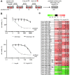
Figure 1
Figure 1. miR-194-5p is downregulated in paclitaxel-resistant ovarian cancer cell lines
(A) Scheme for the establishment of two paclitaxel-resistant sublines, SKOV3ip1-TR and HeyA8-TR, derived from SKOV3ip1 and HeyA8, respectively. These cells were exposed to stepwise increases of paclitaxel until a concentration of 300 nmol/L. (B) In vitro survival assay of ovarian cancer cell lines upon paclitaxel treatment. Growth inhibitory effects of paclitaxel treatment were determined using an MTS assay. Experiments were performed in triplicate. Data are represented as mean ± SE and are obtained from three independent experiments. (C) miRNA microarray. List of miRNAs that exhibited increased (> 2-fold, red columns) or decreased (< 0.5-fold, green columns) expression in both paclitaxel-resistant sublines compared with their corresponding parental cell lines.
Figure 2
Figure 2. miR-194-5p modulates sensitivity to paclitaxel in ovarian cancer cell lines
(A) miRNA qRT-PCR. Cells were transfected with pre-miR-194-5p (miR-194-5p) or control miR (miR-ctrl). Twenty-four hours later, expression of miR-194-5p relative to RNU6B expression was calculated using the 2-ΔΔCT method. Relative fold differences are presented. (B) MTS assay. Twenty-four hours after transfection with miR-194-5p or control miR-ctrl, cells were treated with paclitaxel for 72 hours and cell viability was assessed. Cell viability is shown relative to that in paclitaxel-free conditions. (C) miRNA qRT-PCR. Cells were transfected with anti-miR-194-5p or miR-ctrl for 24 hours. (D) MTS assay. As described in B, cells were transfected with anti-miR-194 or miR-ctrl and cell viability was assessed. Experiments were performed in triplicate. Data are represented as mean ± SE and are obtained from three independent experiments. *P < 0.05; **P < 0.01.
Figure 3
Figure 3. MDM2 is a direct target gene of miR-194-5p and is upregulated in paclitaxel-resistant cells
(A) Schematic illustration of predicted the MDM2 3′-UTR-binding site of miR-194-5p. (B) Luciferase reporter assay. SKOV3ip1-TR cells were co-transfected with miRNA precursor (miR-194-5p or miR-ctrl), luciferase reporter vector containing wildtype or mutant 3′-UTR of MDM2, and Renilla luciferase control vector. Twenty-four hours after incubation, luciferase activity normalized to Renilla activity was measured. Experiments were performed in triplicate. Data are represented as mean ± SE and are obtained from three independent experiments. ***P < 0.001; n.s., not significant. (C and D) Western blot. MDM2 expression between parental cell lines and paclitaxel-resistant sublines (C). After cells were transfected with miR-194-5p or miR-ctrl for 24 hours, cell lysates were collected and MDM2 expression was evaluated. β-actin was used as a loading control. Blots are representative of three experiments (D). (E) Kaplan-Meier survival analysis. Kaplan Meier Plotter database indicated that high MDM2 expression in ovarian cancer patients treated with paclitaxel (n = 632) correlates with poor progression-free survival (P = 0.0034).
Figure 4
Figure 4. MDM2 modulates sensitivity to paclitaxel in ovarian cancer cell lines
(A) Western blot. Cells were transfected with MDM2-siRNA or control-siRNA. Twenty-four hours after incubation, cell lysates were collected, and MDM2 expression was evaluated. (B) MTS assay. Cells were transfected with MDM2-siRNA or control-siRNA. Twenty-four hours after incubation, cells were treated with paclitaxel for 72 hours and cell viability was assessed. (C) Western blot. Cells were transfected with an MDM2 expression plasmid (MDM2(+)) or empty vector. Forty-eight hours after transfection, cell lysates were collected. (D) MTS assay. Forty-eight hours after transfection with an MDM2 expression plasmid (MDM2(+)) or empty vector, cells were treated with paclitaxel for 72 hours and viability was assessed. Blots are representative of three experiments. MTS assays were performed in triplicate. Data are represented as mean ± SE and are obtained from three independent experiments. *P < 0.05, **P < 0.01.
Figure 5
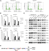
Figure 5. miR-194-5p and its target MDM2 induce G0/G1 cell cycle arrest
(AC) Cell cycle analyses. Paclitaxel-resistant cells transfected with miRNA precursors (miR-194-5p or ctrl-miR) or siRNAs (MDM2-siRNA or ctrl-siRNA) were stained with propidium iodide and analyzed by flow cytometry. Representative flow histograms. P3 = subG1 phase. P4 = G0/G1 phrase. P5 = S phase. P6 = G2/M phase (A). Percentage of cells in subG1, G0/G1, S, and G2/M phase. Paclitaxel-resistant cells transfected with miRNA precursors (B) or siRNAs (C). Data are represented as mean ± SE and are obtained from three independent experiments. (D) Western blot (left, SKOV3ip1-TR cells; right, HeyA8-TR cells). Expression of MDM2 and cell cycle-related proteins in paclitaxel-resistant cells transfected with miRNA precursors or siRNAs. Blots are representative of three experiments. (E) Schematic diagram of the pathway downstream of miR-194-5p. *P < 0.05, ***P < 0.001.

References

    1. Siegel RL, Miller KD, Jemal A. Cancer Statistics, 2017. CA Cancer J Clin. 2017;67:7–30. - PubMed
    1. Bookman MA. Optimal primary therapy of ovarian cancer. Ann Oncol. 2016;27:i158–i162. - PubMed
    1. Cannistra SA. Cancer of the ovary. N Engl J Med. 2004;351:2519–2529. - PubMed
    1. Di Leva G, Garofalo M, Croce CM. MicroRNAs in cancer. Annu Rev Pathol. 2014;9:287–314. - V体育ios版 - PMC - PubMed
    1. Magee P, Shi L, Garofalo M. Role of microRNAs in chemoresistance. Ann Transl Med. 2015;3:332. - "VSports注册入口" PMC - PubMed