Skip to main page content
U.S. flag

An official website of the United States government

Dot gov

The . gov means it’s official. Federal government websites often end in . gov or . mil VSports app下载. Before sharing sensitive information, make sure you’re on a federal government site. .

Https

The site is secure. The https:// ensures that you are connecting to the official website and that any information you provide is encrypted and transmitted securely V体育官网. .

. 2018 Dec 27;37(1):324.
doi: 10.1186/s13046-018-0965-2.

Hepatocellular carcinoma-derived exosomal miRNA-21 contributes to tumor progression by converting hepatocyte stellate cells to cancer-associated fibroblasts (V体育2025版)

Affiliations

Hepatocellular carcinoma-derived exosomal miRNA-21 contributes to tumor progression by converting hepatocyte stellate cells to cancer-associated fibroblasts

Yuan Zhou (VSports手机版) et al. J Exp Clin Cancer Res. .

"VSports app下载" Erratum in

Abstract (V体育ios版)

Background: Hepatocellular carcinoma (HCC) remains a global challenge due to its high morbidity and mortality rates as well as poor response to treatment. The communication between tumor-derived elements and stroma plays a critical role in facilitating cancer progression of HCC. Exosomes are small extracellular vesicles (EVs) that are released from the cells upon fusion of multivesicular bodies with the plasma membrane. There is emerging evidence indicating that exosomes play a central role in cell-to-cell communication. Much attention has been paid to exosomes since they are found to transport bioactive proteins, messenger RNA (mRNAs) and microRNA (miRNAs) that can be transferred in active form to adjacent cells or to distant organs. However, the mechanisms underlying such cancer progression remain largely unexplored VSports手机版. .

Methods: Exosomes were isolated by differential ultracentrifugation from conditioned medium of HCC cells and identified by electron microscopy and Western blotting analysis. Hepatic stellate cells (HSCs) were treated with different concentrations of exosomes, and the activation of HSCs was analyzed by Western blotting analysis, wound healing, migration assay, Edu assay, CCK-8 assay and flow cytometry. Moreover, the different miRNA levels of exosomes were tested by real-time quantitative PCR (RT-PCR) V体育安卓版. The angiogenic ability of activated HSCs was analyzed by qRT-PCR, CCK-8 assay and tube formation assay. In addition, the abnormal lipid metabolism of activated HSCs was analyzed by Western blotting analysis and Oil Red staining. Finally, the relationship between serum exosomal miRNA-21 and prognosis of HCC patients was evaluated. .

Results: We showed that HCC cells exhibited a great capacity to convert normal HSCs to cancer-associated fibroblasts (CAFs). Moreover, our data revealed that HCC cells secreted exosomal miRNA-21 that directly targeted PTEN, leading to activation of PDK1/AKT signaling in HSCs. Activated CAFs further promoted cancer progression by secreting angiogenic cytokines, including VEGF, MMP2, MMP9, bFGF and TGF-β. Clinical data indicated that high level of serum exosomal miRNA-21 was correlated with greater activation of CAFs and higher vessel density in HCC patients. V体育ios版.

Conclusions: Intercellular crosstalk between tumor cells and HSCs was mediated by tumor-derived exosomes that controlled progression of HCC. Our findings provided potential targets for prevention and treatment of live cancer. VSports最新版本.

Keywords: AKT; Angiogenesis; Cancer associated fibroblasts; Exosome; Hepatic stellate cells; Hepatocellular carcinoma; PTEN; miRNA-21 V体育平台登录. .


Conflict of interest statement

Ethics approval and consent to participate

The present study was authorized by the Ethics Committee of Nanjing Drum Tower Hospital. All procedures performed in studies were in accordance with the ethical standards VSports注册入口. All patients and volunteers were anonymous and provided written informed consent.

Consent for publication

Not applicable.

Competing interests

The authors declare that they have no competing interests.

Publisher’s Note

Springer Nature remains neutral with regard to jurisdictional claims in published maps and institutional affiliations.

Figures
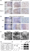
Fig. 1
Fig. 1
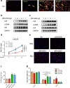
Tumor-derived exosomes activated HSCs. a, b Representative images of α-SMA staining in primary HCC and in orthotopically implanted tumors in mice. c, d, e Exosomes released by different cells were detected by electron microscopy. f Immunoblotting assay of indicated proteins in exosomes from different cancer cells. Each experiment was performed three times independently, and results are presented as mean ± s.d. Student’s t-test was used to analyze the data. (*p < 0.05; **p < 0.01; ***p < 0.001)
Fig. 2
Fig. 2
Tumor-derived exosomes activated HSCs in vitro. a Immunofluorescence imaging showed the delivery of 97H-labeled exosomes (green) to α-SMA-labeled HSCs (red). Yellow arrows represented delivered exosomes. b and c HSCs were activated by HCC cell-derived exosomes in a concentration-dependent manner. CCK8 assay (d), Edu (e and f) and flow cytometry assays (g) of HSCs treated with equal quantities of exosomes derived from different liver cancer cells or blank control. Experiments were performed at least in triplicate, and results are shown as mean ± s.d. Student’s t-test was used to analyze the data (*p < 0.05; **p < 0.01; ***p < 0.001)
Fig. 3
Fig. 3
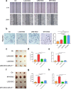
Tumor-derived exosomes activated HSCs in vitro. Wound-healing assays (a) and migration assay (b) of HSCs treated with equal quantities of exosomes derived from different liver cancer cells or blank control. ch Xenograft assays of Huh7 with indicated treatments were performed on nude mice. Representative tumors, tumor volume and number of tumor nudes were shown. Experiments were performed at least in triplicate, and results are shown as mean ± s.d. Student’s t-test was used to analyze the data (NS, not significant; *p < 0.05; **p < 0.01; ***p < 0.001)
Fig. 4
Fig. 4
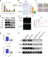
Exosomal miRNA-21 is characteristically secreted by liver cancer cells and mediates HSCs activation. a Expression of exosomal miRNAs from different cancer cells was presented. Wound-healing assay (b), migration assay (c), Western blotting assay (d), immunofluorescence assay (e) and CCK-8 assay (f) of HSCs transfected miRNA-21-mimic or normal control. Representative images were shown. g qPCR of miRNA-21 in HCC-derived treated with miRNA-21 inhibitor exosomes. h Western blotting assay of HSCs transfected miRNA-21 inhibitor. Experiments were performed at least in triplicate, and results are shown as mean ± s.d. Student’s t-test was used to analyze the data (NS, not significant; *p < 0.05; **p < 0.01; ***p < 0.001)
Fig. 5
Fig. 5
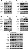
Exosomal miRNA-21 activates HSCs via PTEN/PDK1/AKT signaling axis. a, b Immunoblotting assays of indicated proteins in HSCs treated with control or exosomes from different tumor cells. c-e Western blotting assays of indicated proteins in HSCs with indicated treatments. Each experiment was performed in triplicate, and data are presented as mean ± s.d. Student’s t-test was used to analyze the data (*p < 0.05; **p < 0.01; ***p < 0.001)
Fig. 6
Fig. 6
HCC derived exosomes induced abnormal lipid metabolism. a, b Western blotting assays of lipid metabolism related proteins in HCC patients or HSCs with different stimulations. c Oil Red staining assay showed the abnormal lipid accumulation in HSCs with indicated treatments. Each experiment was performed in triplicate, and data are presented as mean ± s.d. Student’s t-test was used to analyze the data (*p < 0.05; **p < 0.01; ***p < 0.001)
Fig. 7
Fig. 7
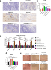
Activated HSCs promote angiogenesis a, b Immunohistochemistry staining of CD31 staining in subcutaneously implanted tumor under different conditions. c qRT-PCR assay indicated gene expression levels of HSCs treated with exosomes derived from different cells in the presence of miRNA-21 inhibitor or AKT inhibitor. Experiments were performed at least in triplicate. d, e The proliferation of HUVECs with exosomes derived from different cells in the presence of miRNA-21 inhibitor or AKT inhibitor was tested by CCK-8 assay and tube formation assay. Data are mean ± SEM from three independent experiments, *p < 0.05, **p < 0.01, ***p < 0.001 by unpaired Student’s t-test
Fig. 8
Fig. 8
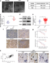
miRNA-21 is associated with HCC progression. ad Exosomes in normal and HCC serums were detected by electron microscopy. e miRNA-21 expression level in serum exosomes from healthy donors and primary HCC patients. Data are presented as mean ± s.d. Student’s t-test was used to analyze the data. **p < 0.01. f In situ hybridization assay of HCC samples and scores of miRNA-21 level. Representative images were shown. g Kaplan–Meier plots of overall survival and disease-free survival of 83 HCC patients, stratified by expression of miRNA-21. Survival data were analyzed by the Kaplan−Meier method and log-rank test. h The correlation analysis between expression of miRNA-21 in serum and expression of miRNA-21 in HCC was detected. i In situ hybridization of miRNA-21 in combination with IHC staining of HSCs markers (FAP) and proliferation markers (Ki67) on serial sections of human HCC tissues. White arrows indicate CAFs; black arrows indicate vessels
Fig. 9
Fig. 9
Proposed schematic diagram of HCC exosomal miR-21-mediating HSCs activation to promote angiogenesis of HCC

References

    1. Singal AG, El-Serag HB. Hepatocellular carcinoma from epidemiology to prevention: translating knowledge into practice. Clin Gastroenterol Hepatol. 2015;13:2140–2151. doi: 10.1016/j.cgh.2015.08.014. - "VSports在线直播" DOI - PMC - PubMed
    1. Bruix J, Gores GJ, Mazzaferro V. Hepatocellular carcinoma: clinical frontiers and perspectives. Gut. 2014;63:844–855. doi: 10.1136/gutjnl-2013-306627. - DOI - PMC - PubMed
    1. Kubo N, Araki K, Kuwano H, Shirabe K. Cancer-associated fibroblasts in hepatocellular carcinoma. World J Gastroenterol. 2016;22:6841–6850. doi: 10.3748/wjg.v22.i30.6841. - DOI - PMC - PubMed
    1. Affo S, Yu LX, Schwabe RF. The role of Cancer-associated fibroblasts and fibrosis in liver Cancer. Annu Rev Pathol. 2017;12:153–186. doi: 10.1146/annurev-pathol-052016-100322. - DOI - PMC - PubMed
    1. Lau EY, Lo J, Cheng BY, Ma MK, Lee JM, Ng JK, Chai S, et al. Cancer-associated fibroblasts regulate tumor-initiating cell plasticity in hepatocellular carcinoma through c-met/FRA1/HEY1 signaling. Cell Rep. 2016;15:1175–1189. doi: 10.1016/j.celrep.2016.04.019. - V体育2025版 - DOI - PubMed

MeSH terms

Substances (VSports手机版)