Skip to main page content (V体育平台登录)
U.S. flag

An official website of the United States government

Dot gov

The . gov means it’s official. Federal government websites often end in . gov or . mil VSports app下载. Before sharing sensitive information, make sure you’re on a federal government site. .

Https

The site is secure. The https:// ensures that you are connecting to the official website and that any information you provide is encrypted and transmitted securely. V体育官网.

. 2018 Sep 6;7(12):e1494108.
doi: 10.1080/2162402X.2018.1494108. eCollection 2018.

Bovine herpesvirus 4-based vector delivering the full length xCT DNA efficiently protects mice from mammary cancer metastases by targeting cancer stem cells (VSports)

Affiliations

Bovine herpesvirus 4-based vector delivering the full length xCT DNA efficiently protects mice from mammary cancer metastases by targeting cancer stem cells

Gaetano Donofrio et al. Oncoimmunology. .

Abstract (VSports注册入口)

Despite marked advancements in its treatment, breast cancer is still the second leading cause of cancer death in women, due to relapses and distal metastases. Breast cancer stem cells (CSCs), are a cellular reservoir for recurrence, metastatic evolution and disease progression, making the development of novel therapeutics that target CSCs, and thereby inhibit metastases, an urgent need. We have previously demonstrated that the cystine-glutamate antiporter xCT (SLC7A11), a protein that was shown to be overexpressed in mammary CSCs and that plays a key role in the maintenance of their redox balance, self-renewal and resistance to chemotherapy, is a potential target for mammary cancer immunotherapy. This paper reports on the development of an anti-xCT viral vaccine that is based on the bovine herpesvirus 4 (BoHV-4) vector, which we have previously showed to be a safe vaccine that can transduce cells in vivo and confer immunogenicity to tumor antigens VSports手机版. We show that the vaccination of BALB/c mice with BoHV-4 expressing xCT (BoHV-4-mxCT), impaired lung metastases induced by syngeneic mammary CSCs both in preventive and therapeutic settings. Vaccination induced T lymphocyte activation and the production of anti-xCT antibodies that can mediate antibody-dependent cell cytotoxicity (ADCC), and directly impair CSC phenotype, self-renewal and redox balance. Our findings pave the way for the potential future use of BoHV-4-based vector targeting xCT in metastatic breast cancer treatment. .

Keywords: Mammary cancer; bovine herpesvirus 4-based vector; cancer stem cell; immunotherapy; xCT V体育安卓版. .


Figures

Figure 1.
Figure 1.
Generation of BoHV-4-mxCT. A) Diagram (not to scale) showing the retargeting event obtained by heat-inducible homologous recombination in SW102 E. coli containing pBAC-BoHV-4-A-TK-KanaGalK-TK, where the Kana/GalK cassette was replaced with the CMV-mxCTgD106 expression cassette flanked by BoHV-4 TK sequences, located in pINT2 shuttle plasmid vector. B) Representative 2-deoxy-galactose resistant colonies tested by HindIII restriction enzyme analysis, agar gel electrophoresis and Southern blotting performed with a specific probe for the mxCT ORF. The 2,650 bp band (blue circle), corresponding to the un-retargeted pBAC-BoHV-4-A-TK-KanaGalK-TK control, has been replaced by a 3156 bp band (red circle) in pBAC-BoHV-4-A-CMV-mxCTgD106ΔTK. C) Representative phase contrast and fluorescent microscopic images of plaque formed by viable reconstituted recombinant BoHV-4-mxCT after the corresponding BAC DNA electroporation into BEK cells expressing cre recombinase (Magnification, 10X). D) Replication kinetics of BoHV-4-mxCT growth on BEK cells compared with the parental BoHV-4-A isolate. The data presented are the means ± SEM of triplicate measurements (P > 0.05 for all time points as measured by Student’s t-test). E) Western immunoblotting of cells infected with BoHV-4-mxCT or the parental BoHV-4-A used as a negative control. The lanes were loaded with different amounts of total protein cell extracts (5, 10 and 15 μl).
Figure 2.
Figure 2.
BoHV-4-mxCT vaccination induces an anti-xCT humoral response. Sera from untreated (white bars), BoHV4-A29- (gray bars), and BoHV-4-mxCT- (black bars) vaccinated mice were tested by ELISA on wells coated with A) full-length mouse xCT protein, or peptides corresponding to mouse xCT extracellular loops B) 1, C) 2, D) 3, E) 4 or F) 6. Graphs show mean ± SEM of sera pooled from 3 independent experiments. In A, antibody concentration was calculated based on a standard curve obtained with a commercial anti-xCT antibody targeting its N-terminal region. G) Representative immunofluorescence images of dissociated 4T1 tumorsphere cells incubated with sera of vaccinated mice. The specific signal (green), was detected using an Alexa Fluor488-conjugated anti-mouse secondary antibody. Nuclei were counterstained with DAPI (blue). Magnification 40X, Scale bar, 40 µm. **, P < 0.01, Student’s t-test. H) ADCC assay performed using CFSE+ 4T1 target cells incubated with 1:50 pooled sera from untreated, BoHV-4-A29- and BoHV-4-mxCT-vaccinated mice and splenocytes from untreated mice as effector cells at different effector/target cells ratios (200:1, 100:1, and 50:1). Results shown are the mean ± SEM of the percentage of ADCC, calculated as in Material and Methods.
Figure 3.
Figure 3.
BoHV-4-mxCT induces antibodies to target CSCs and affects self-renewal and ROS flux. TUBO A-L)- and 4T1 M-P)-derived tumorspheres were incubated for 5 days with medium, sera of untreated, BoHV-4-A29- and BoHV-4-mxCT-vaccinated mice, or with SASP (50 μM). A) Representative images of tumorspheres, magnification 40X, scale bar 40 µm. B) Sphere generating ability reported as tumorsphere number/103 plated cells. C) Sphere diameter measured with the AxioVision 4.8 software. D) Percentage of cells in the different phases of the cell cycle, as measured by FACS analysis using propidium iodide E-F) Aldefluor positivity reported as percentage of positive cells or as representative density plots. G-H) FACS analysis of ROS production, reported as DCF MFI or shown as representative histograms. I-L) Real time PCR of the ferroptosis marker Chac1 or of xCT. All graphs show mean ± SEM from at least three independent experiments. M) Sphere generating ability reported as tumorsphere number/103 plated cells. N) FACS analysis of ROS production, reported as DCF MFI. O) Aldefluor positivity reported as percentage of positive cells. P) Evaluation of glutamate in the supernatants of treated 4T1 tumorspheres or of parental 4T1 cells cultured as monolayers. *, P < 0.05; **, P < 0.01, ***, P < 0.001, Student’s t-test.
Figure 4.
Figure 4.
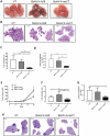
Vaccination with BoHV-4-mxCT decreases mammary cancer lung metastases. A-D) BALB/c mice were vaccinated twice with BoHV-4-mxCT, BoHV-4-A29 or left untreated. One week after the final administration, TUBO-derived tumorspheres were injected into the tail vein of treated mice. 20 days after cell challenge, lungs were removed, sectioned and micrometastasis number was determined in at least 2 H&E sections per mouse. A) Photograph of representative lungs, B) Representative images of lung metastases after H&E staining. Graphs showing C) the % of metastatic area and D) metastases number per square mm (sqmm), measured in mouse lungs from 3 independent experiments. E-H) BALB/c mice were s.c. challenged with 1 × 104 4T1 tumorsphere-derived cells. When the tumors reached 24 mm diameter, mice were vaccinated and boosted 14 days later. E) Subcutaneous tumor diameters were measured at the indicated time points and tumor volume was calculated. Graphs showing F) the % of metastatic area and G) the number of metastases per square mm measured in lungs. H) Representative images of lung metastases after H&E staining. *P < 0.05, **P < 0.01, Student’s t-test.
Figure 5.
Figure 5.
BoHV-4-mxCT induces lung T lymphocyte activation in vaccinated mice. Cytofluorimetric analysis of lung immune infiltrates in untreated (white bars), BoHV4-A29- (gray bars) and BoHV-4-mxCT- (black bars) vaccinated mice. A) Graph shows the percentage ± SEM of CD45+ myeloid cells expressing the markers of mMDSC (CD11b+Ly6C+), neutrophil/gMDSC (CD11b+Ly6G+), macrophage (CD11b+F4/80+), and dendritic cell (CD11bCD11c+), populations. B) Graph shows the percentage ± SEM of CD45+ cells expressing the markers of T (CD3+CD49b), and NK (CD3CD49b+), populations. C) Percentage ± SEM of CD4+ or CD8+ cells among the CD45+CD3+ T cell population. D) Percentage ± SEM of CD69+ cells among total T, CD4+ or CD8+ T, and NK cell populations. E) Percentage ± SEM of PD1+ cells among total T, CD4+ or CD8+ T, and NK cell populations. F) Percentage ± SEM of PDL1+ cells among epithelial (CD45) or myeloid (CD11b+) cells. G-H) Representative density plots showing G) CD3 and CD49b expression on CD45+ cells, and H) CD69 and PD1 expression on CD4+ or CD8+ T lymphocytes . *, P < 0.05; **, P < 0.01, ***, P < 0.001, Student’s t-test.
Figure 6.
Figure 6.
BoHV-4-mxCT vaccination induces degranulation and IFN-γ production in T lymphocytes. A-N) Splenocytes from untreated (white bars), BoHV4-A29- (gray bars), and BoHV-4-mxCT- (black bars), vaccinated mice, were co-cultured with mitomycin C treated 4T1 cells overnight (A-F) or for 5 days (G-N), incubated with FITC-conjugated antibodies against CD107a and b in the presence of Brefeldin A for 4 hours, then stained with antibodies against CD45, CD4, CD8, and CD69 and for intracellular IFN-γ and analyzed by FACS. Graphs show means ± SEM of the percentages of A-C, G-I) CD4+ or D-F, L-N) CD8+ cells expressing A, D, G, L) CD69, B, E, H, M) IFN-γ or C, F, I, N) CD107 from three experiments. O) Splenocytes from untreated, BoHV4-A29 and BoHV-4-mxCT-vaccinated mice were co-cultured with CFSE+ 4T1 cells for 48 hours, then the percentage of 7-AAD+ dead cells among the CFSE+ target populations was analyzed by FACS. Graphs shows mean ± SEM of the percentages of specific lysis, calculated as described in Material and Methods. *, P < 0.05; **, P < 0.01, ***, P < 0.001, Student’s t-test.
Figure 7.
Figure 7.
Depletion of T lymphocytes impairs BoHV-4-mxCT anti-metastatic activity. A, B) BALB/c mice were vaccinated twice with BoHV-4-mxCT, BoHV-4-A29 or left untreated. One week after the final vaccination, TUBO-derived tumorspheres were injected intravenously. Mice were then treated i.p., every 3 days, with either PBS or 200 µg of anti-CD4 (@CD4), anti-CD8 (@CD8) or isotype control antibodies. 20 days after cell challenge, lungs were removed, sectioned and micrometastasis number was determined in H&E sections. Graphs showing means ± SEM of A) the % of metastatic area and B) metastasis number per square mm (sq mm) (n = 5 per group). C) TUBO-derived tumorspheres were incubated for 5 days with medium, sera of untreated, BoHV-4-A29- or BoHV-4-mxCT-vaccinated BALB/c mice, or with IgG purified from sera from BoHV-4-mxCT-vaccinated mice (50 μg/ml@xCT). FACS analysis of MHC class I, MHC class II and Fas, reported as means ± SEM of MFI from three experiments. *, P < 0.05; **, P < 0.01, ***, P < 0.001, Student’s t-test.

References

    1. Siegel RL, Miller KD, Jemal A.. Cancer Statistics, 2017. CA Cancer J Clin. 2017;67:7–30. doi:10.3322/caac.21387. - DOI - PubMed
    1. Colleoni M, Sun Z, Price KN, Karlsson P, Forbes JF, Thurlimann B, Gianni L, Castiglione M, Gelber RD, Coates AS, et al. Annual hazard rates of recurrence for breast cancer during 24 years of follow-up: results from the international breast cancer study group trials I to V. J Clin Oncol. 2016;34:927–935. doi:10.1200/JCO.2015.62.3504. - DOI - PMC - PubMed
    1. Peitzsch C, Tyutyunnykova A, Pantel K, Dubrovska A. Cancer stem cells: the root of tumor recurrence and metastases. Semin Cancer Biol. 2017;44:10–24. doi:10.1016/j.semcancer.2017.02.011. - VSports注册入口 - DOI - PubMed
    1. Baccelli I, Trumpp A. The evolving concept of cancer and metastasis stem cells. J Cell Biol. 2012;198:281–293. doi:10.1083/jcb.201202014. - DOI - PMC - PubMed
    1. Lollini PL, Cavallo F, De Giovanni C, Nanni P. Preclinical vaccines against mammary carcinoma. Expert Rev Vaccines. 2013;12:1449–1463. doi:10.1586/14760584.2013.845530. - DOI - PubMed

Publication types