Skip to main page content
U.S. flag

An official website of the United States government

Dot gov

The . gov means it’s official. Federal government websites often end in . gov or . mil. Before sharing sensitive information, make sure you’re on a federal government site VSports app下载. .

Https

The site is secure. The https:// ensures that you are connecting to the official website and that any information you provide is encrypted and transmitted securely V体育官网. .

. 2018 Oct 30;115(44):E10352-E10361.
doi: 10.1073/pnas.1812125115. Epub 2018 Oct 11.

RPA1 binding to NRF2 switches ARE-dependent transcriptional activation to ARE-NRE-dependent repression

Affiliations

RPA1 binding to NRF2 switches ARE-dependent transcriptional activation to ARE-NRE-dependent repression

"V体育官网" Pengfei Liu et al. Proc Natl Acad Sci U S A. .

"VSports在线直播" Abstract

NRF2 regulates cellular redox homeostasis, metabolic balance, and proteostasis by forming a dimer with small musculoaponeurotic fibrosarcoma proteins (sMAFs) and binding to antioxidant response elements (AREs) to activate target gene transcription. In contrast, NRF2-ARE-dependent transcriptional repression is unreported. Here, we describe NRF2-mediated gene repression via a specific seven-nucleotide sequence flanking the ARE, which we term the NRF2-replication protein A1 (RPA1) element (NRE). Mechanistically, RPA1 competes with sMAF for NRF2 binding, followed by interaction of NRF2-RPA1 with the ARE-NRE and eduction of promoter activity. Genome-wide in silico and RNA-seq analyses revealed this NRF2-RPA1-ARE-NRE complex mediates negative regulation of many genes with diverse functions, indicating that this mechanism is a fundamental cellular process VSports手机版. Notably, repression of MYLK, which encodes the nonmuscle myosin light chain kinase, by the NRF2-RPA1-ARE-NRE complex disrupts vascular integrity in preclinical inflammatory lung injury models, illustrating the translational significance of NRF2-mediated transcriptional repression. Our findings reveal a gene-suppressive function of NRF2 and a subset of negatively regulated NRF2 target genes, underscoring the broad impact of NRF2 in physiological and pathological settings. .

Keywords: MYLK/MLCK; NRF2; RPA1; acute lung injury; transcriptional regulation V体育安卓版. .


Conflict of interest statement

The authors declare no conflict of interest.

VSports在线直播 - Figures

Fig. 1.
Fig. 1.
NRF2 negatively regulates MYLK expression. (A) qRT-PCR analysis of MYLK expression in WT and NRF2−/− cell lines. n = 3. Each gene was normalized to its control (Ctrl); unnormalized results are shown in SI Appendix, Fig. S1A. Data are presented as mean ± SD. *P < 0.05. (B) Immunoblot analysis of WT and NRF2−/− cell lines and lung tissue lysates from C57BL/6J mice. Quantification is shown in SI Appendix, Fig. S1B. (C) Immunoblot analysis of HPAECs treated (16 h) with NRF2 activators SF (5 μM), tBHQ (20 μM), and AsIII (1 μM) and with the NRF2 inhibitor brusatol (40 nM). Quantification is shown in SI Appendix, Fig. S1C. (D) qRT-PCR analysis of gene expression in HPAECs pretreated with NRF2 activators and brusatol as in C. Each gene is normalized to its control (Ctrl). Unnormalized results are shown in SI Appendix, Fig. S1F. (E) TEER of HPAECs pretreated (16 h) with DMSO, tBHQ (20 μM), or brusatol (40 nM), with or without thrombin (1 U/mL) challenge. Data were collected continuously every 30 s during the entire 4.5-h period. n = 4. Data are presented as mean ± SD. (F) Representative immunofluorescence images of polymerized F-actin stained with phalloidin (red) in HPAECs pretreated for 16 h with SF (5 μM) or brusatol (40 nM), followed by a 5-min challenge with thrombin (1 U/mL). Nuclei were labeled with DAPI (blue). (Scale bar: 50 μm.)
Fig. 2.
Fig. 2.
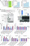
An NRE exists adjacent to the MYLK ARE. (A) Relative basal luciferase activity of A549 WT and NRF2−/− cells transfected with the pGL3-Luc vector containing portions of the human MYLK promoter cloned upstream of the firefly luciferase gene. Cells were cotransfected with Renilla luciferase to normalize firefly luciferase activity to this transfection control. n = 3. Data are presented as mean ± SD. *P < 0.05. (B) Pull-down assay of A549 WT and NRF2−/− cells. The biotinylated DNA probe containing the MYLK ARE WT or mutant (mARE) was incubated with lysates from either A549 WT or NRF2−/− cells. DNA-binding proteins were pulled down using streptavidin beads and detected by immunoblot analysis. (C) ChIP-PCR of A549 WT cells. Rabbit IgG served as a negative control. The expected size of the PCR product was 133 bp. (D and E) Relative luciferase activity of A549 WT and NRF2−/− cells transfected with the pGL4.22-Luc vector containing different AREs. WT and NRF2−/− cells were untreated or pretreated with brusatol (40 nM, 16 h). The sequences of the different AREs are shown in SI Appendix, Fig. S2 A and B. Cells were cotransfected with Renilla luciferase to normalize firefly luciferase activity to this transfection control. Results were further normalized to each untreated control (Ctrl). Unnormalized results are shown in SI Appendix, Fig. S2 C and D. n = 3. Data are presented as mean ± SD. *P < 0.05.
Fig. 3.
Fig. 3.
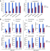
NRE-mediated attenuation of MYLK transcription is ARE-specific. (A) Relative basal luciferase activity of A549 WT and NRF2−/− cells transfected with the pGL4.22-Luc vector containing different response elements with and or without the NRE sequence. The promoter sequences are shown in SI Appendix, Fig. S3A. Cells were cotransfected with Renilla luciferase to normalize firefly luciferase activity to this transfection control. Results were further normalized to the cells transfected with response elements without NRE (WT) for each pair. Unnormalized results are shown in SI Appendix, Fig. S3B. n = 3. Data are presented as mean ± SD. *P < 0.05. (B) Relative luciferase activity of A549 WT and NRF2−/− cells transfected with different response elements with or without the NRE in uninduced (Ctrl) or induced condition [TNFα (20 ng/mL) for 4 h, CoCl2 (0.2 mM) for 16 h]. n = 3. Data are presented as mean ± SD. *P < 0.05.
Fig. 4.
Fig. 4.
Involvement of RPA1-NRE binding in repression of MYLK transcription. (A) Pull-down assay using a biotinylated dsDNA probe in A549-WT cell lysates. The probes used include WT ARE and WT NRE (MYLK-ARE-NRE), mutated ARE and WT NRE of MYLK (MYLK-mARE-NRE), and WT ARE and mutated NRE (MYLK-ARE-mNRE). Proteins identified by SDS/PAGE and silver staining as differentially pulled down (green rectangles, present in MYLK-ARE-NRE only) were identified by mass spectrometry. The probe sequences are shown in SI Appendix, Fig. S4A, and the peptides identified in MYLK-ARE-NRE pull-down are shown in SI Appendix, Fig. S4B. (B) Immunoblotting of biotinylated dsDNA probe pull-down of A549 WT and NRF2−/− cell lysates. (C) Immunoblot analysis of A549 WT and NRF2−/− cells transfected with control siRNA (Ctrl) or four different siRNAs targeting RPA1. Quantification is shown in SI Appendix, Fig. S4E. (D) qRT-PCR analysis of RPA1 and MYLK levels in A549 WT and NRF2−/− cells transfected with control siRNA or RPA1 siRNA. Unnormalized results are shown in SI Appendix, Fig. S4F. n = 3. Data are presented as mean ± SD. *P < 0.05. (E) Relative luciferase activity of A549 WT and NRF2−/− cells transfected with Ctrl siRNA or RPA1 siRNA. Cells were cotransfected with Renilla luciferase to normalize firefly luciferase activity to this transfection control. Results were further normalized to each untreated control (Ctrl). Unnormalized results are shown in SI Appendix, Fig. S4G. n = 3. Data are presented as mean ± SD. *P < 0.05.
Fig. 5.
Fig. 5.
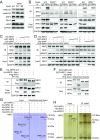
RPA1 competes with sMAFs to directly bind NRF2. (A) Immunoblot analysis of A549 WT and RPA1−/− cells. Quantification is shown in SI Appendix, Fig. S5A. (B) Immunoprecipitation assay using different antibodies with A549 WT, NRF2−/−, and RPA1−/− cell lysates. Rabbit and mouse IgG served as negative controls for NRF2 and RPA1 antibodies, respectively. (C) In vitro pull-down assay of purified GST-NRF2 (1 μg for each group) incubated with His-RPA1 (0, 0.5, 1, and 2 μg, respectively) and His-sMAF (1 μg for each group). (D) Immunoblotting of biotinylated dsDNA probe pull-down of purified GST-NRF2 (10 μg for each group), His-RPA1 (10 μg for each group), and His-sMAF (10 μg for each group); 0, 2.5, 5, and 10 μg of protein was used in the different dose-dependent groups. (E) Immunoprecipitation assay of HEK293 cells transfected with HA-tagged WT NRF2 (HA-NRF2-WT) or Neh1 domain deletion mutant (HA-NRF2ΔNeh1) alone or in combination with Flag-RPA1. (F) In vitro pull-down assay of purified His-RPA1 incubated with GST-NRF2-WT or GST-NRF2ΔNeh1. (G) In vitro pull-down assay and Coomassie blue staining of purified GST-NRF2-WT and His-tagged FL RPA1 (His-RPA1-FL) or RPA1 deletion mutants (His-RPA1-D1, His-RPA1-D2, or His-RPA1-D3). (H) In vitro immunoprecipitation assay and silver staining of purified His-sMAFG alone or in combination with GST-NRF2-WT and His-RPA1.
Fig. 6.
Fig. 6.
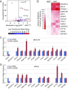
The NRF2-mediated negative transcriptional regulation is a fundamental mechanism controlling the expression of other genes. (A) Volcano plot showing differentially expressed genes harboring ARE-NRE sites for RNA-seq data comparing A549-RPA1+/+ and A549-RPA1−/− cells. The red line indicates −log10 (adjusted P value) = 1.30, which corresponds to an adjusted P value of 0.05. Points are colored according to their log-transformed base mean value. (B) Heatmap representing variance stabilized-transformed distance data from RNA sequencing of A549-RPA+/+ and A549-RPA−/− cells. The presented genes harbor ARE-NRE sites within their promoter regions or the first intron and follow the same expression pattern as MYLK following RPA1 knockout. (C and D) qRT-PCR analysis of candidate gene mRNA expression in Ctrl-siRNA– and RPA1-siRNA–transfected BEAS-2B cells (C) and HPAECs (D). n = 3. Data are presented as mean ± SD. *P < 0.05.
Fig. 7.
Fig. 7.
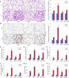
NRF2-driven repression of MYLK expression attenuates inflammatory lung injury. (A, Left) Hematoxylin and eosin staining of lung tissue sections from control (Ctrl; n = 3) and VILI (n = 6) mice. (Scale bar: 50 μm.) (A, Right) Quantification of inflammatory cell infiltration is shown on the right. (B, Left) IHC staining of 8-oxo-dG in lung tissue sections from Ctrl (n = 3) and VILI (n = 6) mice. (Scale bar: 50 μm.) (B, Right) Relative intensity of 8-oxo-dG staining. (C) Total protein concentration in BAL fluid from Ctrl (n = 3) and VILI (n = 6) mice. Data are presented as mean ± SD. *P < 0.05 compared with WT mice; # P < 0.05 compared with Nrf2−/−;Mylk−/− mice. (D) Total BAL cell numbers from Ctrl (n = 3) and VILI (n = 6) mice. Data are presented as mean ± SD. *P < 0.05 compared with WT mice; #P < 0.05 compared with Nrf2−/−;Mylk−/− mice. (E) Percentage of BAL neutrophils in Ctrl (n = 3) and VILI (n = 6) mice. Data are presented as mean ± SD. *P < 0.05 compared with WT mice; #P < 0.05 compared with Nrf2−/−;Mylk−/− mice. (F) Percentage of BAL lymphocytes in Ctrl (n = 3) and VILI (n = 6) mice. Data are presented as mean ± SD. *P < 0.05 compared with WT mice; # P < 0.05 compared with Nrf2−/−;Mylk−/− mice. (G) BAL IL-6 quantification in Ctrl (n = 3) and VILI (n = 6) mice. Data are presented as mean ± SD. *P < 0.05 compared with WT mice; #P < 0.05 compared with Nrf2−/−;Mylk−/− mice. (H) BAL TNFα quantification in Ctrl (n = 3) and VILI (n = 6) mice. Data are presented as mean ± SD. *P < 0.05 compared with WT mice; #P < 0.05 compared with Nrf2−/−;Mylk−/− mice.
Fig. 8.
Fig. 8.
Model of NRF2-RPA1-ARE-NRE–dependent transcriptional repression.

"VSports手机版" References

    1. Tao S, et al. Bixin protects mice against ventilation-induced lung injury in an NRF2-dependent manner. Sci Rep. 2016;6:18760. - PMC - PubMed
    1. Mills EL, et al. Itaconate is an anti-inflammatory metabolite that activates Nrf2 via alkylation of KEAP1. Nature. 2018;556:113–117. - PMC (V体育安卓版) - PubMed
    1. Hayes JD, Dinkova-Kostova AT. The Nrf2 regulatory network provides an interface between redox and intermediary metabolism. Trends Biochem Sci. 2014;39:199–218. - V体育平台登录 - PubMed
    1. Rojo de la Vega M, Chapman E, Zhang DD. NRF2 and the hallmarks of cancer. Cancer Cell. 2018;34:21–43. - PMC - PubMed
    1. Mitsuishi Y, et al. Nrf2 redirects glucose and glutamine into anabolic pathways in metabolic reprogramming. Cancer Cell. 2012;22:66–79. - PubMed

Publication types

V体育2025版 - Substances

LinkOut - more resources (VSports最新版本)