Skip to main page content
U.S. flag

An official website of the United States government

Dot gov

The . gov means it’s official. Federal government websites often end in VSports app下载. gov or . mil. Before sharing sensitive information, make sure you’re on a federal government site. .

Https

The site is secure. The https:// ensures that you are connecting to the official website and that any information you provide is encrypted and transmitted securely V体育官网. .

. 2018 Sep 13;16(9):e2006092.
doi: 10.1371/journal.pbio.2006092. eCollection 2018 Sep.

Refined RIP-seq protocol for epitranscriptome analysis with low input materials

Affiliations

V体育2025版 - Refined RIP-seq protocol for epitranscriptome analysis with low input materials

Yong Zeng et al. PLoS Biol. .

Abstract

N6-Methyladenosine (m6A) accounts for approximately 0. 2% to 0. 6% of all adenosine in mammalian mRNA, representing the most abundant internal mRNA modifications. m6A RNA immunoprecipitation followed by high-throughput sequencing (MeRIP-seq) is a powerful technique to map the m6A location transcriptome-wide. However, this method typically requires 300 μg of total RNA, which limits its application to patient tumors. In this study, we present a refined m6A MeRIP-seq protocol and analysis pipeline that can be applied to profile low-input RNA samples from patient tumors. We optimized the key parameters of m6A MeRIP-seq, including the starting amount of RNA, RNA fragmentation, antibody selection, MeRIP washing/elution conditions, methods for RNA library construction, and the bioinformatics analysis pipeline. With the optimized immunoprecipitation (IP) conditions and a postamplification rRNA depletion strategy, we were able to profile the m6A epitranscriptome using 500 ng of total RNA. We identified approximately 12,000 m6A peaks with a high signal-to-noise (S/N) ratio from 2 lung adenocarcinoma (ADC) patient tumors. Through integrative analysis of the transcriptome, m6A epitranscriptome, and proteome data in the same patient tumors, we identified dynamics at the m6A level that account for the discordance between mRNA and protein levels in these tumors. The refined m6A MeRIP-seq method is suitable for m6A epitranscriptome profiling in a limited amount of patient tumors, setting the ground for unraveling the dynamics of the m6A epitranscriptome and the underlying mechanisms in clinical settings. VSports手机版.


Conflict of interest statement

The method is subjected to a University Health Network patent application with Shiyan Wang, Yong Zeng, and Housheng He as inventors.

Figures

Fig 1
Fig 1. Low/high salt-washing method outperforms competitive elution method.
(A) Schematic diagram of 3 strategies of m6A MeRIP. (B) S/N ratio of GLuc/CLuc was highest in Method II using low/high salt washing. An RNA mixture containing equal amounts of the m6A modified control RNA GLuc, the unmodified control RNA CLuc, and NEB antibody were used for m6A MeRIP. (C) S/N ratio of GLuc/CLuc was further increased in a second round of IP using Method II. S/N ratio of GLuc/CLuc (panel D) and SETD7/GAPDH (panel E) in 3 replicates of 1 round of IP. Data related to this figure can be found in S1 Data. CLuc, unmodified control RNA; GLuc, m6A-modified control RNA; IP, immunoprecipitation; m6A, N6-Methyladenosine; m6A MeRIP, m6A RNA immunoprecipitation followed by high-throughput sequencing; NEB, New England Biolabs; S/N, signal-to-noise.
Fig 2
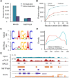
Fig 2. Comparison between MACS- and MeTPeak-based m6A detection pipeline with published data from A549.
(A) m6A peaks detected by MACS and MeTPeak with or without inclusion of duplication reads. (B) m6A peaks detected with increasing sequencing depth. The dashed line represents the overlapping peaks called by both MeTpeak and MACS. (C) Top m6A motifs detected from top 5,000 m6A summit centered at 200-nt peak regions. (D) The location and frequency of the top motif to the summit; ***p < 1 × 10⁻⁴. (E) Example showing the difference between the results of MACS and MeTPeak. Data related to this figure can be found in S1 Data. IP, immunoprecipitation; m6A, N6-Methyladenosine.
Fig 3
Fig 3. Comparison of 3 different m6A antibodies for MeRIP.
(A) S/N ratio of GLuc/CLuc and SETD7/GAPDH with different antibodies. The amount of 32 μg total RNA from human lung cancer cell line A549 with spiked-in control RNA GLuc and CLuc was used for m6A MeRIP using Method II. (B) m6A peak signals of SETD7 transcripts in 3 MeRIP-seq libraries. (C) Overlap of m6A peaks from the SySy, NEB, and Millipore libraries. (D) Number of m6A peaks called by subsampling to different read depths with different antibodies. (E) The percentages of m6A peaks in 5 nonoverlapping transcript segments: TSS; 5’UTR; CDS; stop codon; and 3’UTR. (F) Metagene profiles depicting sequence coverage in windows surrounding the TSS and stop codon demonstrated that m6A peaks were enriched in the vicinity of the stop codon. (G) Top enriched motifs identified in the SySy, NEB, and Millipore libraries. Data related to this figure can be found in S1 Data. CDS, coding sequence; CLuc, unmodified control RNA; GLuc, m6A-modified control RNA; m6A, N6-Methyladenosine; m6A MeRIP, m6A RNA immunoprecipitation followed by high-throughput sequencing; NEB, New England Biolabs; S/N, signal-to-noise; SySy, Synaptic Systems; TSS, transcription start site; UTR, untranslated region.
Fig 4
Fig 4. Optimized m6A MeRIP-seq protocol worked well starting with 2 μg total RNA.
(A) MeRIP efficiency decreases with the reduction of starting RNA amount. (B) At the same sequencing depth, the total number of m6A peaks identified increased with the increase of starting RNA amount. The “unique” and “overlapped with 2 μg” peaks indicate the peak number compared to 2 μg. (C) The average RNA expression level of the transcripts with unique m6A peaks identified in the 32-μg library was significantly lower than that of overlapping m6A peaks; ***p < 1 × 10⁻⁴. (D) Top: pie chart represents the proportion of m6A peaks in each of the 5 nonoverlapping transcript segments in the 2-μg library. Middle: relative enrichment of m6A peaks across the 5 nonoverlapping transcript segments in the 2 μg library. Bottom: top enriched motifs in the 2 μg library. Data related to this figure can be found in S1 Data. CDS, coding sequence; m6A, N6-Methyladenosine; m6A MeRIP, m6A RNA immunoprecipitation followed by high-throughput sequencing; TSS, transcription start site; UTR, untranslated region.
Fig 5
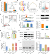
Fig 5. m6A dynamics in ADC tumors.
(A) Transcriptome-wide distribution of m6A sites. (B) Top motif and their distance to summit of m6A peaks; ***p < 1 × 10⁻⁴. (C) Volcano plot for peaks with differential m6A intensity between tumor1 and tumor2. RTumor1 and RTumor2 stand for the ratio of IP over Input for sample tumor1 and tumor2, respectively. Peaks with p < 1 × 10⁻¹⁰ were reassigned to be p = 1 × 10⁻¹⁰. (D) Correlation between the ratios (tumor1/tumor2) at RNA and protein levels for genes with differential m6A peaks. Red dots represent genes with discordance ratio at mRNA and protein levels, and dot size represents m6A odds ratio between tumor1 and tumor2. (E) Top: the IP (orange for tumor1 and cyan for tumor2) and Input (light green) signal at m6A peak (marked by red box) near SLC2A1 stop codon in the 2 tumors. Bottom: ratio between tumor1 and tumor2 at mRNA and protein levels for SLC2A1. (F) Western blotting analysis of tumor1 and tumor2 samples. (G) Real-time PCR analysis of METTL3 and SLC2A1 mRNA expression levels in A549 upon silencing of METTL3 using 2 different siRNAs. *p < 0.05; ***p < 1 × 10⁻⁴. (H) Western blotting analysis of METTL3 and SLC2A1 protein levels in the A549 upon METTL3 knockdown. (I) Real-time PCR analysis of METTL3 mRNA in rescue assays. METTL3 primer recognized both full-length and 1–200AA METTL3 mutant. METTL3 knockdown cells were transfected with either full-length METTL3 (siM3_1+full) or 1–200AA METTL3 mutant overexpressing plasmid (siM3_1+200). siM3_1 is the abbreviation for siMETTL3_1; ***p < 1 × 10⁻⁴. (J) Western blotting analysis of SLC2A1 in rescue assays. Data related to this figure can be found in S1 Data. ADC, adenocarcinoma; CDS, coding sequence; IP, immunoprecipitation; lincRNA, long intergenic noncoding RNA; m6A, N6-Methyladenosine; METTL3, Methyltransferase Like 3; siRNA, small interfering RNA; SLC2A1, Solute Carrier family 2, Facilitated Glucose Transporter member 1; TSS, transcription start site; UTR, untranslated region.

References

    1. Zhao BS, Roundtree IA, He C. Post-transcriptional gene regulation by mRNA modifications. Nat Rev Mol Cell Biol. Nature Publishing Group; 2017;18: 31–42. 10.1038/nrm.2016.132 - DOI - PMC - PubMed
    1. Roignant J-Y, Soller M. m(6)A in mRNA: An Ancient Mechanism for Fine-Tuning Gene Expression. Trends Genet. 2017;33: 380–390. 10.1016/j.tig.2017.04.003 - DOI (VSports最新版本) - PubMed
    1. Meyer KD, Jaffrey SR. Rethinking m(6)A Readers, Writers, and Erasers. Annu Rev Cell Dev Biol. Annual Reviews 4139 El Camino Way, PO Box 10139, Palo Alto, California 94303–0139, USA; 2017;33: annurev–cellbio–100616–060758. 10.1146/annurev-cellbio-100616-060758 - DOI - PMC - PubMed
    1. Boccaletto P, Machnicka MA, Purta E, Piatkowski P, Baginski B, Wirecki TK, et al. MODOMICS: a database of RNA modification pathways. 2017 update. Nucleic Acids Res. 2018;46: D303–D307. 10.1093/nar/gkx1030 - DOI - PMC - PubMed
    1. Desrosiers R, Friderici K, Rottman F. Identification of methylated nucleosides in messenger RNA from Novikoff hepatoma cells. Proc Natl Acad Sci USA. National Academy of Sciences; 1974;71: 3971–3975. - V体育2025版 - PMC - PubMed

Publication types