<code lang="j1Io7gL"></code> VSports在线直播 - Skip to main page content
U.S. flag

An official website of the United States government

Dot gov

The . gov means it’s official. Federal government websites often end in . gov or . mil. Before sharing sensitive information, make sure you’re on a federal government site. VSports app下载.

Https

The site is secure. The https:// ensures that you are connecting to the official website and that any information you provide is encrypted and transmitted securely. V体育官网.

. 2018 Oct;24(10):1339-1350.
doi: 10.1261/rna.064238.117. Epub 2018 Jul 3.

"V体育安卓版" The m6A reader protein YTHDC2 interacts with the small ribosomal subunit and the 5'-3' exoribonuclease XRN1

Affiliations

The m6A reader protein YTHDC2 interacts with the small ribosomal subunit and the 5'-3' exoribonuclease XRN1

"VSports" Jens Kretschmer et al. RNA. 2018 Oct.

Abstract (V体育平台登录)

N6-methyladenosine (m6A) modifications in RNAs play important roles in regulating many different aspects of gene expression. While m6As can have direct effects on the structure, maturation, or translation of mRNAs, such modifications can also influence the fate of RNAs via proteins termed "readers" that specifically recognize and bind modified nucleotides. Several YTH domain-containing proteins have been identified as m6A readers that regulate the splicing, translation, or stability of specific mRNAs. In contrast to the other YTH domain-containing proteins, YTHDC2 has several defined domains and here, we have analyzed the contribution of these domains to the RNA and protein interactions of YTHDC2. The YTH domain of YTHDC2 preferentially binds m6A-containing RNAs via a conserved hydrophobic pocket, whereas the ankyrin repeats mediate an RNA-independent interaction with the 5'-3' exoribonuclease XRN1. We show that the YTH and R3H domains contribute to the binding of YTHDC2 to cellular RNAs, and using crosslinking and analysis of cDNA (CRAC), we reveal that YTHDC2 interacts with the small ribosomal subunit in close proximity to the mRNA entry/exit sites. YTHDC2 was recently found to promote a "fast-track" expression program for specific mRNAs, and our data suggest that YTHDC2 accomplishes this by recruitment of the RNA degradation machinery to regulate the stability of m6A-containing mRNAs and by utilizing its distinct RNA-binding domains to bridge interactions between m6A-containing mRNAs and the ribosomes to facilitate their efficient translation. VSports手机版.

Keywords: N6-methyladenosine (m6A); RNA modification; YTH domain; exoribonuclease; ribosome; translation V体育安卓版. .


Figures

FIGURE 1.
FIGURE 1.
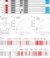
The YTH domain of YTHDC2 preferentially binds m6A-containing RNAs. (A) Schematic view of the defined domains of YTHDC2 (top) and truncations used (middle, bottom). (FL) Full-length YTHDC2, (YTH) YT521-B homology domain, (R3H) RNA binding domain characterized by an R-(X3)-H motif, (ANK) ankyrin repeat. (B) Nine-nucleotide RNAs labeled with 5′-fluorescein were prepared by solid-phase synthesis followed by labeling via click chemistry. Purified RNAs were analyzed by anion exchange HPLC monitored by UV absorbance. (C) The wild-type YTH domain of YTHDC2 (YTH) and the YTH domain of YTHDC2 carrying W1310A or W1360A substitutions were expressed in E. coli and purified. Purified proteins were separated by SDS-PAGE and visualized by Coomassie staining. (D) Anisotropy measurements of a fluorescein-labeled RNA containing either a N6-methyladenosine (m6A) or an unmodified adenosine in the presence of different amounts of the YTH domain of YTHDC2. Data from three independent experiments are shown as mean ± standard deviation. (E) A sequence alignment of YTH domains of the five human YTH domain-containing proteins is shown. Secondary structural features of the YTH domain of YTHDC1 (PBD 4R3I) are shown above the corresponding amino acids. (α) alpha helix, (β) beta sheet, (TT) turn, (η) 310-helix. Amino acids conserved in all YTH domain proteins are highlighted with a red background, and letters denoting amino acids with similar properties are shown in red. The tryptophan residues proposed to contribute to formation of a hydrophobic m6A-binding pocket are indicated. (F) Anisotropy measurements of a fluorescein-labeled RNA containing an N6-methyladenosine (m6A) with different amounts of the wild-type YTH domain of YTHDC2 (YTH) or the YTH domain in which tryptophan 1310 or tryptophan 1360 were substituted for alanine (W1310A and W1360A, respectively). Data from three independent experiments are shown as mean ± standard deviation.
FIGURE 2.
FIGURE 2.
Both the YTH and R3H domains contribute to RNA binding by YTHDC2 and YTHDC2 interacts specifically with the 5′–3′ exoribonuclease XRN1 via its ANK repeats. (A) Cells expressing equal amounts of Flag-tagged versions of full-length YTHDC2, YTHDC2 lacking the R3H domain (ΔR3H), and YTHDC2 lacking the YTH domain (ΔYTH) were treated with 4-thiouridine and crosslinked in vivo. After tandem affinity purification of crosslinked protein–RNA complexes, RNA trimming and 5′ labeling with [32P], complexes were separated by denaturing PAGE, transferred to a nitrocellulose membrane, and radioactively labeled RNAs were detected using autoradiography. Protein eluates were analyzed by western blotting using an anti-Flag antibody. (B) Extracts from HEK293 cells expressing Flag-tagged YTHDC2, YTHDC1, YTHDF1, YTHDF2, YTHDF3, or the Flag tag were used in immunoprecipitation experiments in the presence (+) or absence (−) of RNase A and T1 (RNase). Inputs (1%) and eluates (IP) were analyzed by western blotting using antibodies against XRN1, GAPDH, and the Flag tag. (C) Immunoprecipitation experiments were performed and analyzed as in B in the absence of RNase treatment using extracts prepared from cells expressing Flag-tagged full-length YTHDC2 (YTHDC2), YTHDC2 lacking the R3H domain (ΔR3H), the YTH domain (ΔYTH), or one or both ankyrin repeats (ΔANK1, ΔANK2, ΔANK1+2). All experiments presented in this figure were performed in duplicate or triplicate and representative data are shown.
FIGURE 3.
FIGURE 3.
YTHDC2 is enriched in perinuclear regions and associates with ribosomes. (A) HeLa cells or HEK293 cells expressing Flag-tagged YTHDC2 were fixed, and the localization of YTHDC2 was determined by immunofluorescence using an anti-YTHDC2 antibody (upper panels; HeLa cells) or an anti-Flag antibody (lower panels; HEK cells). Nuclear material was visualized by DAPI staining and an overlay (merge; YTHDC2, green; DAPI, blue) is shown. Scale bar represents 10 µm. (B) Extracts prepared from HeLa cells and a HEK293 cell line expressing FLAG-tagged YTHDF2 were separated by sucrose density gradient centrifugation. The distributions of YTHDC2 and YTHDF2 were determined by western blotting using an anti-YTHDC2 antibody (upper panel) or an anti-Flag antibody (lower panel; YTHDF2-Flag). The absorbance of the fractions at 260 nm was used to generate a profile on which the peaks corresponding to the 40S and 60S ribosomal subunits and 80S monosomes are indicated. All experiments presented in this figure were performed at least in triplicate and representative data are shown.
FIGURE 4.
FIGURE 4.
YTHDC2 contacts the head region of the small ribosomal subunit. (A) HEK293 cells expressing YTHDC2-Flag or the Flag tag were UV crosslinked in vivo. Protein–RNA complexes were tandem affinity purified, and RNAs were trimmed, ligated to adaptors, and labeled at the 5′ end using [32P]. Complexes were separated by PAGE, transferred to a nitrocellulose membrane, and labeled RNAs were visualized by autoradiography. The area excised from the membrane is indicated by red boxes. (B) RNA fragments isolated from the membrane shown in A were used to prepare a cDNA library that was subjected to Illumina sequencing. The obtained sequence reads were mapped to the human genome, and the number of reads per nucleotide mapped to the rDNA sequence is shown above a schematic view of the 47S pre-rRNA transcript containing the sequences of the mature 18S, 5.8S, and 28S rRNAs. (ETS) external transcribed spacer, (ITS) internal transcribed spacer. Asterisks indicate peaks that are present in both YTHDC2-Flag and Flag samples. Three independent CRAC experiments were performed and two representative data sets are presented. (C) Magnified views of the YTHDC2-Flag and Flag CRAC reads mapping close to the 3′ end of the 18S rRNA are shown. Lower panel indicates the number and position of T–C mutations that are introduced as a result of nucleotide mis-incorporation during reverse transcription at sites where a 4-thiouridine is crosslinked to an amino acid. (D) The number of reads in the YTHDC2-Flag A data set mapping to each nucleotide of the 18S rRNA are shown on the 3D structure of the mature 18S rRNA (PDB 4V6X) using a color scale in which the nucleotides to which the maximum number of reads map are shown in red (100%) and nucleotides with lesser numbers of mapped reads are indicated in yellow (above a threshold of 30%). The ribosomal proteins are indicated in pale cyan, the 18S rRNA in gray, and key structural features of the small ribosomal subunit are labeled.

VSports app下载 - References

    1. Adams JM, Cory S. 1975. Modified nucleosides and bizarre 5′-termini in mouse myeloma mRNA. Nature 255: 28–33. - "V体育2025版" PubMed
    1. Alarcón CR, Goodarzi H, Lee H, Liu X, Tavazoie S, Tavazoie SF. 2015. HNRNPA2B1 is a mediator of m6A-dependent nuclear RNA processing events. Cell 162: 1299–1308. - V体育平台登录 - PMC - PubMed
    1. Anger AM, Armache JP, Berninghausen O, Habeck M, Subklewe M, Wilson DN, Beckmann R. 2013. Structures of the human and Drosophila 80S ribosome. Nature 497: 80–85. - PubMed
    1. Atanassov I, Urlaub H. 2013. Increased proteome coverage by combining PAGE and peptide isoelectric focusing: comparative study of gel-based separation approaches. Proteomics 13: 2947–2955. - PMC - PubMed
    1. Bailey AS, Batista PJ, Gold RS, Chen YG, de Rooij DG, Chang HY, Fuller MT. 2017. The conserved RNA helicase YTHDC2 regulates the transition from proliferation to differentiation in the germline. Elife 6: e26116. - PMC - PubMed

Publication types

MeSH terms