Skip to main page content
U.S. flag

An official website of the United States government

Dot gov

The VSports app下载. gov means it’s official. Federal government websites often end in . gov or . mil. Before sharing sensitive information, make sure you’re on a federal government site. .

Https

The site is secure. The https:// ensures that you are connecting to the official website and that any information you provide is encrypted and transmitted securely. V体育官网.

. 2018 Mar 27;9(1):1239.
doi: 10.1038/s41467-018-02977-8.

"VSports app下载" Ikaros family zinc finger 1 regulates dendritic cell development and function in humans

Affiliations

Ikaros family zinc finger 1 regulates dendritic cell development and function in humans (V体育ios版)

Urszula Cytlak et al. Nat Commun. .

"VSports手机版" Abstract

Ikaros family zinc finger 1 (IKZF1) is a haematopoietic transcription factor required for mammalian B-cell development. IKZF1 deficiency also reduces plasmacytoid dendritic cell (pDC) numbers in mice, but its effects on human DC development are unknown. Here we show that heterozygous mutation of IKZF1 in human decreases pDC numbers and expands conventional DC1 (cDC1). Lenalidomide, a drug that induces proteosomal degradation of IKZF1, also decreases pDC numbers in vivo, and reduces the ratio of pDC/cDC1 differentiated from progenitor cells in vitro in a dose-dependent manner. In addition, non-classical monocytes are reduced by IKZF1 deficiency in vivo VSports手机版. DC and monocytes from patients with IKZF1 deficiency or lenalidomide-treated cultures secrete less IFN-α, TNF and IL-12. These results indicate that human DC development and function are regulated by IKZF1, providing further insights into the consequences of IKZF1 mutation on immune function and the mechanism of immunomodulation by lenalidomide. .


Conflict of interest statement

The authors declare no competing financial interests.

"V体育平台登录" Figures

Fig. 1
Fig. 1
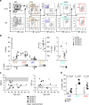
FlowSOM analysis of PB reveals a reduced proportion of pDC but increased cDC1 in a patient carrying IKZF1 mutation. a Minimal spanning tree visualisation of a self-organising map using compensated flow cytometric data from a family B member (B5) compared to a travel control (equal number of events). Data were taken from the lineage (CD3, CD7, CD14, CD16, CD19, CD20)-negative HLA-DR-positive gate. FlowSOM nodes represent clusters of cells. Metaclusters of the nodes, determined by the map, are represented by the background colour of the nodes. Star charts represent the mean marker value of cells in that node with the height of each part corresponding to marker intensity. b Comparison between samples used to generate the map; size of nodes represents proportional number of cells in each node, colour represents proportional differences between samples with red and blue indicating higher or lower numbers in IKZF1 mutation compared to wild type, respectively. c mRNA and d protein expression of IKZF1 in healthy donor monocytes and dendritic cells by NanoString gene expression analysis of FACS sorted cells or intracellular flow cytometry, respectively (n = 3 donors for each experiment). IKZF3 mRNA expression is shown for comparison in c. cDC1/2, conventional dendritic cell 1/2; pDC, plasmacytoid dendritic cell; 14+ CD14+, classical monocyte; 16+ CD16+, non-classical monocyte
Fig. 2
Fig. 2
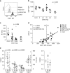
Reduced pDC and non-classical monocytes but expanded cDC1 in the IKZF1 haploinsufficiency cohort. a Flow cytometric quantification of PB monocyte and dendritic cell (DC) subsets showing HLA-DR+CD4 B cells, CD14+ classical (orange) and CD16+ non-classical monocytes (brown), CD123+ plasmacytoid DCs (pDC) (blue), CD141+ conventional DC1 (cDC1) (turquoise) and CD1c+ cDC2 (red) in healthy donor (cont, (control)) and representative individual with IKZF1 mutation. b Absolute counts of monocytes and DCs in 20 individuals from 4 affected families compared to n = 32 healthy donors. Bars show mean ± 95% confidence interval (CI). Histograms show subset-specific p-values for each family compared to healthy donors by two-tailed Mann–Whitney U-test with significance set at p < 0.05. c Absolute pDC or cDC1 counts plotted against the age of the individual. Grey zones represent the normal range of healthy controls (n = 32), black lines represent linear regression analysis with p = 0.77 and p = 0.86, respectively. d Proportion of cells expressing CD56 in healthy donors (n = 5) and individuals carrying IKZF1 mutation (n = 6). Bars represent mean ± 95% CI. The p-values derived from two-tailed Mann–Whitney U-test
Fig. 3
Fig. 3
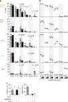
Dose-dependent reduction of IKZF1 protein and pDCs in patients receiving lenalidomide treatment. a Intracellular flow cytometric quantification (mean fluorescence intensity (MFI)) of IKZF1 protein levels in B cells (identified as SSClowLinDR+CD2 cells) of a family G member carrying IKZF1 mutation, patient on lenalidomide treatment, a healthy donor and isotype control. b Intracellular IKZF1 level (MFI) in healthy donors (n = 5), patients tested at the end of a week off treatment with specified dose of lenalidomide (n = 16) (5, 10, 15 or 25 mg daily) and patients taking lenalidomide (Len) on the day of analysis (n = 3) (15 mg daily). Bars represent mean ± s.d. *p = 0.036 by two-tailed Mann–Whitney U-test versus healthy donor controls. c Summary of absolute B-cell counts in whole blood from healthy donors (n = 4) (Cont), patients tested at the end of a week off treatment with specified dose of lenalidomide (n = 16) (5 or 10, 15 or 25 mg daily) and patients taking lenalidomide on the day of analysis (n = 3) (15 mg daily). Bars represent mean ± s.d. *p = 0.006 Mann–Whitney U-test versus healthy donor controls. d IKZF1 protein levels (MFI) plotted against absolute plasmacytoid dendritic cell (pDC) counts in affected (n = 3) and unaffected (n = 1) members of family G (white or grey open circles, respectively), patients after a week off lenalidomide (n = 22) (black circles) or taking lenalidomide on the day of analysis (n = 3) (black diamond) or healthy donors (n = 4) (grey circles). Data from two patients taking pomalidomide (4 mg daily) are displayed (grey triangles). Black line represents linear regression analysis of lenalidomide-treated patients only. Inclusion of family G members and healthy controls in the linear regression analysis resulted in r2 = 0.6541, p < 0.0001. Inclusion of pomalidomide-treated patients resulted in r2 = 0.6419, p < 0.0001. Dashed line represents IKZF1 MFI = 680 dividing group R1 (IKZF1 MFI > 680) from group R2 (IKZF1 MFI < 680). e PB absolute monocyte and DC counts in healthy controls (C) compared to patients from group R1 and R2. The p-values from two-tailed Mann–Whitney U-test of R2 versus healthy donors
Fig. 4
Fig. 4
Reduced production of TNF, IL-12 and IFN-α in IKZF1 deficiency. a Intracellular flow cytometric analysis of cytokine production (% positive cells) in PB CD14+ monocytes (mono), CD14 monocytes and dendritic cell (DC) subsets (cDC2, cDC1, pDC) from n = 5 individuals carrying IKZF1 mutation (black bars) or n = 8 healthy donors (grey bars) following 14 h of stimulation of PBMCs with a cocktail of TLR agonists (CpG, poly(I:C), CL075, LPS). Histograms represent mean and bars represent s.e.m. Dots represent individuals. The p-values derived from Mann–Whitney U-test of IKZF1 mutation versus healthy donors. b Analysis repeated on healthy donor PBMCs in the presence of increasing concentrations of lenalidomide (0, 0.1, 1 or 10 μM). Black dots represent mean, bars represent s.e.m. and lines represent individual data points from n = 3 donors in each condition. c IL-12 or IFN-α production from cDC2 or pDC, respectively, from healthy donor PBMCs stimulated with TLR cocktail (TLR) and without (solid bars) or with (unfilled bars) the addition of exogenous IFN-α and without (grey) or with (black) anti-CD303 and anti-CD304 antibodies
Fig. 5
Fig. 5
Impaired in vitro pDC development in IKZF1 deficiency. a Phenotypic flow cytometric analysis of dendritic cell (DC) subsets generated after 22 days of culture in vitro, compared to PB counterparts. Found within the LineageHLA-DR+ gate were CD14+CD11c+ monocytes, CD141+CLEC9A+ cDC1s, CD11c+CD1c+ cDC2s and CD123+CD303+CD304+ pDC. b IKZF1 protein levels, determined by flow cytometric analysis and IKZF1 mean fluorescence intensity (MFI), in LinDR+ cells after 22 days of culture in the presence of a lenalidomide titration. c Summary of the proportion of in vitro generated LinDR+ cells identified as CD14+ monocytes (orange), cDC2 (red), cDC1 (turquoise) and pDC (blue). Bars represent mean and s.e.m. of triplicate wells, dots represent single cultures. d Ratio of total number of LinDR+ cells generated in vitro per input CD34+ cell in the presence of increasing concentrations of lenalidomide. e Lenalidomide dose response of the cDC1 to pDC ratio of in vitro generated cells. Grey zone indicates published human in vivo plasma Cmax of lenalidomide (450–600 ng/ml or 1.7–2.3 μM). Dots represent mean, bars represent s.e.m. and lines represent individual cultures

V体育2025版 - References

    1. Milner JD, Holland SM. The cup runneth over: lessons from the ever-expanding pool of primary immunodeficiency diseases. Nat. Rev. Immunol. 2013;13:635–648. doi: 10.1038/nri3493. - V体育平台登录 - DOI - PubMed
    1. Collin M, Bigley V, Haniffa M, Hambleton S. Human dendritic cell deficiency: the missing ID? Nat. Rev. Immunol. 2011;11:575–583. doi: 10.1038/nri3046. - "V体育官网入口" DOI - PubMed
    1. Kapsenberg ML. Dendritic-cell control of pathogen-driven T-cell polarization. Nat. Rev. Immunol. 2003;3:984–993. doi: 10.1038/nri1246. - DOI - PubMed
    1. Sapoznikov A, et al. Perivascular clusters of dendritic cells provide critical survival signals to B cells in bone marrow niches. Nat. Immunol. 2008;9:388–395. doi: 10.1038/ni1571. - DOI - PubMed
    1. Reboldi A, et al. IgA production requires B cell interaction with subepithelial dendritic cells in Peyer’s patches. Science. 2016;352:aaf4822. doi: 10.1126/science.aaf4822. - DOI - PMC - PubMed

Publication types

VSports注册入口 - MeSH terms