Skip to main page content
U.S. flag

An official website of the United States government

Dot gov

The . gov means it’s official. Federal government websites often end in . gov or VSports app下载. mil. Before sharing sensitive information, make sure you’re on a federal government site. .

Https

The site is secure V体育官网. The https:// ensures that you are connecting to the official website and that any information you provide is encrypted and transmitted securely. .

. 2018 Jan 11;8(1):e3178.
doi: 10.1038/cddis.2017.367.

"VSports在线直播" Influence of c-Src on hypoxic resistance to paclitaxel in human ovarian cancer cells and reversal of FV-429

Affiliations

Influence of c-Src on hypoxic resistance to paclitaxel in human ovarian cancer cells and reversal of FV-429

Qinglong Guo et al. Cell Death Dis. .

Abstract

SRC family kinase was documented to have vital roles in adjusting cancer cell malignant behaviors. To date, the role of c-Src, a member of SRC family kinase, in resistance to paclitaxel in human ovarian cancer cells under hypoxia has not been investigated. In the present study, we discovered that hypoxic environment suppressed paclitaxel-induced G2/M phase arrest and blockade of c-Src improved ovarian cancer cells' sensitivity to paclitaxel. FV-429, a derivative of natural flavonoid wogonin, could suppress gene expression and activation of c-Src, followed by deteriorated Stat3 nuclear translocation and its binding to HIF-1α, resulting in paclitaxel resistance reversal through G2/M arrest potentiation VSports手机版. Our study demonstrated that c-Src contributed to hypoxic microenvironment-rendered paclitaxel resistance in human epithelial ovarian cancer cells by G2/M phase arrest deterioration, and through c-Src suppression, FV-429 was capable of reversing the resistance by blocking c-Src/Stat3/HIF-1α pathway. .


"VSports注册入口" Conflict of interest statement

The authors declare no conflict of interest.

Figures (VSports)

Figure 1
Figure 1
Hypoxia deteriorated G2/M arrest induced by paclitaxel. (a) Cell growth inhibition of paclitaxel treatment under normoxia and hypoxia assessed by MTT assay. Data had been statistically analyzed by Microsoft Excel 2013 and expressed as means±S.D. for three independent experiments. (b) The influence of paclitaxel treatment on cell cycle under normoxia and hypoxia. Summary of the percentage of cells at G0/G1, S and G2/M phase was performed. (c) After 12, 24 and 36 h hypoxic incubation, cyclin A and HIF-1α expression detected by western blottings. Protein expression change was represented by densitometric analysis. The results are representative of three independent experiments and expressed as means±S.D., *P<0.05 and **P<0.01, compared with the control groups
Figure 2
Figure 2
c-Src contributed to the resistance to paclitaxel under hypoxia. (a) The cell growth inhibition of paclitaxel combined dasatinib assessed by MTT assay. Data had been statistically analyzed by Microsoft Excel 2013 and expressed as means±S.D. for three independent experiments. (b) Cell growth inhibition of paclitaxel on c-Src siRNA transfected cells assessed by MTT assay. Data had been statistically analyzed by Microsoft Excel 2013 and expressed as means±S.D. for three independent experiments. (c) The influence of c-Src siRNA transfection on cell cycle with paclitaxel treatment under hypoxia. Summary of the percentage of cells at G0/G1, S and G2/M phase was performed. (d) The influence of c-Src siRNA transfection on the expression of cylin B1, p-cdc25c, cdc25c, p-CDK1 and CDK1 with paclitaxel treatment under hypoxia. Protein expression change was represented by densitometric analysis. The results are representative of three independent experiments and expressed as means±S.D., **P<0.01, compared with normoxia control groups and #P<0.05 and ##P<0.01, compared with the hypoxia control groups
Figure 3
Figure 3
Mild doses of FV-429 enhanced paclitaxel-induced G2/M phase arrest under hypoxia. (a) The influence of FV-429 on cell growth inhibition assessed by MTT assay. Data had been statistically analyzed by Microsoft Excel 2013 and expressed as means±S.D. for three independent experiments. (b) Percentage of non-apoptotic cells induced by 5, 10 and 20 μM FV-429 detected by Annexin V/PI double staining assay. Data had been statistically analyzed by Microsoft Excel 2013 and expressed as means±S.D. for three independent experiments. (c) The influence of FV-429 on cell growth inhibition of paclitaxel under hypoxia assessed by MTT assay. Data had been statistically analyzed by Microsoft Excel 2013 and expressed as means±S.D. for three independent experiments. (d) The influence of FV-429 on cell cycle with paclitaxel treatment under hypoxia. Summary of the percentage of cells at G0/G1, S and G2/M phase was performed. (e) The influence of FV-429 on the expression of cylin B1, p-cdc25c, cdc25c, p-CDK1 and CDK1 with paclitaxel treatment under hypoxia. Protein expression change was represented by densitometric analysis. The results are representative of three independent experiments and expressed as means±S.D., **P<0.01, compared with normoxia control groups and ##P<0.01, compared with the hypoxia control groups
Figure 4
Figure 4
FV-429 suppressed c-Src/Stat3/HIF-1α pathway and c-Src mRNA expression. (a) The influence of FV-429 on p-Src (Tyr 416), c-Src, p-Stat3 (Tyr 705), Stat3 and HIF-1α expression with paclitaxel treatment under hypoxia. Protein expression change was represented by densitometric analysis. The results are representative of three independent experiments and expressed as means±S.D., **P<0.01, compared with normoxia control groups and ##P<0.01, compared with the hypoxia control groups. (b) The influence of FV-429 on mRNA expression of CSK, STAT3 and HIF-1α detected by electrophoresis on 3% agarose gel. RNA expression change was represented by densitometric analysis. The results are representative of three independent experiments and expressed as means±S.D., **P<0.01, compared with normoxia control groups and ##P<0.01, compared with the hypoxia control groups. (c) The influence of FV-429 on mRNA expression of CSK, STAT3 and HIF-1α detected by reverse transcriptase PCR. Data had been statistically analyzed and displayed in column charts as means±S.D. for three independent experiments by Graphpad Prism 6.0c, **P<0.01, compared with the normoxia control groups and ##P<0.01, compared with the hypoxia control groups
Figure 5
Figure 5
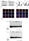
FV-429 inhibited Stat3 nuclear translocation and binding to HIF-1α. (a) The influence of FV-429 on nuclear expression of Stat3 and total expression of HIF-1α under hypoxia. Protein expression change was represented by densitometric analysis. The results are representative of three independent experiments and expressed as means±S.D., **P<0.01, compared with the normoxia control groups and ##P<0.01, compared with the hypoxia control groups. (b) The influence of FV-429 on Stat3 nuclear translocation examined by immunofluorescence (× 600). (c) The influence of FV-429 on binding ability of Stat3 to HIF-1α under hypoxia assessed by EMSA
Figure 6
Figure 6
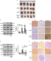
Antixenografted tumor effect and reversal mechanism of FV-429 combined with paclitaxel in vivo. (a) Image of the resected xenografted tumors. (b) Expression of cylin B1, p-cdc25c, cdc25c, p-CDK1 and CDK1 in hypoxic regions of xenografted tumors detected by western blottings. Protein expression change was represented by densitometric analysis. The results are representative of three independent experiments and expressed as means±S.D., *P<0.05 and **P<0.01, compared with the control groups. (c) Expression of ki67, cylin A and cylin B1 in hypoxic regions of xenografted tumors detected by immunohistochemistry (× 400). (d) Expression of p-Src (Tyr 416), c-Src, p-Stat3 (Tyr 705), Stat3 and HIF-1α in normoxic and hypoxic regions of xenografted tumors detected by western blottings. Protein expression change was represented by densitometric analysis. The results are representative of three independent experiments and expressed as means±S.D., **P<0.01, compared with the normoxia control groups and ##P<0.01, compared with the hypoxia control groups. (e) Expression of c-Src, Stat3 and HIF-1α in normoxic and hypoxic regions detected by immunohistochemistry (× 400). Arrows referred to Stat3 nuclear translocation
Figure 7
Figure 7
Toxicological assessment. (a) Body weight change of xenografted nude mice by days. Data had been statistically analyzed by Microsoft Excel 2013 and expressed as means±S.D. (n=5). (b) The influence of FV-429 and paclitaxel single or combined treatment on the heart, liver, spleen, lung and kidney was examined by hematoxylin and eosin staining (× 200)

References

    1. Lee H, Lee T, Lee N, Yang GE, Lee C, Lee J et al. Src activates HIF-1α not through direct phosphorylation of HIF-1a-specific prolyl-4 hydroxylase 2 but through activation of the NADPH oxidase/Rac pathway. Carcinogenesis 2011; 32: 703–712. - PubMed (V体育安卓版)
    1. Ng KP, Manjeri A, Lee KL, Huang W, Tan SY, Chuah CTH et al. Physiologic hypoxia promotes maintenance of CML stem cells despite effective BCR-ABL1 inhibition. Blood 2015; 123: 3316–3326. - V体育平台登录 - PubMed
    1. Semenza GL. Hypoxia-inducible factors in physiology and medicine. Cell 2012; 148: 399–408. - PMC - PubMed
    1. Simon MC, Keith B. The role of oxygen availability in embryonic development and stem cell function. Nat Rev 2008; 9: 285–296. - PMC - PubMed
    1. Zhao L, Li W, Zhou YX, Zhang Y, Huang SL, Xu XF et al. The overexpression and nuclear translocation of Trx-1 during hypoxia confers on HepG2 cells resistance to DDP, and GL-V9 reverses the resistance by suppressing the Trx-1/Ref-1 axis. Free Radic Biol Med 2015; 82: 29–41. - PubMed

"VSports最新版本" Publication types

MeSH terms