Skip to main page content
U.S. flag

An official website of the United States government

Dot gov

The . gov means it’s official. Federal government websites often end in . gov or . mil. Before sharing sensitive information, make sure you’re on a federal government site VSports app下载. .

Https

The site is secure. The https:// ensures that you are connecting to the official website and that any information you provide is encrypted and transmitted securely V体育官网. .

. 2017 Jun 29;129(26):3452-3464.
doi: 10.1182/blood-2017-03-773341. Epub 2017 May 17.

Inhibiting the osteocyte-specific protein sclerostin increases bone mass and fracture resistance in multiple myeloma

Affiliations

Inhibiting the osteocyte-specific protein sclerostin increases bone mass and fracture resistance in multiple myeloma

VSports - Michelle M McDonald et al. Blood. .

Abstract

Multiple myeloma (MM) is a plasma cell cancer that develops in the skeleton causing profound bone destruction and fractures. The bone disease is mediated by increased osteoclastic bone resorption and suppressed bone formation. Bisphosphonates used for treatment inhibit bone resorption and prevent bone loss but fail to influence bone formation and do not replace lost bone, so patients continue to fracture. Stimulating bone formation to increase bone mass and fracture resistance is a priority; however, targeting tumor-derived modulators of bone formation has had limited success. Sclerostin is an osteocyte-specific Wnt antagonist that inhibits bone formation. We hypothesized that inhibiting sclerostin would prevent development of bone disease and increase resistance to fracture in MM. Sclerostin was expressed in osteocytes from bones from naive and myeloma-bearing mice. In contrast, sclerostin was not expressed by plasma cells from 630 patients with myeloma or 54 myeloma cell lines. Mice injected with 5TGM1-eGFP, 5T2MM, or MM1. S myeloma cells demonstrated significant bone loss, which was associated with a decrease in fracture resistance in the vertebrae. Treatment with anti-sclerostin antibody increased osteoblast numbers and bone formation rate but did not inhibit bone resorption or reduce tumor burden. Treatment with anti-sclerostin antibody prevented myeloma-induced bone loss, reduced osteolytic bone lesions, and increased fracture resistance. Treatment with anti-sclerostin antibody and zoledronic acid combined increased bone mass and fracture resistance when compared with treatment with zoledronic acid alone. This study defines a therapeutic strategy superior to the current standard of care that will reduce fractures for patients with MM VSports手机版. .


Figures

Figure 1.
Figure 1.
Sclerostin is expressed by osteocytes but not myeloma cells. (A) Gene expression profiling using DNA microarray analysis of memory B cells (MBCs [n = 5]), in vitro–generated PPCs (n = 5), normal BMPCs (n = 10), malignant plasma cells from patients with newly diagnosed MM (n = 630) and relapsed/refractory MM (RMM [n = 82]), as well as human myeloma cell lines (HMCLs [n = 54]). Gray dots represent the absence of expression, and black dots represent the presence of expression according to the presence-absence calls with negative probesets (PANP) algorithm. (B) RNA-seq analysis of DKK1 and SOST of MBCs (n = 4), PPCs (n = 4), BMPCs (n = 10), MM cells (n = 263), and HMCLs (n = 19). DKK1 (204602_at) is expressed by the majority of malignant plasma cell samples from previously untreated and relapsed myeloma patients. In contrast, SOST (223869_at) expression is absent in all normal and malignant plasma cells. (C) Immunohistochemical staining for CD138 (top panel) and sclerostin (bottom panel) counter-stained with hematoxylin in bone marrow of naive mice and mice bearing 5TGM1-eGFP myeloma cells. Slides were scanned on Scanscope CS2 (Aperio) up to original magnification ×40, and images were captured by using Aperio Imagescope at digital magnification ×20. Scale bar represents 50 μm. (D) Sclerostin protein was measured in media conditioned by MM1.S, OPM2 human myeloma cells lines, 5TGM1 murine myeloma cells, and primary osteocytes (n = 4; data are mean ± 1 standard error of the mean [SEM]; ****P < .0001). (E) Density plot highlighting the osteocyte-enriched expression of secreted Wnt signaling antagonists (n = 6). (F) Tumor burden significantly alters the expression of the secreted Wnt signaling pathway antagonists Sost, Dkk1, and Frzb (n = 6; data are mean ± 1 standard deviation [SD]; multiple comparison–adjusted P value *P < .05; **P < .01; ****P < .0001). (G) Immunohistochemical staining for sclerostin in osteocytes from naive and 5TGM1-bearing mice; arrow heads denote positive-stain osteocytes with positive-stained canaliculi (inset). Slides were scanned on Scanscope CS2 (Aperio) up to original magnification ×40, and images were captured by using Aperio Imagescope at digital magnification ×40. Scale bar represents 50 µm.
Figure 2.
Figure 2.
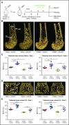
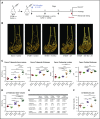
Treatment with anti-sclerostin antibody prevents myeloma-induced bone loss in the 5TGM1 murine model of myeloma. (A) Schematic depicting study design for 5TGM1 studies. (B) 3D microCT reconstructions of distal femora representing each treatment group in experiment (Expt) 2. (C) Dot plot of the ratio of trabecular bone volume to total volume (BV/TV) from distal femora of experiment 1 (n = 5-6) and experiment 2 (n = 8) (data are mean ± 1 SEM). (D) 3D microCT reconstructions of L4 vertebrae representing L4 from each treatment group in experiment 2. (E) Dot plot of trabecular BV/TV from lumbar vertebra 2 (L2) from experiment 1 (n = 6) and lumbar vertebra 4 (L4) from experiment 2 (n = 8) (data are mean ± 1 SEM; *P < .05; **P < .01; ***P < .001; ****P < .0001). BM, bone marrow; CB, cortical bone; GP, growth plate; i.v., intravenous; s.c., subcutaneous; Scl, sclerostin; TB, trabecular bone.
Figure 3.
Figure 3.
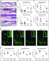
Treatment with anti-sclerostin antibody prevents 5TGM1 myeloma-induced suppression of bone formation. (A) Tartrate-resistant acid phosphatase (TRAP)/hematoxylin-stained histologic sections from representative mice from each treatment group showing osteoblasts on the endocortical bone surface (inset, blue arrows) and osteoclasts reacted for TRAP and stained red (white arrows); scale bar represents 200 µm. Slides were scanned on a Scanscope CS2 (Aperio) up to original magnification ×40, and images were captured by using an Aperio Imagescope at digital magnification ×20. (B) Dot plots of osteoclast surface/bone surface (Oc.S/BS) and number of osteoclasts/bone surface (N.Oc/BS) (data are mean ± 1 SEM; *P < .05; **P < .001). (C) Dot plots of osteoblast surface/bone surface (Ob.S/BS) and number of osteoblasts/bone surface (N.Ob/BS) (n = 8) (data are mean ± 1 SEM; *P < .05; **P < .001). (D) Representative sections from each group showing mineralized bone surfaces labeled with calcein. Images were captured on a Leica DMI 5500 at ×2.5 by using LAS X software (Leica); scale bar represents 500 µm. Double-label bone surfaces were used to measure mineral apposition rate (inset: red arrows show the distance between 2 labels separated by a fixed time interval). Images were captured on a Leica DMI 5500 at ×40 using LAS X software (Leica). Scale bar represents 20 µm. (E) Dot plots of MAR, mineralizing surface (MS), and BFR (n = 5; data are mean ± 1 SEM; *P < .05; **P < .01; #P < .05 (Student t test); ##P < .01 (Student t test). ES, endosteal bone surface; PS, periosteal bone surface.
Figure 4.
Figure 4.
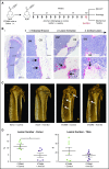
Treatment with anti-sclerostin antibody reduces formation of osteolytic bone lesions. (A) Schematic describing the study design for investigations of bone lesions in 5T2MM-bearing mice. (Bi) TRAP/hematoxylin-stained histologic section of a tibia from a 5T2MM-bearing mouse. Slides were scanned on a Scanscope CS2 (Aperio) up to original magnification ×40, and images were captured by using an Aperio Imagescope at digital magnification ×2. Scale bar represents 500 µm; black arrows point to cortical lesions. (Bii) Higher magnification (×12 and ×30) images that demonstrate the temporal development of CB lesions in the 5T2MM model. (1) Initial erosion of the endosteal surface of the CB by red TRAP-positive osteoclasts (black arrows) adjacent to tumor cells in the BM. (2) Formation of a CB lesion (blue dotted line) by osteoclasts (black arrows). (3) A cortical lesion that has penetrated the cortex with the tumor (blue dotted line) extending into the surrounding soft tissue. (Top panel) scale bar represents 100 µm; (bottom panel) scale bar represents 50 µm. (C) Representative 3D reconstruction of tibia from each treatment group showing full cortex penetration lesions (white arrows) in the 5T2MM+ control and 5T2MM+ anti-sclerostin antibody groups. (D) Dot plots of lesion number per bone for femora and tibiae (n = 7-8 per group; data are mean ± 1 SEM; *P < .05).
Figure 5.
Figure 5.
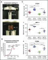
Treatment with anti-sclerostin antibody prevents loss of bone mechanical strength in myeloma-burdened bones. (A) Images demonstrating the positioning of L4 vertebrae for compression testing between the load cell and the custom-designed jig in the Instron 5944 load frame. (B) Representative load displacement curve of an L4 vertebra from the naive control group showing the outcomes of stiffness, yield point, and maximum load at the first point of failure. (C) Dot plots of maximum load for L4 vertebrae from the (i) 5TGM1-eGFP, (ii) MM1.S, and (iii) 5T2MM models of myeloma (5TGM1: n = 8; MM1.S: n = 5-9; 5T2MM: n = 6-8). Data are mean ± 1 SEM; *P < .05; **P < .01; ***P < .001; ****P < .0001; #P < .05 (Student t test).
Figure 6.
Figure 6.
Combination treatment with anti-sclerostin antibody and ZA is superior in preventing bone loss in the 5TGM1 murine model of myeloma. (A) Schematic describing the study design for investigations of bone loss in 5TGM1-eGFP–bearing mice treated with both anti-sclerostin antibody and ZA. (B) Examples of 3D microCT reconstructions of distal femora from each treatment group. (C) Dot plots of BV/TV trabecular thickness and number and cortical bone thickness in femora for each treatment group (n = 8 per group). Data are mean ± 1 SEM; *P < .05; **P < .01; ***P < .001; ****P < .0001. (D) Dot plot of L4 vertebra BV/TV for each treatment group (data are mean ± 1 SEM; **P < .01; ***P < .001; ****P < .0001; #P < .05 [Student t test]). (E) Structural parameters from microCT scans of L4 vertebrae (n = 8 per group). Data are mean ± 1 SEM; aP < .001; bP < .01; dP < .05 compared with naive plus control mice; eP < .001; fP < .05 compared with 5TGM1 plus control mice; gP < .001 compared with 5TGM1 plus ZA; hP < .001 compared with 5TGM1 plus anti-sclerostin antibody. (F) Dot plot of maximum load from L4 vertebra for each treatment group (n = 8 per group). Data are mean ± 1 SEM; **P < .01; ***P < .001; #P < .05 [Student t test].
Figure 7.
Figure 7.
Treatment with anti-sclerostin antibody does not have an impact on tumor burden. (A) Representative flow cytometry plots of 5TGM1-eGFP control-treated bone marrow and 5TGM1-eGFP anti-sclerostin antibody–treated bone marrow showing the percentage of 5TGM1-eGFP+ cells in the sample (black box). (B) Dot plots of bone marrow tumor burden as a percent of total bone marrow cells (% GFP+) in experiments 1 and 2 of the 5TGM1 study (n = 6 [experiment 1]; n = 8 [experiment 2]). Data are mean ± 1 SEM. (C) Dot plots of spleen tumor burden as a percent of total cells (% GFP+) in experiments 1 and 2 of the 5TGM1 study (n = 6 [experiment 1]; n = 8 [experiment 2]). Data are mean ± 1 SEM. (D) Dot plots of bone marrow and spleen tumor burden (% idiotype+) in the 5T2MM study (n = 6-7). Data are mean ± 1 SEM. (E) Dot plots of whole body tumor burden (total flux p/s) and bone marrow percent GFP+ cells in MM1.S experiment 1 (n = 9 per group). Data are mean ± 1 SEM; *P < .05. (F) Dot plots of whole body tumor burden (total flux p/s) at 3 weeks and bone marrow percent GFP+ cells in MM1.S experiment 2 (n = 8-9 for whole body tumor; n = 5-9 for bone marrow tumor). Data are mean ± 1 SEM.

VSports在线直播 - References

    1. Becker N. Epidemiology of multiple myeloma. Recent Results Cancer Res. 2011;183:25-35. - PubMed
    1. Röllig C, Knop S, Bornhäuser M. Multiple myeloma. Lancet. 2015;385(9983):2197-2208. - PubMed
    1. Kyle RA, Gertz MA, Witzig TE, et al. Review of 1027 patients with newly diagnosed multiple myeloma. Mayo Clin Proc. 2003;78(1):21-33. - PubMed
    1. Avet-Loiseau H, Attal M, Moreau P, et al. Genetic abnormalities and survival in multiple myeloma: the experience of the Intergroupe Francophone du Myélome. Blood. 2007;109(8):3489-3495. - V体育ios版 - PubMed
    1. Melton LJ III, Kyle RA, Achenbach SJ, Oberg AL, Rajkumar SV. Fracture risk with multiple myeloma: a population-based study. J Bone Miner Res. 2005;20(3):487-493. - PubMed (VSports)

MeSH terms