Skip to main page content
U.S. flag

An official website of the United States government

Dot gov

The . gov means it’s official VSports app下载. Federal government websites often end in . gov or . mil. Before sharing sensitive information, make sure you’re on a federal government site. .

Https

The site is secure. The https:// ensures that you are connecting to the official website and that any information you provide is encrypted and transmitted securely V体育官网. .

. 2017 May 4;36(18):2577-2588.
doi: 10.1038/onc.2016.414. Epub 2017 Feb 13.

Downregulation of miR-218 contributes to epithelial-mesenchymal transition and tumor metastasis in lung cancer by targeting Slug/ZEB2 signaling

Affiliations

"V体育官网入口" Downregulation of miR-218 contributes to epithelial-mesenchymal transition and tumor metastasis in lung cancer by targeting Slug/ZEB2 signaling

VSports在线直播 - Z-M Shi et al. Oncogene. .

Abstract

Epithelial-mesenchymal transition (EMT) has been recognized as a key element of cell migration and invasion in lung cancer; however, the underlying mechanisms are not fully elucidated. Recently, emerging evidence suggest that miRNAs have crucial roles in control of EMT and EMT-associated traits such as migration, invasion and chemoresistance. Here, we found that miR-218 expression levels were significantly downregulated in lung cancer tissues compared with adjacent non-cancerous tissues, and the levels of miR-218 were significantly associated with histological grades and lymph node metastasis. Overexpression of miR-218 inhibited cell migration and invasion as well as the EMT process. Of particular importance, miR-218 was involved in the metastatic process of lung cancer cells in vivo by suppressing local invasion and distant colonization. We identified Slug and ZEB2 as direct functional targets of miR-218. Inverse correlations were observed between miR-218 levels and Slug/ZEB2 levels in cancer tissue samples. In addition, overexpression of miR-218 in H1299 increased chemosensitivity of cells to cisplatin treatment through suppression of Slug and ZEB2 VSports手机版. These findings highlight an important role of miR-218 in the regulation of EMT-related traits and metastasis of lung cancer in part by modulation of Slug/ZEB2 signaling, and provide a potential therapeutic strategy by targeting miR-218 in NSCLC. .


V体育官网 - Conflict of interest statement

The authors declare no conflict of interest.

Figures

Figure 1
Figure 1
MiR-218 was downregulated and associated with advanced clinical stage and NSCLC metastasis. (a) The relative mRNA levels of miR-218 were detected by qRT–PCR and normalized against an endogenous control (U6 RNA) in 60 pairs of lung cancer tissues. (b) Relative expression levels of miR-218 in different stages of cancer tissues. (c) qRT–PCR analysis of miR-218 expression levels in 60 pairs of primary NSCLC tissues and their corresponding lymph node metastases. Data represent mean±s.d. of three replicates. *Indicated significant difference at P<0.05; **Indicated significant difference at P<0.01.
Figure 2
Figure 2
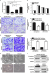
MiR-218 negatively regulated the migration, invasion and EMT of NSCLC cells. (a) The expression levels of miR-218 were detected by qRT–PCR in human lung cancer cells (A549, H1299, PC9 and SPCA-1). (b) The levels of cell migration and invasion in indicated human lung cancer cells (A549, H1299, PC9 and SPCA-1) were analyzed using the Transwell chambers assay with or without coated Matrigel. (c) MiR-218 overexpression decreased cell migration and invasion activities in H1299 cells. Indicated cells were subjected to migration and invasion assay. Scale bar, 20 μm. (d) Anti-miR-218 inhibitor increased cell migration and invasion activities in A549 cells. Indicated cells were subjected to migration and invasion assay. Scale bar, 20 μm. (e) Phase contrast images of H1299 and A549 cells in indicated treatment. H1299 transfected with miR-218 underwent morphological change from a spindle-shaped to a rounded or cobblestone-like shaped. In contrast, more rounded A549 cells became spindle-shaped after the treatment with anti-miR-218 inhibitor. (f) Western blot analysis of EMT markers E-cadherin and Vimentin were shown in H1299 and A549 cells, respectively. (g) Immunofluorescence images of EMT markers, E-cadherin and Vimentin were shown in H1299 and A549 cells, respectively. Data represent mean±s.d. of three replicates. *Indicated significant difference at P<0.05; **Indicated significant difference at P<0.01.
Figure 2
Figure 2
MiR-218 negatively regulated the migration, invasion and EMT of NSCLC cells. (a) The expression levels of miR-218 were detected by qRT–PCR in human lung cancer cells (A549, H1299, PC9 and SPCA-1). (b) The levels of cell migration and invasion in indicated human lung cancer cells (A549, H1299, PC9 and SPCA-1) were analyzed using the Transwell chambers assay with or without coated Matrigel. (c) MiR-218 overexpression decreased cell migration and invasion activities in H1299 cells. Indicated cells were subjected to migration and invasion assay. Scale bar, 20 μm. (d) Anti-miR-218 inhibitor increased cell migration and invasion activities in A549 cells. Indicated cells were subjected to migration and invasion assay. Scale bar, 20 μm. (e) Phase contrast images of H1299 and A549 cells in indicated treatment. H1299 transfected with miR-218 underwent morphological change from a spindle-shaped to a rounded or cobblestone-like shaped. In contrast, more rounded A549 cells became spindle-shaped after the treatment with anti-miR-218 inhibitor. (f) Western blot analysis of EMT markers E-cadherin and Vimentin were shown in H1299 and A549 cells, respectively. (g) Immunofluorescence images of EMT markers, E-cadherin and Vimentin were shown in H1299 and A549 cells, respectively. Data represent mean±s.d. of three replicates. *Indicated significant difference at P<0.05; **Indicated significant difference at P<0.01.
Figure 3
Figure 3
MiR-218 targets Slug and ZEB2. (a) The miR-218 binding site predicted in the 3′-UTR regions of Slug/ZEB2 mRNAs. Mutant constructs were generated at the seed regions of Slug/ZEB2 3′-UTRs as indicated in red. 3′-UTR fragments of Slug/ZEB2 mRNAs containing wild-type or mutant of the miR-218 binding sequences were cloned into the downstream of the luciferase gene in pMIR-vector. (b) H1299 cells were transfected with pMIR reporter vectors containing either wild-type or mutant Slug/ZEB2 3′-UTRs (indicated as pMIR-Slug/ZEB2-WT and pMIR-Slug/ZEB2-MT) with either miR-NC or miR-218. Luciferase activities were determined 48 h after transfection. (c) The protein levels of Slug and ZEB2 in H1299 and A549 cells were examined by western blot analysis. (d) The expression levels of Slug or ZEB2 in normal tissues and human NSCLC specimens were determined by qRT–PCR analysis, and fold changes were obtained by the ratios of Slug or ZEB2 to glyceraldehyde 3-phosphate dehydrogenase (GAPDH) levels. (e) Spearman′s correlation analysis was used to determine the correlations between the expression levels of Slug or ZEB2 and miR-218 in human NSCLC specimens. Data represent mean±s.d. of three replicates. *Indicated significant difference at P<0.05; **Indicated significant difference at P<0.01.
Figure 4
Figure 4
Effect of miR-218 on EMT and drug resistance through Slug/ZEB2 pathway. (a) Image showing the morphological changes of H1299 cells expressing miR-218 mimics or miR-NC or miR-218 in combination with Slug or ZEB2 overexpression. Overexpression of Slug or ZEB2 partially reversed the morphological change caused by ectopic expression of miR-218 in H1299 cells. (b) H1299 cells stably expressing miR-NC or miR-218, or miR-218 in combination with Slug or ZEB2 overexpression were conducted by western blot analysis. (c) Indicated cells were subjected to Matrigel invasion assay. Scale bar, 20 μm. (d and e) H1299 cells stably expressing miR-NC or miR-218, miR-218 in combination with Slug or ZEB2 overexpression were pretreated with 5 μm cisplatin for indicated time points, then subjected to CCK-8 Assay, and apoptosis analysis by flow cytometry (f). Data represent mean±s.d. of three replicates. *Indicated significant difference at P< 0.05 compared with miR-NC control; **At P<0.01 compared with miR-NC control; #At P<0.05 compared with miR-218 plus Slug or ZEB2 overexpression.
Figure 5
Figure 5
The role of miR-218 in tumor growth in vivo. (a) H1299/miR-NC, H1299/miR-218, A549/miR-NC-inhibitor and A549/miR-218 inhibitor cells (5 × 10⁶ cells) were dispersed in 100 μl of serum-free RPMI 1640 medium, and subcutaneously injected into both sides of posterior flanks of the nude mice (n=6). Tumors were measured every 3 days after they were apparently detectable at day 12. Tumor volumes were calculated using the following formula: volume=0.5 × (length × width2). (b) Tumor growth assay and average weights of tumors from indicated groups (bar=2 mm). (c) The total proteins were extracted from xenografts and subjected to western blot analysis to test levels of Slug and ZEB2 as indicated. Glyceraldehyde 3-phosphate dehydrogenase (GAPDH) levels were used as the internal control. Data represent mean±s.d. of three replicates. *Indicated significant difference at P<0.05; **Indicated significant difference at P<0.01.
Figure 6
Figure 6
The role of miR-218 in tumor growth and metastasis in vivo. (a) We established an orthotopic lung cancer xenograft model in nude mice. The trimmed tumor pieces of 1 mm in diameter were transplanted into lung tissues by surgical orthotopic implantation. The fluorescent areas in lungs of mice indicated were tumor tissues with H1299-red fluorescent protein (RFP) cells 7 weeks after the transplantation. (b) We counted the incidence of lung metastasis in the H1299/miR-NC and H1299/miR-218 xenografts based on the fluorescent areas showing the metastatic foci in the lung tissues of the mice. (c) Hematoxylin and eosin (H&E) staining was conducted, and arrowheads indicated metastatic tumor nodules. Magnification, × 40; scale bar, 200 μm. (d) In situ hybridization analysis showed miR-218 expression was not detected in the majority of miR-NC tissues with blue staining, and very high levels of miR-218 in miR-218-expressing tissues with strong brown signals in the cytoplasm. The expression levels of Slug and ZEB2 were analyzed in tumor tissues using immunohistochemistry. Magnification, × 400; scale bar, 50 μm.
Figure 7
Figure 7
Schematic diagram. TGF-β1 signaling promoted EMT, tumor growth and metastasis by altering expression levels of downstream genes including miR-218. In cancer cells, activated TGF-β1 inhibited miR-218 expression, which in turn activated expression of the key EMT promoting transcription factors Slug and ZEB2 that repressed the expression of E-cadherin, and induced the expression of Vimentin, which in turn promotes EMT and metastasis of disseminated tumor cells in lung.

VSports - References

    1. Siegel R, Naishadham D, Jemal A. Cancer statistics, 2013. CA Cancer J Clin 2013; 63: 11–30. - PubMed
    1. Heist RS, Engelman JA. SnapShot: non-small cell lung cancer. Cancer Cell 2012; 21: 448 e442. - PubMed
    1. Scagliotti GV, Parikh P, von Pawel J, Biesma B, Vansteenkiste J, Manegold C et al. Phase III study comparing cisplatin plus gemcitabine with cisplatin plus pemetrexed in chemotherapy-naive patients with advanced-stage non-small-cell lung cancer. J Clin Oncol 2008; 26: 3543–3551. - PubMed
    1. Wu YC, Tang SJ, Sun GH, Sun KH. CXCR7 mediates TGFbeta1-promoted EMT and tumor-initiating features in lung cancer. Oncogene 2015; 35: 2123–2132. - "V体育平台登录" PubMed
    1. Zhang J, Tian XJ, Zhang H, Teng Y, Li R, Bai F et al. TGF-beta-induced epithelial-to-mesenchymal transition proceeds through stepwise activation of multiple feedback loops. Sci Signal 2014; 7: ra91. - "V体育2025版" PubMed

Publication types

MeSH terms