Skip to main page content
U.S. flag

An official website of the United States government

Dot gov

The . gov means it’s official. Federal government websites often end in . gov or . mil. Before sharing sensitive information, make sure you’re on a federal government site. VSports app下载.

Https

The site is secure V体育官网. The https:// ensures that you are connecting to the official website and that any information you provide is encrypted and transmitted securely. .

. 2017 Jan 31;8(5):8342-8355.
doi: 10.18632/oncotarget.14184.

MicroRNA-1908-5p contributes to the oncogenic function of the splicing factor SRSF3

Affiliations

MicroRNA-1908-5p contributes to the oncogenic function of the splicing factor SRSF3

Hye Ree Kim et al. Oncotarget. .

Abstract

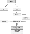
Serine/arginine (SR)-rich proteins that contain RS domains and SR repeats have diverse cellular functions including transcription, polyadenylation, translation, and RNA export. The splicing factor SRSF3, also termed SRp20, is the smallest member of the SR protein family and is a known proto-oncogene. Although it is implicated in the malignant phenotypes of various cancer cells, the molecular mechanism underlying SRSF3-mediated cancer progression is still obscure VSports手机版. We investigated here the oncogenic functions of SRSF3 in osteosarcoma U2OS cells. Knockdown of SRSF3 inhibited proliferation, clonogenicity, and metastatic potential including migration and invasion. It also decreased the level of miR-1908 independent of its host gene FADS1. Although FADS1 was not associated with SRSF3-mediated malignant properties, overexpression of miR-1908-5p increased cell proliferation, migration, and invasion, suggesting that miR-1908-5p is responsible for the oncogenic functions of SRSF3. Knockdown of SRSF3 decreased the expression of miR-1908-5p by inhibiting transactivation of NF-κB. We observed that miR-1908-5p downregulated NF-κB inhibitor interacting Ras-like 2 (NKIRAS2), a negative regulator of the NF-κB pathway by directly binding to the 3'UTR of NKIRAS2 mRNA. Consistent with overexpression of miR-1908-5p, knockdown of NKIRAS2 diminished the expression level of IκB-β and provoked translocation of NF-κB into the nucleus where it transcriptionally activates its target genes including miR-1908-5p expression, thus elevating the proliferation and metastatic potential. Taken together, our results demonstrate that SRSF3 confers the malignant characteristics on cancer cells via the SRSF3/miR-1908-5p/NKIRAS2 axis. .

Keywords: FADS1; NF-κB; NKIRAS2; SRSF3; miR-1908-5p V体育安卓版. .


Conflict of interest statement

CONFLICTS OF INTEREST

The authors declare no competing financial interests.

"V体育官网入口" Figures

Figure 1
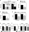
Figure 1. SRSF3 contributes to the malignant properties of U2OS cells
A. U2OS cells were transfected with control (CTRL) or SRSF3-specific siRNA. The expression level of SRSF3 was determined by western blot analysis and GAPDH was used as a loading control. B. The equal number of transfected cells was resuspended into 12-well plates and cellular proliferation was assessed by counting the number of viable cells at every 24 h. C. For the clonogenic assay, transfected cells were plated into 6-well plates and cultured for more than 2 weeks. Clonogenic activity was assessed by counting the number of colonies. D. The migratory and invasive abilities were assessed by a wound healing assay and Matrigel invasion assay, respectively, as described in Materials and Methods. All experiments are performed more than three times and data represent the mean ± S.D. Asterisk (*) indicates statistical significance of p<0.05 as determined by Student's t-test.
Figure 2
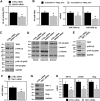
Figure 2. Knockdown of SRSF3 decreased the expression of miR-1908 and its host gene, FADS1
A. Microarray data showing decreased expression of FADS1 in SRSF3-silenced U2OS cells with three different siRNAs. B. U2OS cells were transfected with control (CTRL) or SRSF3-specific siRNA for 48 h and then the expression level of FADS1 mRNA was determined by RT-qPCR analysis. C-D. The levels of pri-miR-1908 and mature miR-1908-3p/5p were assessed by RT-qPCR analysis using pri-miR-1908-specific primers and TaqMan miRNA primers, respectively. E. Cells were transfected with control (CTRL) or FADS1-specific siRNA for 48 h and then the levels of pri-miR-1908 and mature miR-1908-3p/5p were assessed by RT-qPCR as described above. F. Cells were transfected with control (CTRL) or Sp1-specific siRNA for 48 h. The expression level of FADS1 mRNA was determined by RT-qPCR analysis and the levels of pri-miR-1908 and mature miR-1908 (3p/5p) were assessed by RT-qPCR analysis as described above. All experiments are performed more than three times and data represent the mean ± S.D. Asterisk (*) indicates statistical significance of p<0.05 as determined by Student's t-test.
Figure 3
Figure 3. miR-1908-5p increased proliferation and metastatic potential
A. U2OS cells were transfected with control (Con-miR) or pre-miR-1908-5p for 24 h and then resuspended into 12-well plates. The proliferation rate was determined by counting the number of viable cells at every 24 h. B-C. The invasive (B) and migratory (C) activities of miR-1908-5p-overexpressed cells were assessed by a wound healing assay and Matrigel invasion assay, respectively. All experiments are performed more than three times and data represent the mean ± S.D. Asterisk (*) indicates statistical significance of p<0.05 as determined by Student's t-test.
Figure 4
Figure 4. NF-κB is involved in SRSF3-regulated miR-1908 expression
A. U2OS cells were transfected with control (CTRL) or SRSF3-specific siRNA. Transfected cells were resuspended into 12-well plates and then transfected with the NF-κB reporter vector. Transactivation of NF-κB was assessed by calculating the luciferase activity. B. To check whether NF-κB regulates the expression of miR-1908, cells were treated with the NF-κB-specific inhibitor BAY11-7082 (10 μM) for 24 h. The level of miR-1908-3p/5p was determined by RT-qPCR analysis using TaqMan primers. C. Cells were transfected with control (CTRL) or SRSF3-specific siRNA for 48 h and then whole cell lysates were prepared. Western blot analyses were performed using adequate specific antibodies. D. Cells were transfected as described above and then cellular fractionation was performed. Western blot analysis was performed to determine the localization of NF-κB (p65) and α-tubulin and lamin B was checked for cytoplasmic and nuclear markers, respectively. E-F. Cells were transfected with control (CTRL) or TAK1-specific siRNA and then the levels of TAK1 and phosphorylated IKKα/β and NF-κB were assessed by Western blot analysis. The level of miR-1908-5p was determined by RT-qPCR analysis. G-H. The protein and mRNA expression levels of NF-κB target genes including HIF1-α, cyclin D1, and slug, were determined by western blot and RT-qPCR analysis, respectively. All experiments are performed more than three times and data represent the mean ± S.D. Asterisk (*) indicates statistical significance of p<0.05 as determined by Student's t-test.
Figure 5
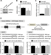
Figure 5. NKIRAS2 is a novel target of miR-1908-5p
A-B. U2OS cells were transfected with control (Con-miR) or pre-miR-1908-5p for 48 h and then the level of NKIRAS2 protein and mRNA was assessed by western blot and RT-qPCR analysis, respectively. C. To check direct interaction between miRNA and target mRNA, Ago2 immunoprecipitation (IP) was performed using an Ago2-specifc antibody. Cytoplasmic lysates were prepared from control (CTRL) or pre-miR-1908-5p-transfectected cells and used for Ago2-IP. The mRNAs bound to miRNA-Ago2 were isolated and the level of NKIRAS2 mRNA was assessed by RT-qPCR analysis. D. To verify miR-1908-5p suppressed NKIRAS2, luciferase vectors harboring wild-type and mutated miR-1908-5p-binding sequences were manufactured. Luciferase activity was assessed as described in Materials and Methods. All experiments are performed more than three times and data represent the mean ± S.D. Asterisk (*) indicates statistical significance of p<0.05 as determined by Student's t-test.
Figure 6
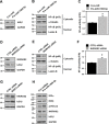
Figure 6. Downregulation of NKIRAS2 by miR-1908-5p is implicated in the oncogenic functions of SRSF3
A-F. U2OS cells were transfected with control (CTRL) or SRSF3-specific siRNA (A-C). In the case of miRNA, cells were transfected with control (Con-miR) or pre-miR-1908-5p (D-F). The level of IκB-β was determined by western blot analysis (A and C). To assess the cytoplasmic and nuclear localization of NF-kB, cellular fractionation was performed and the level of NF-κB (p65) in cytoplasmic and nuclear fraction was determined by western blot analysis (B and D). The level of α-tubulin and lamin B was checked for cytoplasmic and nuclear markers, respectively. Transcriptional activity of NF-κB was assessed by checking the luciferase activity as described in Materials and Methods (C and F). G. U2OS cells were transfected with control (CTRL) or SRSF3-specific siRNA and the level of NKIRAS2 and IκB-β was determined by western blot analysis. H. U2OS cells were transfected with control (Con-miR) or pre-miR-1908-5p. After 48 h post-transfection, IKKα/β activation and expression of NKIRAS2 and IκB-β were assessed by western blot analysis.
Figure 7
Figure 7. Downregulation of NKIRAS2 by miR-1908-5p is implicated in the oncogenic functions of SRSF3
A. To check the effect of miR-1908-5p/NKIRAS2 on the anchorage-independent proliferation, U2OS cells were transfected with pre-miR-1908-5p or NKIRAS2-specific siRNA for 24 h. Equal numbers of transfected cells were plated in attachment or ultra-low attachment plates. After 48 h post-incubation, cellular viability was calculated by the MTS assay. B-D. U2OS cells were transfected with control (CTRL) or NKIRAS2-specific siRNA for 48 h. Cellular proliferation was calculated by counting viable cells every 24 h (B). Invasive and migratory abilities were assessed by Matrigel invasion (C) and wound healing assays (D), respectively. All experiments are performed more than three times and data represent the mean ± S.D. Asterisk (*) indicates statistical significance of p<0.05 as determined by Student's t-test.
Figure 8
Figure 8. Proposed model for the role of miR-1908-5p in the oncogenic function of SRSF3

References

    1. Guo J, Jia J, Jia R. PTBP1 and PTBP2 impaired autoregulation of SRSF3 in cancer cells. Sci Rep. 2015;5:14548. doi: 10.1038/srep14548. - DOI - PMC - PubMed
    1. Corbo C, Orru S, Salvatore F. SRp20: an overview of its role in human diseases. Biochem Biophys Res Commun. 2013;436:1–5. doi: 10.1016/j.bbrc.2013.05.027. - DOI - PubMed
    1. Long JC, Caceres JF. The SR protein family of splicing factors: master regulators of gene expression. Biochem J. 2009;417:15–27. doi: 10.1042/BJ20081501. - DOI - PubMed
    1. Jia R, Li C, McCoy JP, Deng CX, Zheng ZM. SRp20 is a proto-oncogene critical for cell proliferation and tumor induction and maintenance. Int J Biol Sci. 2010;6:806–26. - PMC - PubMed
    1. Zahler AM, Lane WS, Stolk JA, Roth MB. SR proteins: a conserved family of pre-mRNA splicing factors. Genes Dev. 1992;6:837–47. - PubMed

MeSH terms