Skip to main page content
U.S. flag

An official website of the United States government

Dot gov

The . gov means it’s official. Federal government websites often end in . gov or . mil VSports app下载. Before sharing sensitive information, make sure you’re on a federal government site. .

Https

The site is secure. The https:// ensures that you are connecting to the official website and that any information you provide is encrypted and transmitted securely V体育官网. .

. 2017 Jan 3;127(1):383-401.
doi: 10.1172/JCI83822. Epub 2016 Dec 5.

Cardiac myofibroblast engulfment of dead cells facilitates recovery after myocardial infarction

VSports手机版 - Cardiac myofibroblast engulfment of dead cells facilitates recovery after myocardial infarction

Michio Nakaya et al. J Clin Invest. .

Abstract

Myocardial infarction (MI) results in the generation of dead cells in the infarcted area VSports手机版. These cells are swiftly removed by phagocytes to minimize inflammation and limit expansion of the damaged area. However, the types of cells and molecules responsible for the engulfment of dead cells in the infarcted area remain largely unknown. In this study, we demonstrated that cardiac myofibroblasts, which execute tissue fibrosis by producing extracellular matrix proteins, efficiently engulf dead cells. Furthermore, we identified a population of cardiac myofibroblasts that appears in the heart after MI in humans and mice. We found that these cardiac myofibroblasts secrete milk fat globule-epidermal growth factor 8 (MFG-E8), which promotes apoptotic engulfment, and determined that serum response factor is important for MFG-E8 production in myofibroblasts. Following MFG-E8-mediated engulfment of apoptotic cells, myofibroblasts acquired antiinflammatory properties. MFG-E8 deficiency in mice led to the accumulation of unengulfed dead cells after MI, resulting in exacerbated inflammatory responses and a substantial decrease in survival. Moreover, MFG-E8 administration into infarcted hearts restored cardiac function and morphology. MFG-E8-producing myofibroblasts mainly originated from resident cardiac fibroblasts and cells that underwent endothelial-mesenchymal transition in the heart. Together, our results reveal previously unrecognized roles of myofibroblasts in regulating apoptotic engulfment and a fundamental importance of these cells in recovery from MI. .


Conflict of interest statement

The authors have declared that no conflict of interest exists.

Figures (V体育安卓版)

Figure 1
Figure 1. Cardiac myofibroblasts engulf dead cells ex vivo.
(A) Ex vivo phagocytosis assay of apoptotic cells. Cardiac macrophages and myofibroblasts isolated from WT mice that underwent MI were exposed to apoptotic cells (green) (n = 3). The numbers of engulfed apoptotic cells per cardiac macrophage (Mac) and myofibroblast (Myo) are shown. Images were taken at 12–15 randomly selected fields. Arrows indicate engulfed apoptotic cells. Scale bar: 100 μm. Original magnification, ×40. (B) mRNA expression levels of Il6 or Tgfb1 in myofibroblasts treated with LPS after apoptotic engulfment. Cardiac myofibroblasts cocultured with (+) or without (−) apoptotic thymocytes (Apo) for 2 hours were treated with (+) or without (−) LPS at 1 μg/ml for 24 hours (n = 4). (C) αSMA-positive myofibroblasts did not express CD45. The cells collected from infarcted mouse hearts as myofibroblasts were harvested by treatment with accutase and immediately stained with antibodies for αSMA and CD45. Error bars represent the mean ± SEM. (A) **P < 0.01, unpaired 2-tailed Student’s t test. (B) ***P < 0.001, 1-way ANOVA followed by Newman-Keuls analysis.
Figure 2
Figure 2. Cardiac myofibroblasts engulf dead cells in vivo.
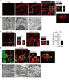
(A) Representative LV sections from WT mice that underwent MI were double stained with TUNEL (green) and anti-αSMA antibody (red) (n = 4). The area indicated by a white square on the merged image is enlarged. Kymograph along the white dashed line in the enlarged image is shown. The area indicated by a white square on the enlarged image was further enlarged, and a 3D image in a white square on the enlarged image is shown. White arrows indicate myofibroblasts containing an apoptotic cell. Scale bars: 30 μm. (B) Transmission electron micrographs of engulfment of an apoptotic cell by a myofibroblast. The areas in yellow and red boxes within a myofibroblast (orange dotted line) are enlarged in the middle and right panels. Well-developed rough-surfaced endoplasmic reticulum and intracellular actin filaments (arrowheads) can be seen in the myofibroblast. MN, myofibroblast nucleus; AN apoptotic nucleus; rER, rough-surfaced endoplasmic reticulum; Mt, mitochondria. Scale bar: 1 μm. (C) Quantification of engulfed TUNEL+ apoptotic cells by macrophages (CD68+) or myofibroblasts (αSMA+) on the sections from mouse hearts 3 days after MI (n = 4). The number of engulfed apoptotic cells per square millimeter of CD68+ macrophage or αSMA+ myofibroblast area is shown. Scale bars: 50 μm. (D) Representative infarcted LV sections from WT mice with EGFP-labeled cardiomyocytes were stained with anti-αSMA antibody (red) (n = 4). Scale bars: 30 μm. (E) Transmission electron micrographs of engulfment of a necrotic cell by a myofibroblast. The areas in black and red dotted boxes are enlarged in middle and right panels. Necrotic nucleus (NN) was observed in the myofibroblast. Arrowheads indicate single-membrane structure. Scale bar: 1 μm. Error bars represent the mean ± SEM. (C) **P < 0.01, unpaired 2-tailed Student’s t test.
Figure 3
Figure 3. MFG-E8 expression is increased in the heart after MI.
(A) mRNA expression levels of apoptotic cell recognition–related genes in hearts of sham-operated mice (white bars, n = 3) and in infarcted (black bars, n = 5) or remote areas (gray bars, n = 5) of mouse hearts 3 days after MI. (B) Western blot images of the long (black arrowhead) or short (white arrowhead) form of MFG-E8 in LV after sham (Sh) operation or MI. Western blot data were quantified (n = 3). Error bars represent the mean ± SEM. (A) *P < 0.05; **P < 0.01; ***P < 0.001, 1-way ANOVA followed by Newman-Keuls analysis. (B) *P < 0.05; **P < 0.01, unpaired 2-tailed Student’s t test.
Figure 4
Figure 4. MFG-E8 is expressed in cardiac myofibroblasts after MI.
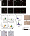
(A and B) MFG-E8 expression colocalizes with vimentin-positive (A) or αSMA-positive (B) myofibroblasts in LV 3 days after MI (n = 4). White squares on merged images mark the magnified areas. Arrows indicate merged cells. Scale bars: 50 μm (lower magnification); 20 μm (higher magnification). (C) Cardiac myofibroblasts from WT or MFG-E8 KO mice were costained with anti–MFG-E8 and anti-vimentin or anti-αSMA antibodies (n = 4). Numbers in quadrants indicate the percentages of cells. (D) Cardiac myofibroblasts were stained with anti–MFG-E8 antibody without (−) or with saponin (+) permeabilization (n = 3). The staining profile (purple) and its control (without primary antibody; green) are shown. (E) Staining of heart sections from non-MI patients and patients with MI by anti–MFG-E8 antibody (n = 4). Arrows indicate MFG-E8 signals. Percentages of MFG-E8–positive cells in each area are shown in the graph. Scale bars: 30 μm. (F) Serial heart sections of patients with MI stained with anti–MFG-E8 antibody or anti-αSMA antibody. Arrows indicate double-positive myofibroblasts. Scale bar: 10 μm. Error bars represent the mean ± SEM. (E) **P < 0.01, unpaired 2-tailed Student’s t test.
Figure 5
Figure 5. MFG-E8–dependent engulfment by cardiac myofibroblasts has a protective effect on heart after MI.
(A) Cardiac myofibroblasts from WT and MFG-E8 KO mice were exposed to fluorescently labeled apoptotic cells in the absence (−) or presence of recombinant MFG-E8 (rMFG-E8). The number of engulfed apoptotic cells per myofibroblast was determined (n = 4). Scale bars: 100 μm. (B) The staining profile of cardiac myofibroblasts with PE–anti-integrin αv antibody is shown in red (n = 3). The blue histogram represents the control. (C) Integrin β5 (red) and vimentin (green) staining of infarcted area of heart sections from WT mice 3 days after MI (n = 3). A square on the first column marks the areas shown at a higher magnification. White arrows indicate vimentin-positive myofibroblasts merged with integrin β5. The percentages of integrin β5–positive fibroblasts in remote or infarct area of hearts after MI is shown in the graph. Scale bars: 50 μm (lower magnification); 20 μm (higher magnification). (D) Decreased survival rate of MFG-E8 KO mice (sham, n = 8; MI, n = 38) compared with that of WT mice (sham, n = 11; MI, n = 36) after MI. Kaplan-Meier survival analysis using a log-rank test. ###P < 0.001. (E) PicroSirius red staining of the cardiac sections of WT and MFG-E8 KO mice at day 3 after MI. The collagen volume fraction (CVF) was determined by counting collagen-depositing areas (WT, n = 4; KO, n = 5). Scale bars: 100 μm. Error bars represent the mean ± SEM. (A) *P < 0.05; ***P < 0.001, 1-way ANOVA followed by Newman-Keuls analysis. (C and E) ***P < 0.001; ##P < 0.01, unpaired 2-tailed Student’s t test.
Figure 6
Figure 6. MFG-E8–dependent engulfment by cardiac myofibroblasts influences the conditions of heart after MI.
(A) TUNEL-positive nuclei in the border zone of hearts in WT (n = 4) and MFG-E8 KO (n = 3) mice at day 3 after MI. The percentages indicate the ratio of TUNEL-positive nuclei to DAPI-positive nuclei. Scale bars: 100 μm. (B) Representative LV sections from WT mice that underwent MI were double-stained with anti-TNNI3 (green) and anti-αSMA (red) antibodies (n = 5). The numbers of TNNI3-positive cardiomyocytes per square millimeter of αSMA-positive area in infarcted hearts of WT and MFG-E8 KO mice are shown in the graph. Scale bar: 20 μm. (C) mRNA expression levels of inflammatory genes 3 days after sham operation or MI in the hearts of WT or MFG-E8 KO mice (sham, n = 4; MI, n = 5). (D) Echocardiographic measurements of WT mice (n = 7) or MFG-E8 KO mice (n = 5) at 4 weeks after MI. Ejection fraction (EF) and fractional shortening (FS) are shown. (E) Ratio of heart weight (HW) to body weight (BW) of WT mice (n = 7) or MFG-E8 KO mice (n = 5) at 4 weeks after MI. (F) Representative heart sections of WT mice (n = 6) or MFG-E8 KO mice (n = 5) at 4 weeks after MI stained with Masson’s trichrome. The ratios of the fibrotic area to the LV area were quantitatively estimated and are shown in the graph. Scale bars: 1 mm. Error bars represent the mean ± SEM. (A, B, DF) *P < 0.05; **P < 0.01; #P < 0.05, unpaired 2-tailed Student’s t test. (C) *P < 0.05; ***P < 0.001; #P < 0.05; ##P < 0.01, 1-way ANOVA followed by Newman-Keuls analysis.
Figure 7
Figure 7. MFG-E8 is produced in myofibroblasts derived from resident cardiac fibroblasts or cells that underwent EndMT.
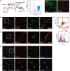
(A) Schematic representation of the experimental design. LV sections from infarcted MFG-E8 KO mice (CD45.2+) transplanted with the bone marrow cells of WT mice (CD45.1+) were stained with anti-CD45.1 (green) and anti–MFG-E8 (red) antibodies (n = 3). Scale bar: 50 μm. FACS dot plot of peripheral blood cells for recipient-type CD45.2-APC and donor-type CD45.1-FITC is shown. (B and C) LV sections from WT mice on day 3 after MI were stained with anti–MFG-E8 (red) and anti-PDGFRβ (green) (B) or anti–MFG-E8 (red) and anti–Wt-1 (green) (C) antibodies (n = 3). White squares on the merged images (first columns) mark the areas shown at a higher magnification. Higher-magnification images are shown on the right side of lower-magnification images. Scale bars: 50 μm (lower magnification); 20 μm (higher magnification). (D) LV sections from WT mice on day 3 after MI were stained with anti–MFG-E8 (red), anti-CD31 (green), and anti-vimentin (blue) antibodies. The first column is the merged image of MFG-E8, CD31, and vimentin. The magnified views of the white boxed region on the low-magnification merged images are displayed in a vertical row. The insets (lower panels) show the details of the MFG-E8, vimentin, and CD31 immunoreactive myofibroblasts. Scale bars: 50 μm (lower magnification); 20 μm (higher magnification). White arrows indicate merged cells. (E) Cardiac myofibroblasts isolated from WT mice on day 3 after MI were stained with anti–MFG-E8, FITC-anti-αSMA, and PerCP/Cy5.5-anti-CD31 antibodies and analyzed by flow cytometry (n = 4). Representative data are shown (AE).
Figure 8
Figure 8. MFG-E8 is produced in myofibroblasts via the SRF-dependent pathway.
(A) Acta2, Adam12, and Mfge8 mRNA expression levels in rat neonatal cardiac fibroblasts treated with TGF-β1 (24, 48, and 72 hours, n = 5). (B) Acta2 and Mfge8 mRNA expression levels in rat neonatal cardiac fibroblasts treated with TGF-β1 (10 ng/ml) in the absence or presence of CCG-1423 (1 or 10 μM, n = 5 or 6). (C) Acta2, Mfge8, and Srf mRNA expression levels in HUVECs treated with or without TGF-β2 (10 ng/ml) for 72 hours in the presence of siRNA against SRF (n = 4 or 5). (D and E) Acta2 and Mfge8 mRNA expression levels in cardiac myofibroblasts, which were treated with or without CCG-1423 (1 or 10 μM) for 24 hours (D) or treated with siRNA against SRF (E) (n = 4 or 5). Decreased Srf mRNA expression in myofibroblasts treated with siRNA against SRF was confirmed in E (n = 4 or 5). Error bars represent the mean ± SEM. (A–D) *P < 0.05; **P < 0.01; ***P < 0.001, 1-way ANOVA followed by Newman-Keuls analysis. (E) *P < 0.05; ***P < 0.001, unpaired 2-tailed Student’s t test.
Figure 9
Figure 9. MFG-E8 administration after MI improves cardiac function in vivo.
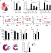
(A) Schematic representation of MFG-E8 intramyocardial injection. (B) TUNEL-positive nuclei in border zone of PBS- or MFG-E8–administered mice (3.2 μg) (sham, n = 5; MI, n = 7) 3 days after MI. (C) MFG-E8 intramyocardial injection (1.6 or 3.2 μg) decreased expression of the upregulated inflammatory genes at the infarct (Inf), not remote (Rem), areas of hearts 3 days after MI (n = 4 each). (D) Temporal changes in echocardiographic parameters (LV end-diastolic internal diameter [LVIDd], LV end-systolic internal diameter [LVIDs], interventricular-septal thickness at end-diastole [IVSTd], ejection fraction, and fractional shortening) of PBS-treated (sham, n = 7, black-dotted lines; MI, n = 10, black lines) or MFG-E8–treated mice (sham, n = 7, red-dotted lines; MI; n = 7, red lines) 2, 6, 8, and 10 weeks after MI. (E) Hemodynamic parameters (dP/dtmax, -dP/dtmin, and Tau) of PBS-treated (sham, n = 3; MI, n = 7) or MFG-E8–treated mice (sham, n = 5; MI, n = 7) 10 weeks after MI. (F) Heart weight to body weight ratio of PBS-treated (sham, n = 7; MI, n = 10) or MFG-E8–treated mice (sham, n = 7; MI, n = 9) 10 weeks after MI. (G) Representative Masson’s trichrome–stained heart sections of PBS-administered (n = 6) or MFG-E8–administered mice (n = 5) 10 weeks after MI. Scale bars: 1 mm. Error bars represent the mean ± SEM. *P < 0.05; **P < 0.01; ***P < 0.001, 1-way ANOVA followed by Newman-Keuls analysis.
Figure 10
Figure 10. Schematic model depicting origins and functions of MFG-E8–producing myofibroblasts appearing at the infarcted area after MI.
Resident fibroblasts and cells that underwent EndMT differentiate into MFG-E8–producing myofibroblasts upon inflammation. MFG-E8 was produced during the differentiation via the SRF-dependent pathway. The myofibroblasts not only produce extracellular matrix, but also engulf dead cells via an MFG-E8/integrin αvβ5–dependent pathway, and attenuate inflammation, similarly to macrophages.

References

    1. Roger VL, et al. Heart disease and stroke statistics--2012 update: a report from the American Heart Association. Circulation. 2012;125(1):e2–e220. doi: 10.1161/CIR.0b013e31823ac046. - DOI (V体育官网入口) - PMC - PubMed
    1. Yellon DM, Hausenloy DJ. Myocardial reperfusion injury. N Engl J Med. 2007;357(11):1121–1135. doi: 10.1056/NEJMra071667. - DOI (V体育ios版) - PubMed
    1. Olivetti G, et al. Apoptosis in the failing human heart. N Engl J Med. 1997;336(16):1131–1141. doi: 10.1056/NEJM199704173361603. - DOI - PubMed
    1. Whelan RS, Kaplinskiy V, Kitsis RN. Cell death in the pathogenesis of heart disease: mechanisms and significance. Annu Rev Physiol. 2010;72:19–44. doi: 10.1146/annurev.physiol.010908.163111. - "VSports手机版" DOI - PubMed
    1. Frangogiannis NG. Regulation of the inflammatory response in cardiac repair. Circ Res. 2012;110(1):159–173. doi: 10.1161/CIRCRESAHA.111.243162. - DOI (V体育2025版) - PMC - PubMed

Publication types

MeSH terms