Skip to main page content
U.S. flag

An official website of the United States government

Dot gov

The . gov means it’s official. Federal government websites often end in . gov or . mil. Before sharing sensitive information, make sure you’re on a federal government site VSports app下载. .

Https

The site is secure. The https:// ensures that you are connecting to the official website and that any information you provide is encrypted and transmitted securely V体育官网. .

. 2016 Sep 8;1(14):e89014.
doi: 10.1172/jci.insight.89014.

Multiparametric profiling of non-small-cell lung cancers reveals distinct immunophenotypes

Affiliations

V体育平台登录 - Multiparametric profiling of non-small-cell lung cancers reveals distinct immunophenotypes

Patrick H Lizotte et al. JCI Insight. .

Abstract

BACKGROUND. Immune checkpoint blockade improves survival in a subset of patients with non-small-cell lung cancer (NSCLC), but robust biomarkers that predict response to PD-1 pathway inhibitors are lacking. Furthermore, our understanding of the diversity of the NSCLC tumor immune microenvironment remains limited. METHODS. We performed comprehensive flow cytometric immunoprofiling on both tumor and immune cells from 51 NSCLCs and integrated this analysis with clinical and histopathologic characteristics, next-generation sequencing, mRNA expression, and PD-L1 immunohistochemistry (IHC). RESULTS. Cytometric profiling identified an immunologically "hot" cluster with abundant CD8+ T cells expressing high levels of PD-1 and TIM-3 and an immunologically "cold" cluster with lower relative abundance of CD8+ T cells and expression of inhibitory markers. The "hot" cluster was highly enriched for expression of genes associated with T cell trafficking and cytotoxic function and high PD-L1 expression by IHC. There was no correlation between immunophenotype and KRAS or EGFR mutation, or patient smoking history, but we did observe an enrichment of squamous subtype and tumors with higher mutation burden in the "hot" cluster VSports手机版. Additionally, approximately 20% of cases had high B cell infiltrates with a subset producing IL-10. CONCLUSIONS. Our results support the use of immune-based metrics to study response and resistance to immunotherapy in lung cancer. FUNDING. The Robert A. and Renée E. Belfer Family Foundation, Expect Miracles Foundation, Starr Cancer Consortium, Stand Up to Cancer Foundation, Conquer Cancer Foundation, International Association for the Study of Lung Cancer, National Cancer Institute (R01 CA205150), and the Damon Runyon Cancer Research Foundation. .


Figures

Figure 1
Figure 1. Clinical characteristics of NSCLC data set.
Major immune cell lineages profiled from 51 NSCLC patients are depicted as the percentage of live cells and arranged by increasing percentage of CD8+ T cells. Colored tile tracks above indicate smoking status, histological subtype, mutant KRAS or EGFR, and PD-L1 IHC.
Figure 2
Figure 2. Clustering of NSCLC data set.
Unbiased hierarchical clustering of 51 NSCLC samples and, where available, matched normal lung (top row). Immune parameters measured by multicolor flow cytometry are listed. Tiles are shaded by percentage of expression of markers.
Figure 3
Figure 3. Immune cell lineages by clinical features.
Percentages of total CD45+ cells based on smoking history (A), histological subtype (B), oncogene status (C), and PD-L1 immunohistochemical scoring (D) of tumor cells (TC+) and immune cells (IC+) of major immune cell lineages that vary significantly between tumors are presented. Data for bar graphs were calculated using unpaired Student’s t test. *P < 0.05; **P < 0.01; ***P < 0.001. Mean with SD. Two-way ANOVA: smoker vs. never smoker, P = 0.4207; adenocarcinoma vs. squamous, P = 0.0362; adenocarcinoma vs. normal lung, P = 0.6034; squamous vs. normal lung, P = 0.0332; EGFR vs. KRAS, P = 0.1901; KRAS vs. neither, P = 0.9915; EGFR vs. neither, P = 0.2636; TC+ vs. IC+, P = 0.9990; TC+ vs. negative, P = 0.0630; IC+ vs. negative, P = 0.1667..
Figure 4
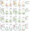
Figure 4. T cell expression of inhibitory receptors by clinical features.
Percentage of expression of inhibitory receptors PD-1, TIM-3, and CTLA -4 by CD4+ T cells (left) and CD8+ T cells (right) based on smoking history (A), histological subtype (B), oncogene status (C), and PD-L1 immunohistochemical scoring (D) of tumor cells (TC+) and immune cells (IC+). Data for bar graphs were calculated using unpaired Student’s t test with. *P < 0.05; **P < 0.01; ***P < 0.001. Mean with SD. Two-way ANOVA CD4+ T cells: smoker vs. never smoker, P = 0.2314; adenocarcinoma vs. squamous, P < 0.0001; adenocarcinoma vs. normal lung, P < 0.0001; squamous vs. normal lung, P < 0.0001; EGFR vs. KRAS, P = 0.2450; KRAS vs. neither, P = 0.1272; EGFR vs. neither, P = 0.0619; TC+ vs. IC+, P = 0.1038; TC+ vs. negative, P < 0.001; IC+ vs. negative, P = 0.3626. Two-way ANOVA CD8+ T cells: smoker vs. never smoker, P = 0.0433; adenocarcinoma vs. squamous, P = 0.0017; adenocarcinoma vs. normal lung, P = 0.0083; squamous vs. normal lung, P < 0.0001; EGFR vs. KRAS, P = 0.0759; KRAS vs. neither, P = 0.9649; EGFR vs. neither, P = 0.0972; TC+ vs. IC+, P = 0.2742; TC+ vs. negative, P < 0.0001; IC+ vs. negative, P = 0.0025.
Figure 5
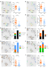
Figure 5. NSCLCs align into immunologically “hot” and “cold” clusters.
The t-distributed stochastic neighbor embedding (t-SNE) algorithm assigned NSCLC cases into 2 clusters (dotted ovals). t-SNE plots are identical by NSCLC case coordinate (i.e., each dot is a case and is in the same place in all 10 plots). Percentage of CD8+ T cells of CD3+ lymphocytes (A), percentage of PD-1 expression on CD8+ T cells (B), percentage of TIM-3 expression on CD8+ T cells (C), and percentage of FOXP3+ Tregs of CD4+ T cells (D), with gradient color coding of blue (low) to red (high). Percentage of PD-L1 expression on tumor cells by IHC (E), percentage of PD-L1 expression on immune cells by IHC (F), histological subtype (G), oncogene status (H), and smoking status (I) are overlaid on t-SNE plots. (J) Mutation burden is shown with gradient color coding of blue (low) to red (high). Vertical scatter plot statistics are analyzed using unpaired Student’s t test and stacked bar graphs are analyzed by Fisher’s exact test. **P < 0.01; ***P < 0.001. Mean with SD. Light gray circles on t-SNE plots indicate data not available. Notable examples are indicated by arrows and reference case numbers.
Figure 6
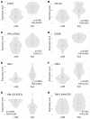
Figure 6. “Hot” cluster is enriched for CTL/Th1-associated genes.
Normalized mRNA expression of signature genes is presented for “hot” and “cold” clusters. CXCL9 (A), CXCL10 (B), IFN-γ (C), granzyme B (D), IDO1 (E), STAT1 (F), and TIM-3 (H) are upregulated and GM-CSF (G) is downregulated in the “hot” cluster relative to the “cold” cluster. In violin plots, horizontal lines depict medians, with narrow shaded boxes representing the first-to-third interquartile range and vertical lines representing the lower-to-upper adjacent value range. P values were calculated with unpaired Student’s t test.
Figure 7
Figure 7. Limited CD8+ T cell markers predict NSCLC immunophenotypes.
(A) NSCLC cases were reanalyzed using t-distributed stochastic neighbor embedding (t-SNE) algorithm based on only 3 parameters: percentage of CD8+ T cells of CD3+ lymphocytes, percentage of TIM-3 expression on CD8+ T cells, and percentage of PD-1 expression on CD8+ T cells. The structure of “hot” and “cold” clusters matched what was observed for multiparameter clustering. (B) Proposed model of immunotherapy-favorable immunophenotype of NSCLC based on limited T cell–intrinsic factors.
Figure 8
Figure 8. B cell abundance and phenotype in NSCLC.
(A) Flow cytometry profiling of NSCLCs revealed a small but reproducible population of IL-10+ B cells in tumor tissue but not normal lung. (B and C) CD19+ B cells were sorted from tumor and normal lung and analyzed by single-cell RNA sequencing. IL-10–producing B cells were present in tumors but not normal lung and display a unique transcriptional profile representative of plasma B cells, oxidative metabolism, and MYC activation.

References

    1. Borghaei H, et al. Nivolumab versus docetaxel in advanced nonsquamous non-small-cell lung cancer. N Engl J Med. 2015;373(17):1627–1639. - PMC - PubMed
    1. Topalian SL, et al. Safety, activity, and immune correlates of anti-PD-1 antibody in cancer. N Engl J Med. 2012;366(26):2443–2454. - PMC - PubMed
    1. Garon EB, et al. Pembrolizumab for the treatment of non-small-cell lung cancer. N Engl J Med. 2015;372(21):2018–2028. - PubMed
    1. EER Stat Fact Sheets: Lung and Bronchus Cancer. National Cancer Institute. VSports app下载 - http://seer.cancer.gov/statfacts/html/lungb.html Accessed August 30, 2016.
    1. Rizvi NA, et al. Activity and safety of nivolumab, an anti-PD-1 immune checkpoint inhibitor, for patients with advanced, refractory squamous non-small-cell lung cancer (CheckMate 063): a phase 2, single-arm trial. Lancet Oncol. 2015;16(3):257–265. - VSports手机版 - PMC - PubMed

Publication types