Skip to main page content
U.S. flag

An official website of the United States government

Dot gov

The . gov means it’s official. Federal government websites often end in . gov or . mil VSports app下载. Before sharing sensitive information, make sure you’re on a federal government site. .

Https

The site is secure. The https:// ensures that you are connecting to the official website and that any information you provide is encrypted and transmitted securely V体育官网. .

. 2016 May 8;6(8):1105-18.
doi: 10.7150/thno.14700. eCollection 2016.

MiR675-5p Acts on HIF-1α to Sustain Hypoxic Responses: A New Therapeutic Strategy for Glioma

Affiliations

MiR675-5p Acts on HIF-1α to Sustain Hypoxic Responses: A New Therapeutic Strategy for Glioma

VSports最新版本 - Alessia Lo Dico et al. Theranostics. .

"VSports最新版本" Abstract

Hypoxia is a common feature in solid tumours. In glioma, it is considered the major driving force for tumour angiogenesis and correlates with enhanced resistance to conventional therapies, increased invasiveness and a poor prognosis for patients. Here we describe, for the first time, that miR675-5p, embedded in hypoxia-induced long non-coding RNA H19, plays a mandatory role in establishing a hypoxic response and in promoting hypoxia-mediated angiogenesis. We demonstrated, in vitro and in vivo, that miR675-5p over expression in normoxia is sufficient to induce a hypoxic moreover, miR675-5p depletion in low oxygen conditions, drastically abolishes hypoxic responses including angiogenesis VSports手机版. In addition, our data indicate an interaction of miR675-5p, HIF-1α mRNA and the RNA Binding Protein HuR in hypoxia-induced responses. We suggest the modulation of miR675-5p as a new therapeutic option to promote or abolish hypoxia induced angiogenesis. .

Keywords: Angiogenesis; HuR; VHL V体育安卓版. ; glioma; hypoxia; miRNA675; optical imaging. .


Conflict of interest statement

Competing Interests: The authors declare no competing interest.

Figures

Figure 1
Figure 1
U251 glioma cells and HUVEC endothelial cells as suitable models for hypoxia studies. (A) ELISA assay for HIF-1α performed in U251 and HUVEC nuclear extracts after 6h of hypoxia. Data are expressed as Absorbance (ABS) values at 450nm. (B) Real-time PCR performed on U251 and HUVEC cells 6 hours post-hypoxia. Data were normalized for β-actin, and ΔΔct is expressed as fold of induction (FOI) of analyzed genes in hypoxia vs normoxia. The dashed line indicates control sample in normoxia. (C) ELISA assay for VEGF levels in supernatants from both cell lines after 6 hours of hypoxia. Data are expressed as pg/ml of soluble VEGF. (D) Left panel: Real time-PCR for lncRNA H19, after 6h of hypoxia, normalized for β-actin. Data are expressed as FOI of hypoxia-treated cells compared to normoxia samples. Right panel: Real time-PCR for H19's microRNAs, miR675-3p and miR675-5p, after 6h of hypoxia. Data were normalized for RNU48. Data are expressed as FOI compared to scramble-treated cells. Values are presented as the mean ± SD. Hypoxia vs normoxia * p<0.05; **p<0.01;* **p<0.001.
Figure 2
Figure 2
Gain and loss of function suggests a critical role of miR675-5p on hypoxia modulation. (A) ELISA for HIF-1α nuclear level in U251 and HUVECs transfected with miR675-5p inhibitor and exposed to hypoxia for 6h. Data are expressed as ABS values at 450nm. (B) Real-time PCR from U251 and HUVECs exposed to hypoxia after miR675-5p inhibitor or scramble control transfection. Data were normalized for β-actin and ΔΔct is expressed as fold of induction (FOI) of indicated genes after inhibitor transfection compared to scramble control (dashed line). (C) ELISA assay for VEGF level in supernatant from both cell lines transfected with miRNA inhibitor or negative control and exposed to hypoxia for 6h. Data are expressed as pg/ml of soluble VEGF. Values are presented as the mean ± SD. Inhibitor vs scramble * p<0.05; **p<0.0; ***p<0.001. (D) HIF-1α nuclear level analyzed by ELISA assay in U251 and HUVEC cells after 18h of miR675-5p mimic transfection in normoxia. Data are expressed as ABS values at 450nm. (E) Real-time PCR performed on U251 and HUVEC cells after miR675-5p mimic-transfection. Data were normalized for β-actin, and ΔΔct is expressed as fold of induction (FOI) in cells transfected with mimic compared to control (dashed line). (F) ELISA assay for VEGF level in supernatant from both cell lines 18h after mimic transfection. Data are expressed as pg/ml of soluble VEGF. Values are presented as the mean ± SD. Mimic vs scramble * p<0.05; **p<0.01;***p<0.001.
Figure 3
Figure 3
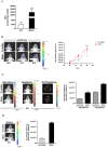
miR675-5p in vivo is able to enhance hypoxia establishment and neo-angiogenesis. (A) Luciferase in vitro assay performed on U251-HRE lysated cells after 18h of miR675-5p mimic-transfection. Data are represented as RLU (luminescent counts normalized to protein content) of mimic-treated cells compared to scramble-treated cells. Mimic vs scramble ***p<0.001. (B) Left panel: Representative (n=5) 2D bioluminescent (rainbow scale) of U251-HRE-mCherry tumours in scramble and miR675-5p mimic-treated mice (one of 5 mice) at day 12 (pre-treatment), day 15 (after two doses) and day 19 (end of treatment). Images are presented with the same scale bar. Right panel: Graphical representation of Luciferase activity over time during treatment. Data are presented as average radiance (photons/s/cm^2/steradian). (C) Left panel: Representative 2D images of mice co-injected with HypoxiSense 680 (BlueHot scale) and IntegriSense 750 (YellowHot scale) and axial images of IntegriSense750 co-registered with CT scan. All scans were performed at the final time point (day 19). Right panel: Quantification of HypoxiSense680 and IntegriSense750 fluorescence by ROI analysis for scramble and mimic-treated mice. Data are expressed as average radiance efficiency [(photons/s/cm^2/steradian)/(μW/cm^2)]. (D) Left panel: Representative (n=5) 2D mCherry fluorescent signal (rainbow scale) of U251-HRE-mCherry tumours in scramble and miR675-5p mimic-treated mice at day 19. Right panel: Graphical representation of mCherry activity at the end of treatment. Data are presented as average radiance efficiency [(photons/s/cm^2/steradian)/(μW/cm^2). Images are presented with the same scale bar. Mimic-treated mice vs scramble- treated mice * p<0.05; **p<0.01; ***p<0.001.
Figure 4
Figure 4
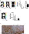
miR675-5p inhibitor counteracts hypoxia-mediated angiogenesis in vivo. (A) Left Panel: Representative (n=5) 2D images of scramble or miR675-5p inhibitor-treated mice co-injected with HypoxiSense 680 (BlueHot scale) and IntegriSense 750 (YellowHot scale) and axial images of IntegriSense750 co registered with CT scan. All scans were performed at the final time point (day 28). Right panel: Quantification of HypoxiSense 680 and IntegriSense 750 fluorescence by ROI analysis for scramble and miR675-5p inhibitor-treated mice (considering five mice for each treatment). Data are expressed as average radiance efficiency [(photons/s/cm^2/steradian)/(μW/cm^2)]. (B) Left Panel: Representative (n=5) 2D mCherry fluorescent signal (rainbow scale) of U251-HRE-mCherry tumours in scramble and inhibitor-treated mice (one of five mouse for each treatment) at day 28. Images are presented with the same scale bar. Right panel: Graphical representation of mCherry activity at the end of treatment considering five mice for each treatment. Data are presented as the average radiance efficiency [(photons/s/cm^2/steradian)/(μW/cm^2). Images are presented with the same scale bar. Inhibitor-treated mice vs scramble-treated mice * p<0.05; **p<0.01; ***p<0.001. (C) Immunoistochemistry for HIF-1α staining in scramble and miR675-inhibitor-treated group (scale bar is represented).
Figure 5
Figure 5
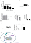
miR675-5p regulates HIF-1α mRNA stability by VHL/HuR axis. (A) Real-time PCR for VHL was done on U251 cells 18h after transfection with 5pM, 15pM, 30pM and 60pM of miR675-5p mimic. The dashed line indicates the control sample (scramble-treated). Values are presented as the mean ± SD. Mimic vs scramble * p<0.05; **p<0.01. (B) Western Blot for VHL and β-actin from U251 cells transfected with miR675-5p mimic (5pM) or scramble negative control. (C) Expression of VHL-regulated miRNA was validated by Luminescence assay kit, a secondary reporter was used for transfection normalization. Data are expressed as Relative Luciferase Activity in pGL3-pVHL-WT construct transfected with miR675-5p or miR21-5p and their relative scrambles. pGL3-control is represented as dashed line. The mean ± SD of three independent experiment is shown, and each sample was assayed in triplicate. (D) Left panel: Real-time PCR for HIF-1α performed on U251 and HUVEC cells 6 hours post-hypoxia. Data were normalized for β-actin, and ΔΔCt is expressed as fold of induction (FOI) of HIF-1α in hypoxia vs normoxia. Right panel: Real-time PCR performed on U251 and HUVEC cells 6 hours post-hypoxia and after miRNA675-5p inhibitor transfection. Data were normalized for β-actin, and ΔΔCt is expressed as fold of induction (FOI) of HIF-1α in hypoxia in inhibitor vs normoxia. (E) Left panel: Western Blot for HuR and β-actin from U251 cells transfected with miR675-5p inhibitor (5pM) or scramble negative control after 6h of hypoxia. Right panel: RNA-immunoprecipitation for HuR. Real time PCR for VEGF and HIF-1α were performed starting from HuR co-immunoprecipitated RNA in different culture conditions. Ct value was normalized to the Input RNA Ct value. Data are expressed as fold enrichment above the input percentage and presented as the mean ± SD. Inhibitor vs scramble negative control ***p<0.001. (F) Molecular mechanism of miRNA 675-5p in hypoxia condition. The dashed line indicates our experimental results.
Figure 6
Figure 6
Time lines represent the schedule of i.v. administration for n=5 mice. Upper panel: mir675-5p mimic/scramble administration. Lower panel: miR675-5p inhibitor/scramble administration.
Figure 6
Figure 6
Time lines represent the schedule of i.v. administration for n=5 mice. Upper panel: mir675-5p mimic/scramble administration. Lower panel: miR675-5p inhibitor/scramble administration.

References

    1. Vaupel P, Harrison L. Tumor hypoxia: causative factors, compensatory mechanisms, and cellular response. Oncologist. 2004;9(Suppl 5):4–9. - "V体育ios版" PubMed
    1. Folkman J. Angiogenesis in cancer, vascular, rheumatoid and other disease. Nat Med. 1995;1:27–31. - "VSports手机版" PubMed
    1. Mongiardi MP. Angiogenesis and hypoxia in glioblastoma: a focus on cancer stem cells. CNS Neurol Disord Drug Targets. 2012;11:878–83. - PubMed
    1. Jensen RL. Hypoxia in the tumorigenesis of gliomas and as a potential target for therapeutic measures. Neurosurg Focus. 2006;20:E24. - PubMed
    1. Zhong H, De Marzo AM, Laughner E, Lim M, Hilton DA, Zagzag D. et al. Overexpression of hypoxia-inducible factor 1alpha in common human cancers and their metastases. Cancer Res. 1999;59:5830–5. - PubMed

"VSports手机版" Publication types