Skip to main page content
U.S. flag

An official website of the United States government

Dot gov

The . gov means it’s official VSports app下载. Federal government websites often end in . gov or . mil. Before sharing sensitive information, make sure you’re on a federal government site. .

Https

The site is secure. The https:// ensures that you are connecting to the official website and that any information you provide is encrypted and transmitted securely V体育官网. .

. 2016 May 15;76(10):2954-63.
doi: 10.1158/0008-5472.CAN-15-2121. Epub 2016 Mar 15.

The EGF Receptor Promotes the Malignant Potential of Glioma by Regulating Amino Acid Transport System xc(-) (VSports app下载)

Affiliations

The EGF Receptor Promotes the Malignant Potential of Glioma by Regulating Amino Acid Transport System xc(-) (V体育官网入口)

"V体育平台登录" Kenji Tsuchihashi et al. Cancer Res. .

Abstract

Extracellular free amino acids contribute to the interaction between a tumor and its microenvironment through effects on cellular metabolism and malignant behavior. System xc(-) is composed of xCT and CD98hc subunits and functions as a plasma membrane antiporter for the uptake of extracellular cystine in exchange for intracellular glutamate VSports手机版. Here, we show that the EGFR interacts with xCT and thereby promotes its cell surface expression and function in human glioma cells. EGFR-expressing glioma cells manifested both enhanced antioxidant capacity as a result of increased cystine uptake, as well as increased glutamate, which promotes matrix invasion. Imaging mass spectrometry also revealed that brain tumors formed in mice by human glioma cells stably overexpressing EGFR contained higher levels of reduced glutathione compared with those formed by parental cells. Targeted inhibition of xCT suppressed the EGFR-dependent enhancement of antioxidant capacity in glioma cells, as well as tumor growth and invasiveness. Our findings establish a new functional role for EGFR in promoting the malignant potential of glioma cells through interaction with xCT at the cell surface. Cancer Res; 76(10); 2954-63. ©2016 AACR. .


Conflict of interest statement

Conflict of interest: None of the authors have any relevant conflicts of interest pertaining to the studies and data in this manuscript.

Figures

Figure 1
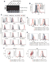
Figure 1. EGFR promotes surface xCT expression in glioma cells
(A) Reverse transcription (RT) and PCR analysis with primers targeted to exons 5 and 16 of the human CD44 gene as well as to the human glyceraldehyde-3-phosphate dehydrogenase (GAPDH) gene. The breast cancer cell line MDA-MB-468 was examined as a positive control for CD44v expression. (B) Flow cytometric analysis of surface xCT expression in the indicated cell lines. Staining of T98G cells with an isotype control antibody is also shown. (C) Flow cytometric analysis of surface expression of the indicated proteins in xCTlow (U87MG and Becker for EGFR, IGFR1, and integrin α6) and xCThigh (T98G) cells. (D) Flow cytometric analysis of surface expression of EGFR and xCT in T98G cells transfected with control or EGFR siRNAs. mRFI, mean value for relative fluorescence intensity. (E) Flow cytometric analysis of surface expression of EGFR and xCT in T98G cells transfected with a control siRNA or an siRNA targeted to the 5′ untranslated region (5′UTR) of EGFR mRNA as well as with an expression vector for EGFR lacking the 5′UTR or with the corresponding empty vector (Mock). (F) Flow cytometric analysis of surface expression of EGFR and xCT in MGG18 and GB2 cells transfected with control or EGFR siRNAs.
Figure 2
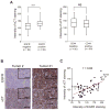
Figure 2. Expression of EGFR is correlated with that of xCT in human gliomas
(A) Box-and-whisker plots of xCT immunostaining intensity for glioma specimens that were negative (n = 84) or positive (n = 56) for EGFR staining (left panel) or negative (n = 80) or positive (n = 60) for CD44 staining (right panel). ***p < 0.001; NS, not significant (Student’s t test). (B) Immunohistochemical analysis of EGFR and xCT in human glioma specimens. Tumor 21 shows more intense staining for both EGFR and xCT compared with tumor 2. Scale bars, 100 μm (main panels) or 20 μm (insets). (C) Scatter plot for the intensity of immunostaining for EGFR and xCT in 56 specimens of human EGFR-expressing glioma. Pearson’s correlation coefficient is indicated by r.
Figure 3
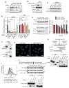
Figure 3. Intracellular domain of EGFR interacts with xCT and thereby promotes its cell surface expression
(A) Immunoblot analysis of EGFR, xCT, and β-actin (loading control) in T98G cells transfected with control, EGFR, or xCT siRNAs for 72 h or the indicated times. (B) Immunoblot analysis of total or phosphorylated (p) forms of EGFR and ERK in T98G cells that had been incubated in the absence or presence of EGF (50 ng/ml) or gefitinib (1 or 2 μM) for 30 min. (C) Immunoblot analysis of xCT in T98G cells treated with gefitinib (1 or 2 μM) or dimethyl sulfoxide (DMSO) vehicle) for the indicated times. (D) Quantitative RT-PCR analysis of EGFR and xCT mRNAs in T98G cells transfected with EGFR or xCT siRNAs for the indicated times. ***p < 0.001 (Student’s t test). (E) Immunoblot analysis of xCT in T98G cells transfected with control or EGFR siRNAs for 36 h and then exposed to cycloheximide (CHX, 100 μg/ml) for the indicated times (left panels). The xCT/β-actin band intensity ratios relative to the corresponding value for time zero were determined as means ± SD from three independent experiments (right panel). *p < 0.05, **p < 0.01 (Student’s t test). (F) T98G cell lysates were subjected to immunoprecipitation (IP) with antibodies to EGFR or to xCT or with control immunoglobulin G (IgG). The resulting precipitates, as well as 5% of the original cell lysates (Input), were subjected to immunoblot analysis with antibodies to xCT and to EGFR. (G) T98G cells were subjected to a PLA with control IgG (left panel) or antibodies to EGFR and to xCT (right panel). Red dots represent PLA signals. Scale bars, 20 μm. (H) Schematic representation of full-length (WT) and mutant forms of human EGFR. EGFRvIII lacks the amino acid sequence encoded by exons 2 to 7 of the EGFR gene. ΔN-term lacks the extracellular region of EGFR and consists of amino acid residues 621 to 1186, whereas ΔC-term consists of residues 1 to 684 and lacks most of the intracellular domain (upper panel). Lysates of HEK293T cells expressing FLAG-tagged WT or mutant forms of EGFR were subjected to immunoprecipitation with antibodies to xCT, and the resulting precipitates, as well as the original cell lysates (Input), were subjected to immunoblot analysis with antibodies to FLAG or to xCT (lower panel). (I) Flow cytometric analysis of surface xCT expression in T98G cells transfected with an siRNA targeted to the 5′UTR of EGFR mRNA as well as with an expression vector for ΔN-term or ΔC-term mutants of EGFR lacking the 5′UTR sequence or with the corresponding empty vector (Mock). (J) Schematic representation of full-length (FL) and mutant forms of human xCT (upper panel). Lysates of HEK293T cells expressing FLAG-tagged EGFR and hemagglutinin epitope (HA)–tagged full-length or mutant forms of xCT were subjected to immunoprecipitation with antibodies to HA. The resulting precipitates, as well as the original cell lysates (Input), were subjected to immunoblot analysis with antibodies to FLAG and to HA (lower panel).
Figure 4
Figure 4. Sulfasalazine disrupts redox status and thereby reduces cell viability in EGFR-expressing glioma cells
(A) Flow cytometric analysis of surface expression of EGFR and xCT in U87MG cells stably expressing EGFR (U87MG-EGFR cells) as well as in parental U87MG cells. (B) Measurement of 15N2-cystine uptake in U87MG and U87MG-EGFR cells. Data are means ± SD from five separate experiments. *p < 0.05 (Student’s t test). See also Supplementary Table S1 for raw data. (C) Analysis of GSH and total glutathione (GSH + 2GSSG) content in U87MG and U87MG-EGFR cells. Data are means ± SD from five independent experiments.*p < 0.05, ***p < 0.001 (Student’s t test). (D) U87MG cells were injected into the left hemisphere and U87MG-EGFR cells were injected into the right hemisphere of NOD/SCID mice. After 2 weeks, the brain with embedded tumors was dissected and serial sections of mouse brain harboring separate tumors formed by U87MG cells (arrowheads) and U87MG-EGFR cells (arrows) were subjected to immunohistochemical staining with antibodies to human vimentin (left panel) as well as to imaging mass spectrometry of GSH (right panel). Scale bar in the right panel, 200 μm. The color scale indicates peak intensity levels at a mass/charge (m/z) ratio of 306.07 (GSH). (E) Glioma cell lines were incubated with the indicated concentrations of sulfasalazine for 60 h and then assayed for cell viability. Data are means ± SD from six independent experiments. ***p < 0.001 (Student’s t test). (F) U87MG-EGFR and T98G cells were incubated for 24 h with or without 400 or 600 μM sulfasalazine, respectively and in the absence or presence of 50 μM Trolox, 1 mM NAC, or 250 μM glutamate (Glu), after which the intracellular ROS level was measured on the basis of dichlorofluorescein (DCF) fluorescence. Data are means ± SD from five independent experiments. ***p < 0.001 (Student’s t test). (G) U87MG-EGFR and T98G cells were incubated for 60 h as in (F) and then assayed for cell viability. Data are means ± SD from five independent experiments. ***p < 0.001 (Student’s t test). (H) T98G cells transfected with an expression vector for xCT or the corresponding empty vector (Mock) were also transfected with control or EGFR (#1) siRNAs for 72 h and then assayed for cell viability (left panel). Data are means ± SD from five independent experiments. ***p < 0.001 (Student’s t test). Cell lysates were also subjected to immunoblot analysis of EGFR and xCT (right panel). (I) Volume of subcutaneous tumors formed in nude mice by U87MG or U87MG-EGFR cells at 14 days after cell injection and treatment with saline (Control), sulfasalazine (250 mg/kg), cisplatin (CDDP, 2 mg/kg), or sulfasalazine (250 mg/kg) plus cisplatin (2 mg/kg). Data are means ± SD for four or five animals per group. *p < 0.05, ***p < 0.001 (Student’s t test).
Figure 5
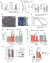
Figure 5. Glutamate release mediated by xCT promotes glioma cell migration
(A) U87MG and U87MG-EGFR cells transfected (or not) with xCT or EGFR (#1) siRNAs were cultured in glutamate-free medium for 8 h in the absence or presence of 1 μM gefitinib, after which the culture supernatants were assayed for glutamate. Data are means ± SD from four independent experiments. **p < 0.01 (Student’s t test). (B) MGG18 and GB2 cells transfected with control or EGFR (#1) siRNAs were cultured in glutamate-free medium for 8 h, after which the culture supernatants were assayed for glutamate. Data are means ± SD from four independent experiments. *p < 0.05, ***p < 0.001 (Student’s t test). (C) T98G cells were subjected to a scratch assay in the absence or presence of 200 μM sulfasalazine or 250 μM glutamate for the indicated times and then imaged by phase-contrast microscopy (left panel). The migratory distances of the cells at each time point are presented as means ± SD from three independent experiments (right panel). **p < 0.01 versus the corresponding value for nontreated cells (Student’s t test). (D) U87MG-EGFR cells were assayed for migration toward 5% FBS, EGF (10 ng/ml), Amphiregulin (20 ng/ml), or TGF-α (5 ng/ml) in the absence or presence of 200 μM sulfasalazine or 250 μM glutamate. Data are means ± SD from three independent experiments. *p < 0.05, **p < 0.01, ***p < 0.001 (Student’s t test). (E) MGG18 and GB2 cells were assayed for matrix invasion toward 5% FBS in the absence or presence of 200 μM sulfasalazine or 250 μM glutamate. Data are means ± SD from three independent experiments. *p < 0.05, **p < 0.01, ***p < 0.001 (Student’s t test). (F) Schematic representation of the organotypic brain slice culture system. (G) Immunofluorescence analysis of human vimentin for detection of matrix-invading glioma cells in a mouse brain slice. Brain slices implanted with U87MG-EGFR tumor pieces were cultured for 14 days in the absence or presence of EGF (10 ng/ml) or 100 μM sulfasalazine. The white dashed lines indicate the edge of each tumor piece. Scale bars, 100 μm. (H) Quantification of matrix invasion determined as in (G). The distance between the edge of the tumor piece and the corresponding invasion front formed by the matrix-invading tumor cells was measured. Data are means ± SD from three independent experiments. *p < 0.05, **p < 0.01 (Student’s t test). (I) Kaplan-Meier survival curves for mice with brain tumors derived from implanted U87MG-EGFR cells stably expressing control or xCT shRNAs (n = 5 for each group). **p < 0.01 (log-rank test).

"V体育安卓版" References

    1. Al-Zhoughbi W, Huang J, Paramasivan GS, Till H, Pichler M, Guertl-Lackner B, et al. Tumor macroenvironment and metabolism. Semin Oncol. 2014;41:281–95. - VSports注册入口 - PMC - PubMed
    1. Cantor JR, Sabatini DM. Cancer cell metabolism: one hallmark, many faces. Cancer Discov. 2012;2:881–98. - VSports最新版本 - PMC - PubMed
    1. Zhang W, Trachootham D, Liu J, Chen G, Pelicano H, Garcia-Prieto C, et al. Stromal control of cystine metabolism promotes cancer cell survival in chronic lymphocytic leukaemia. Nat Cell Biol. 2012;14:276–86. - VSports app下载 - PMC - PubMed
    1. Ganapathy V, Thangaraju M, Prasad PD. Nutrient transporters in cancer: relevance to Warburg hypothesis and beyond. Pharmacol Ther. 2009;121:29–40. - PubMed
    1. Sato H, Shiiya A, Kimata M, Maebara K, Tamba M, Sakakura Y, et al. Redox imbalance in cystine/glutamate transporter-deficient mice. J Biol Chem. 2005;280:37423–9. - PubMed

MeSH terms