Skip to main page content
U.S. flag

An official website of the United States government

Dot gov

The . gov means it’s official. Federal government websites often end in . gov or . mil VSports app下载. Before sharing sensitive information, make sure you’re on a federal government site. .

Https

The site is secure. The https:// ensures that you are connecting to the official website and that any information you provide is encrypted and transmitted securely. V体育官网.

. 2015 Dec 4:6:8792.
doi: 10.1038/ncomms9792.

Fibrocyte-like cells mediate acquired resistance to anti-angiogenic therapy with bevacizumab

Affiliations

Fibrocyte-like cells mediate acquired resistance to anti-angiogenic therapy with bevacizumab

Atsushi Mitsuhashi et al. Nat Commun. .

Abstract

Bevacizumab exerts anti-angiogenic effects in cancer patients by inhibiting vascular endothelial growth factor (VEGF). However, its use is still limited due to the development of resistance to the treatment. Such resistance can be regulated by various factors, although the underlying mechanisms remain incompletely understood. Here we show that bone marrow-derived fibrocyte-like cells, defined as alpha-1 type I collagen-positive and CXCR4-positive cells, contribute to the acquired resistance to bevacizumab. In mouse models of malignant pleural mesothelioma and lung cancer, fibrocyte-like cells mediate the resistance to bevacizumab as the main producer of fibroblast growth factor 2. In clinical specimens of lung cancer, the number of fibrocyte-like cells is significantly increased in bevacizumab-treated tumours, and correlates with the number of treatment cycles, as well as CD31-positive vessels. Our results identify fibrocyte-like cells as a promising cell biomarker and a potential therapeutic target to overcome resistance to anti-VEGF therapy. VSports手机版.


Conflict of interest statement (V体育安卓版)

Y V体育安卓版. N. has received research support from Chugai and Taiho Pharmaceuticals. The remaining authors declare no competing financial interests.

Figures

Figure 1
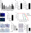
Figure 1. Continuous treatment with bevacizumab induces acquired resistance in vivo.
(a) The survival curve of tumour-bearing mice treated with bevacizumab (bev) beginning from seven days after tumour cell injection. (left) Human MPM cell lines (Y-MESO-14 and EHMES-10) were injected orthotopically. (right) Human lung cancer cell lines (PC14PE6 and A549) were injected via the tail vein. (b) The weights of Y-MESO-14 intrathoracic tumours from the control and bevacizumab (bev)-treated groups at different time points. (c) Representative images of sections from Y-MESO-14 intrathoracic tumours stained for CD31. The tumours were harvested at different time points from the control and bevacizumab (bev)-treated groups. Scale bar, 200 μm. (d) Quantitative evaluation of MVD (quantified as the total number of CD31+ structures/HPF) in each tumour of mice (n=5 per group) studied in c. The data are shown as the means±s.e.m *P<0.05 by a one-way ANOVA.
Figure 2
Figure 2. Host-derived FGF2 is upregulated in the intrathoracic Y-MESO-14 tumours resistant to bevacizumab treatment.
(a) A comparison of mouse mRNA expression of pro-angiogenic factors in the intrathoracic Y-MESO-14 tumours treated with or without bevacizumab (bev). The fold-changes in mRNA expression of the bevacizumab-treated tumours compared with control tumours are shown. *P<0.05 by the Mann–Whitney-U test. The changes in the amounts of mouse (b) FGF2 (n=8 per group) and (c) VEGF (n=6 per group) in the pleural effusion of tumour (Y-MESO-14)-bearing mice treated with or without bevacizumab (bev). *P<0.05 by the Mann–Whitney-U-test. (d) Representative images of FGF2-positive cells (green) in the sections from Y-MESO-14 intrathoracic tumours treated with or without bevacizumab (bev). Scale bar, 100 μm. (e) The number of FGF2-positive cells in the Y-MESO-14 tumours treated with bevacizumab (bev) compared with the control group (n=9 per group). *P<0.01 by the Mann–Whitney-U-test. (f) The survival curves of the tumour (Y-MESO-14)-bearing mice treated with bevacizumab (bev), BGJ-398 (an FGFR inhibitor), or both. Bevacizumab was continuously given from day 7 after tumour cell injection. BGJ-398 was given from days 14–28. (g) Representative images of the CD31-positive vessels in the tumours harvested from each group. Scale bar, 200 μm. (h) Evaluation of microvessel density (MVD, quantified as the total number of CD31+ structures/HPF) in the tumours from each group (n=12 per group). *P<0.01 by a one-way ANOVA. (i) Representative images of the CD31-positive vessels in the tumours harvested from mice treated with bevacizumab (bev) in combination with FGF2 Ab or control IgG. Scale bar, 200 μm. (j) Evaluation of MVD in each group (n=12 per group). *P<0.01 by the Mann–Whitney-U-test. All data are shown as the means±s.e.m.
Figure 3
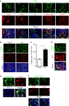
Figure 3. The identification of collagen type I+/CXCR4+ cells as FGF2-producing cells in the intrathoracic tumours produced by Y-MESO-14 cells.
(a) Representative images of tumour sections from mice treated with bevacizumab. FGF2 (green) was co-stained along with CD68, F4/80, Gr-1, collagen type I or CXCR4 (red). Scale bar, 100 μm. (b) Double staining of collagen type I and CXCR4 in tumours from mice treated with or without bevacizumab (bev). Scale bar, 200 μm. (c) The number of double-positive cells in the tumours was compared between the control- and bevacizumab (bev)-treated groups (n=20 per group). The data are shown as the means±s.e.m. *P<0.01 by the Mann–Whitney-U-test. (d) Representative images of double staining for collagen type I and CD45 in Y-MESO-14 tumours treated with bevacizumab. Scale bar, 100 μm. (e) Representative images of triple staining for collagen type I, CXCR4 and FGF2 in the tumours treated with bevacizumab. Scale bar, 50 μm. (f) Representative images of double staining for collagen type I (green) and CD68 or F4/80 (red) in Y-MESO-14 tumours treated with bevacizumab. Scale bar, 100 μm.
Figure 4
Figure 4. The recruitment of bone marrow-derived cells in bevacizumab-resistant tumour.
Mice were given a bone marrow transplant (BMT) from GFP transgenic mice before cancer cell inoculation. (a) Representative images of GFP-positive bone marrow cells (green) recruited into the control or bevacizumab (bev)-treated tumours. Scale bar, 200 μm. (b) The percentages of GFP-positive areas in the tumours were compared between the control- and bevacizumab (bev)-treated groups (n=8 per group). The data are shown as the means±s.e.m. *P<0.01 by the Mann–Whitney-U-test. (c) Representative images of triple staining for GFP, collagen type I and CXCR4 in the tumours treated with bevacizumab. Scale bar, 50 μm. (d) Representative images of double staining for GFP and FGF2 in the tumours treated with bevacizumab. Scale bar, 100 μm.
Figure 5
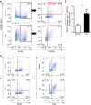
Figure 5. The isolation of mouse fibrocyte-like cells from Y-MESO-14 tumours by flow cytometry.
(a) Representative analysis of the detection of fibrocyte-like cells. The tumour tissues were harvested and single-cell suspended from control- or bevacizumab (bev)-treated mice on day 28. Fibrocyte-like cells were detected as CD45+/collagen type I+/CXCR4+ population. Each samples contained tumour tissues from two mice. (b) The comparison of the percentage of CD45+/collagen type I+/CXCR4+ cells between control- and bevacizumab (bev)-treated group (n=3 in each group, each samples contained tumour tissues from two mice). *P<0.05 by Student's t-test. The data are shown as the means±s.e.m. (c) MDSCs or Macrophages were detected as CD45+/Gr-1+ cells or CD45+/F4/80+ cells, respectively. Each samples contained tumour tissues from two mice. Data are the representative of two experiments with similar results.
Figure 6
Figure 6. The isolation and characterization of mouse fibrocyte-like cells from normal mouse lungs or Y-MESO-14 tumours.
(a) Fibroblasts (Fb) and fibrocyte-like cells (Fc) were isolated from the normal mouse lungs, and their mRNA expression levels were compared (n=3 per group). *P<0.01 by Student's t-test. (b,c) Fibrocyte-like cells were isolated from control- or bevacizumab (bev)-resistant tumours. (b) A comparison of the number of isolated fibrocyte-like cells (n=6 per group). (c) A comparison of the mRNA expression in isolated fibrocyte-like cells (n=6 per group). **P<0.05 by the Mann–Whitney-U-test. All data are shown as the means±s.e.m.
Figure 7
Figure 7. The involvement of CXCL12-CXCR4 axis in the fibrocyte-like cell-mediated angiogenesis.
(a) Assessment of hypoxic lesion in the tumour with immunohistochemistry using anti-CA9 antibody. (left) representative images of the hypoxic lesion stained in brown in the tumour produced by Y-MESO-14 cells treated with or without bevacizumab (bev). Tumours were harvested 28 days after the tumour cell-injection. Scale bar, 200 μm. (right) Evaluation of CA9-positive area (n=6 per group). *P<0.05 by the Mann–Whitney-U-test. (b) (left) the relative mRNA expression of human (tumour cell) CXCL12 in control- or bevacizumab (bev)-treated Y-MESO-14 tumour tissues (n=5 per group). *P<0.01 by the Mann–Whitney-U-test. Right, the mRNA expression of CXCL12 in Y-MESO-14 cells cultured under normoxia or chemical hypoxia induced by deferoxamine (DFX) in vitro (n=3 per group). *P<0.01 by Student's t-test. (c) Representative images of the migrated fibrocyte-like cells in vitro. Fibrocyte-like cells were cultured with or without a CXCR4 inhibitor (AMD3100) in the upper chamber, and those that migrated towards the lower chamber were stained. The lower chamber contained Y-MESO-14 cells cultured under normoxia or chemical hypoxia. Scale bar, 200 μm. (d) The number of fibrocyte-like cells that migrated under each condition. *P<0.05 by a one-way ANOVA. All in vitro data are representative of duplicate experiments with similar results. (eg) The effect of CXCR4 inhibition in addition to bevacizumab treatment in vivo. Thoracic tumours (Y-MESO-14) were harvested from mice treated with bevacizumab (bev) or bevacizumab in combination with AMD3100 (AMD). (e) A comparison of the tumour weights. (f) A comparison of MVD. (f) A comparison of the number of fibrocyte-like cells (collagen type 1+/CXCR4+ cells) (n=6–7 per group). *P<0.01 by the Mann–Whitney-U-test. All in vivo and in vitro data are shown as the means±s.e.m.
Figure 8
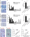
Figure 8. The recruitment of fibrocyte-like cells in the surgically resected human lung cancer specimens.
(a) Representative images of fibrocyte-like cells that were double-positive for CD45 (red) and FSP-1 (brown), in the tumour tissues of patients treated with neoadjuvant chemotherapy containing bevacizumab (bev), chemotherapy without bev or with no prior therapy. Scale bar, 100 μm. (b) The number of CD45+/FSP-1+ cells in the individual tumour samples. (c) The average number of CD45+/FSP-1+ cells in each group. *P<0.01 by a one-way ANOVA. (d) Representative images of the FGF2-positive cells (brown) in each group. Scale bar, 100 μm. (e) The number of FGF2-positive cells in the individual tumour samples. (f) The average number of FGF2-positive cells in each group. *P<0.01 by a one-way ANOVA. (g,h) Representative images of the double staining for (g) FSP-1 (red) or (h) CD68 (red) and FGF2 (brown) in the tumour from patient no. 1. (g) The arrows indicate double-positive cells. (h) The arrow indicates a CD68 single-positive cell, and the arrowhead indicates a FGF2 single-positive cell. Scale bars, 100 μm (top), 50 μm (bottom). (i,j) The correlation of the number of CD45+/FSP-1+ cells in the tumour and (i) the cycles of bevacizumab used and (j) the vessel length in the tumour. Each dot represents an individual tumour sample from patient nos 1–10. The correlation was estimated by Spearman's correlation and a linear regression analysis (the best-fit line is indicated together with the 95% confidence bands). The red dot indicates patient no. 1, who underwent surgical resection after developing obvious clinical resistance to bevacizumab.

References

    1. Folkman J. Angiogenesis: an organizing principle for drug discovery? Nat. Rev. Drug Discov. 6, 273–286 (2007). - PubMed
    1. Carmeliet P. & Jain R. K. Molecular mechanisms and clinical applications of angiogenesis. Nature 473, 298–307 (2011). - PMC - PubMed
    1. Ferrara N., Gerber H. P. & LeCouter J. The biology of VEGF and its receptors. Nat. Med 9, 669–676 (2003). - PubMed
    1. Ferrara N. & Davis-Smyth T. The biology of vascular endothelial growth factor. Endocr. Rev. 18, 4–25 (1997). - PubMed
    1. Sandler A. et al.. Paclitaxel-carboplatin alone or with bevacizumab for non-small-cell lung cancer. N. Engl. J. Med. 355, 2542–2550 (2006). - PubMed

Publication types

"VSports注册入口" MeSH terms

Substances

Associated data