<acronym id="aZuM6iTo"></acronym>
Skip to main page content
U.S. flag

An official website of the United States government

Dot gov

The . gov means it’s official. Federal government websites often end in . gov or . mil VSports app下载. Before sharing sensitive information, make sure you’re on a federal government site. .

Https

The site is secure V体育官网. The https:// ensures that you are connecting to the official website and that any information you provide is encrypted and transmitted securely. .

. 2015 Nov 24:5:17122.
doi: 10.1038/srep17122.

Structures of the CDK12/CycK complex with AMP-PNP reveal a flexible C-terminal kinase extension important for ATP binding (VSports)

Affiliations

Structures of the CDK12/CycK complex with AMP-PNP reveal a flexible C-terminal kinase extension important for ATP binding

Sarah E Dixon-Clarke et al. Sci Rep. .

Abstract

Cyclin-dependent kinase 12 (CDK12) promotes transcriptional elongation by phosphorylation of the RNA polymerase II C-terminal domain (CTD). Structure-function studies show that this activity is dependent on a C-terminal kinase extension, as well as the binding of cyclin K (CycK). To better define these interactions we determined the crystal structure of the human CDK12/CycK complex with and without the kinase extension in the presence of AMP-PNP. The structures revealed novel features for a CDK, including a large β4-β5 loop insertion that contributes to the N-lobe interaction with the cyclin VSports手机版. We also observed two different conformations of the C-terminal kinase extension that effectively open and close the ATP pocket. Most notably, bound AMP-PNP was only observed when trapped in the closed state. Truncation of this C-terminal structure also diminished AMP-PNP binding, as well as the catalytic activity of the CDK12/CycK complex. Further kinetic measurements showed that the full length CDK12/CycK complex was significantly more active than the two crystallised constructs suggesting a critical role for additional domains. Overall, these results demonstrate the intrinsic flexibility of the C-terminal extension in CDK12 and highlight its importance for both ATP binding and kinase activity. .


Figures
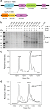
Figure 1
Figure 1. Soluble expression of CDK12 requires CycK.
(a) Schematic showing the domain organisation of human CDK12 and CycK (RS, arginine-serine-rich; PRM, proline-rich motifs). (b) Small scale nickel-affinity purifications from 3 mL baculoviral expression of hexahistidine-tagged proteins. Results are shown for a subset of experiments using the indicated CDK12 constructs as well as CycK11–267 and CycL143–320. (c) Deconvoluted intact mass spectra for CDK12715–1052 purified following co-expression with CycK11–267. The two mass peaks represent the native protein as well as a larger species containing a single phosphorylation on CDK12 Thr893. (d) Deconvoluted intact mass spectra for CDK12715–1038 obtained before and after treatment with recombinant CAK from Candida albicans. The shorter CDK12 construct is essentially unphosphorylated until CAK treatment.
Figure 2
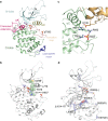
Figure 2. Three distinct structural states of the CDK12/CycK complex.
(a) Ribbon representation overview of the complex structures of CDK12715–1038/CycK11–267 and CDK12715–1052/CycK11–267. CDK12 is coloured green, except for the C-terminal kinase extension shown in pink. CycK is coloured light brown. The C-terminal kinase extension, including αK, adopts distinct packing conformations in the different chains in the CDK12715–1052/CycK11–267 structure. AMP-PNP, included in all crystallisations, is observed only in chain A of this structure where its binding is stabilized by the C-terminal extension. All CDK12 subunits were phosphorylated on Thr893 (shown by sticks). (b) Superposition of the CDK12 chains from available crystal structures. In addition to changes in the C-terminal region, there are subtle differences in the packing of the glycine-rich loop, the β4-β5 loop and the tilt of the αC helix. (c) Superposition of the CycK chains from available crystal structures. The apo-structure of CycK (PDB ID: 2I53) shows differences to the complex structures in the region of the H4’ helix in the second cyclin box as well as in the N and C-termini.
Figure 3
Figure 3
Structural features of the active CDK12 subunit (a) Ribbon representation highlighting selected structural features of the CDK12715–1052 subunit bound to AMP-PNP (PDB ID 4CXA, chain A). (b) CDK12 adopts an active conformation as shown by the correct alignment of the hydrophobic spine (indicated residues shown as spheres). (c) Phosphorylation at CDK12 Thr893 helps to stabilize the active kinase conformation. Shown are the hydrogen bond interactions of pThr893 with Arg882 (activation loop) and Arg858 (catalytic loop) as well as the nearby interaction of Arg773 (PITAIRE motif, αC helix) with CycK Glu108. (d) Ovarian cancer-associated mutations that impair CDK12 activity are mapped onto the structure and are predicted to destabilize the protein fold. Sites of mutation are indicated as spheres. CycK is omitted for clarity.
Figure 4
Figure 4
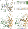
Side chain interactions in the CDK12/CycK interface (a) Binding of the CDK12 β4-β5 loop to CycK. Shown is a superposition of the two complexes in the asymmetric unit of the CDK12715–1052/CycK11–267 structure (CDK12 chain A and CycK chain B are coloured green and light brown, respectively, whereas chains C and D are coloured gray). A structure-based sequence alignment (right panel) reveals the insertion in the β4-β5 loop of CDK12 and CDK13 as well as the sequence divergence across this region and the interacting H5 helix of CycK. The positions of displayed residues participating in hydrophobic (ϕ) and hydrogen bond (*) interactions are marked under the alignment. (b) Hydrophobic interactions define the core of the protein-protein interface. (c) Electrostatic interactions cluster outside the core interface.
Figure 5
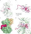
Figure 5. Alternative conformations of the CDK12 C-terminal extension.
(a) Superposition of the two CDK12715–1052 subunits in the asymmetric unit (PDB ID: 4CXA). An asterisk marks the point at which the two chains diverge following Leu1025. In chain A (green and bright pink), the C-terminal extension folds in front of the ATP pocket, whereas in chain C (gray and pale pink) the C-terminus extends across the back of the kinase domain. (b) Superposition reveals that the αK helix is stably formed in both chains despite their alternative packing arrangements. (c) Molecular surface representation of chains A and B highlighting the enclosure of the bound AMP-PNP molecule by the C-terminal kinase extension. Coloured as in Fig. 2. (d) Selected interactions of the C-terminal kinase extension packing at the front of the ATP pocket (CDK12 chain A).
Figure 6
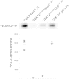
Figure 6. CDK12 truncations show diminished activity against the RNA Pol II CTD.
Comparison of in vitro kinase activity against a GST-CTD substrate by various CDK12 complexes and CDK9/Cyclin T1 (FL denotes full length proteins). A representative autoradiograph detecting 32P-GST-CTD product is shown together with a graphic representation of 3 biological replicates of each phosphorylation reaction.
Figure 7
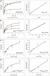
Figure 7. Determination of CDK12 kinetic parameters.
(a) Full length CDK12/CycK1 activity was measured against varying concentrations of ATP over 3 time points (15 min, 30 min and 60 min). Velocity and Lineweaver-Burke plots are shown (left and right panels, respectively). KmATP was determined to be 2 μM +/− 0.2 μM ATP (S.D.) based on two independent experiments using three different preparations of the full length enzyme. (b) Equivalent ATP titrations and plots for the CDK12715–1052/CycK11–267 complex. KmATP was determined to be 25.5 μM +/− 0.01 μM ATP (S.D.) based on two independent experiments. (c) Full length CDK12/CycK1 activity was measured against varying concentrations of GST-CTD substrate using 10 μM ATP and a reaction time of 15 min. Experiments were performed in triplicate (velocity plot, left panel) and the average values plotted in a Lineweaver-Burke plot (right panel). KmCTD was determined to be 0.3 μM +/− 0.06 μM (S.D.). (d) Equivalent GST-CTD titrations for the CDK12715–1052/CycK11–267 complex. KmCTD was determined to be 2 μM +/− 0.7 μM (S.D.). Differences in the Km values between the full length and truncated CDK12 complexes suggest that other domains within the full length protein may contribute to substrate binding and turnover.

References

    1. Malumbres M. et al. Cyclin-dependent kinases: a family portrait. Nat Cell Biol 11, 1275–6 (2009). - PMC - PubMed
    1. Lim S. & Kaldis P. Cdks, cyclins and CKIs: roles beyond cell cycle regulation. Development 140, 3079–93 (2013). - PubMed
    1. Mikolcevic P. et al. Cyclin-dependent kinase 16/PCTAIRE kinase 1 is activated by cyclin Y and is essential for spermatogenesis. Mol Cell Biol 32, 868–79 (2012). - VSports app下载 - PMC - PubMed
    1. Davidson G. et al. Cell cycle control of wnt receptor activation. Dev Cell 17, 788–99 (2009). - PubMed
    1. Mikolcevic P., Rainer J. & Geley S. Orphan kinases turn eccentric: a new class of cyclin Y-activated, membrane-targeted CDKs. Cell Cycle 11, 3758–68 (2012). - V体育官网 - PMC - PubMed

Publication types

MeSH terms

Associated data