Skip to main page content
U.S. flag

An official website of the United States government

Dot gov

The . gov means it’s official. Federal government websites often end in . gov or . mil VSports app下载. Before sharing sensitive information, make sure you’re on a federal government site. .

Https

The site is secure. The https:// ensures that you are connecting to the official website and that any information you provide is encrypted and transmitted securely. V体育官网.

. 2015 Nov 10:5:16386.
doi: 10.1038/srep16386.

Resveratrol inhibits epithelial-mesenchymal transition of retinal pigment epithelium and development of proliferative vitreoretinopathy

Affiliations

Resveratrol inhibits epithelial-mesenchymal transition of retinal pigment epithelium and development of proliferative vitreoretinopathy

Keijiro Ishikawa et al. Sci Rep. .

Abstract

Proliferative vitreoretinopathy (PVR) is a serious complication of retinal detachment and ocular trauma, and its recurrence may lead to irreversible vision loss. Epithelial to mesenchymal transition (EMT) of retinal pigment epithelial (RPE) cells is a critical step in the pathogenesis of PVR, which is characterized by fibrotic membrane formation and traction retinal detachment. In this study, we investigated the potential impact of resveratrol (RESV) on EMT and the fibrotic process in cultured RPE cells and further examined the preventive effect of RESV on PVR development using a rabbit model of PVR. We found that RESV induces mesenchymal to epithelial transition (MET) and inhibits transforming growth factor-β2(TGF-β2)-induced EMT of RPE cells by deacetylating SMAD4. The effect of RESV on MET was dependent on sirtuin1 activation. RESV suppressed proliferation, migration and fibronectin synthesis induced by platelet-derived growth factor-BB or TGF-β2. In vivo, RESV inhibited the progression of experimental PVR in rabbit eyes. Histological findings showed that RESV reduced fibrotic membrane formation and decreased α-SMA expression in the epiretinal membranes. These results suggest the potential use of RESV as a therapeutic agent to prevent the development of PVR by targeting EMT of RPE. VSports手机版.


Figures

Figure 1
Figure 1. RESV induces MET and inhibits TGF-β2 induced EMT by deacetylating SMAD4.
After 1 h pretreatment of RESV at 0, 25, 50 and 100 μM, the cells were stimulated with or without TGF-β2 at 10 ng/ml. (A) mRNA expression of E-cadherin and α-SMA are shown as relative fold to control normalized to GAPDH. NS, not significant. *P < 0.05. Data are presented as mean ± SEM. n = 4/group. (B) Western blot analysis of E-cadherin, α-SMA and the housekeeping protein GAPDH in the cell lysates of RPE cells. An expanded image of the western blot of E-cadherin and its molecular size are shown in Supplementary Figure 6. (C) Triple immunofluorescence staining for α-SMA and E-cadherin in RPE cells. Nuclei are stained blue. Scale bar: 10 μm. (D) Western blot (WB) analysis with acetyl-lysine (Ac-lysine) and SMAD4 in anti-SMAD4 immunoprecipitates (IP) shows a time course of TGF-β2 (10 ng/ml)-induced SMAD4 acetylation. (E) After 1 h pretreatment with RESV at 50 μM, the cells were stimulated with or without TGF-β2 at 10 ng/ml for 30 min. WB analysis of Ac-lysine and SMAD4 in anti-SMAD4 IP from the cells.
Figure 2
Figure 2. Facilitatory role of RESV on MET is dependent on SIRT1.
After transfection with control siRNA, SIRT1 siRNA, empty vector, and SIRT1-encoding vector, RPE cells were incubated for 48 h. (A) mRNA expression of SIRT1, E-cadherin and α-SMA are shown as relative fold to control siRNA normalized to GAPDH. **P < 0.01. *P < 0.05. Data are presented as mean ± SEM. n = 4/group. (B) Western blot analysis of SIRT1, E-cadherin, α-SMA and GAPDH in the cell lysates of RPE cells. An expanded image of the western blot of E-cadherin is shown in Supplementary Figure 6. (C) After transfection with control siRNA and SIRT1 siRNA, the cells were incubated with RESV at 50 μM for 48 h. mRNA expression of SIRT1, E-cadherin and α-SMA are shown as relative fold to control siRNA normalized to GAPDH. **P < 0.01. *P < 0.05. Data are presented as mean ± SEM. n = 4/group. (D) Western blot analysis of SIRT1, E-cadherin, α-SMA and GAPDH in the cell lysates of RPE cells. An expanded image of the western blot of E-cadherin is shown in Supplementary Figure 6.
Figure 3
Figure 3. RESV inhibits cell proliferation, migration and fibronectin synthesis.
After 1 h pretreatment with RESV at 0, 25, 50 and 100 μM, the cells were stimulated with or without PDGF-BB at 50 ng/ml for 48 h. (A) BrdU incorporations were measured to assay proliferation. NS, not significant. **P < 0.01. Data are presented as mean ± SEM. n = 4/group. (B) In the migration assay, the areas of the cells (stained by calcein AM) that migrated into the detection zone (white dotted circle) were measured. NS, not significant. **P < 0.01. Data are presented as mean ± SEM. n = 4/group. (C) Immunofluorescence staining of fibronectin in RPE cells with or without 10 ng/ml TGF-β2 stimulation for 12 h. Scale bar: 10 μm.
Figure 4
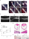
Figure 4. RESV inhibits progression of experimental PVR in rabbits.
(A) Ocular fundus photographs of normal eye and eyes with PVR after treatment with PBS and RESV at 2 mM. PVR classification was based on the extent of PVR in the lesion (arrows). Details of PVR grading procedures for Grades 1-5 for the rabbit PVR model are described in the Methods section. (B) Image of optical coherence tomography taken from normal eye, eyes with PVR after treatment with PBS and RESV at 2 mM. (C) Progression of PVR stages in PVR model of rabbits injected with PBS, RESV at 1 mM, and RESV at 2 mM. **P < 0.01. *P < 0.05. Data are presented as mean ± SEM. n = 6/group. (D) Section of a whole eye stained with hematoxylin and eosin (H&E) in a rabbit injected with PBS or RESV at 2 mM. Scale bars: 1 mm. Boxed region in the sections is shown at higher magnification. The epiretinal fibrotic membranes are indicated by yellow dotted circles. Scale bars: 200 μm. (E) Immunoreactivity to α-SMA in the fibrotic membranes in the eyes injected with PBS and RESV at 2 mM. Scale bars: 100 μm.

References

    1. Ryan S. J. The pathophysiology of proliferative vitreoretinopathy in its management. Am J Ophthalmol 100, 188–193 (1985). - PubMed
    1. Constable I. J. & Nagpal M. In Retina 5th edn, Vol. 3 (eds Ryan S. J. et al.) Ch. 107, p. 1806–1825 (Elsevier, 2013).
    1. Tosi G. M., Marigliani D., Romeo N. & Toti P. Disease pathways in proliferative vitreoretinopathy: an ongoing challenge. J Cell Physiol 229, 1577–1583 (2014). - PubMed
    1. Hiscott P., Sheridan C., Magee R. M. & Grierson I. Matrix and the retinal pigment epithelium in proliferative retinal disease. Prog Retin Eye Res 18, 167–190 (1999). - PubMed
    1. Strauss O. The retinal pigment epithelium in visual function. Physiol. Rev. 85, 845–881 (2005). - "VSports注册入口" PubMed

Publication types

"V体育官网入口" MeSH terms