"VSports最新版本" Skip to main page content
U.S. flag

An official website of the United States government

Dot gov

The . gov means it’s official VSports app下载. Federal government websites often end in . gov or . mil. Before sharing sensitive information, make sure you’re on a federal government site. .

Https

The site is secure. The https:// ensures that you are connecting to the official website and that any information you provide is encrypted and transmitted securely V体育官网. .

. 2015 Sep 15;6(27):23135-56.
doi: 10.18632/oncotarget.5218.

V体育官网入口 - p53-regulated autophagy is controlled by glycolysis and determines cell fate

Affiliations

p53-regulated autophagy is controlled by glycolysis and determines cell fate

Lei Duan et al. Oncotarget. .

"V体育安卓版" Abstract

The tumor suppressor p53 regulates downstream targets that determine cell fate. Canonical p53 functions include inducing apoptosis, growth arrest, and senescence VSports手机版. Non-canonical p53 functions include its ability to promote or inhibit autophagy and its ability to regulate metabolism. The extent to which autophagy and/or metabolic regulation determines cell fate by p53 is unclear. To address this, we compared cells resistant or sensitive to apoptosis by the p53 activator Nutlin-3a. In resistant cells, glycolysis was maintained upon Nutlin-3a treatment, and activated p53 promoted prosurvival autophagy. In contrast, in apoptosis sensitive cells activated p53 increased superoxide levels and inhibited glycolysis through repression of glycolytic pathway genes. Glycolysis inhibition and increased superoxide inhibited autophagy by repressing ATG genes essential for autophagic vesicle maturation. Inhibiting glycolysis increased superoxide and blocked autophagy in apoptosis-resistant cells, causing p62-dependent caspase-8 activation. Finally, treatment with 2-DG or the autophagy inhibitors chloroquine or bafilomycin A1 sensitized resistant cells to Nutlin-3a-induced apoptosis. Together, these findings reveal novel links between glycolysis and autophagy that determine apoptosis-sensitivity in response to p53. Specifically, the findings indicate 1) that glycolysis plays an essential role in autophagy by limiting superoxide levels and maintaining expression of ATG genes required for autophagic vesicle maturation, 2) that p53 can promote or inhibit autophagy depending on the status of glycolysis, and 3) that inhibiting protective autophagy can expand the breadth of cells susceptible to Nutlin-3a induced apoptosis. .

Keywords: Nutlin-3a; autophagy; glycolysis; p53. V体育安卓版.


Conflict of interest statement

CONFLICTS OF INTEREST

The authors declare that they have no conflict of interest.

Figures
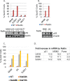
Figure 1
Figure 1. Cell lines sensitive or resistant to Nutlin-induced apoptosis
A. and B. The indicated cell lines were treated with vehicle (NT) or Nutlin (10 μM) for three days and analyzed for apoptosis (Sub-G1). The average percentage of apoptotic cells from triplicate was presented as a graph with standard deviation indicated (upper). Lysates of the cells treated with vehicle or Nultin for 24 hours were immunoblotted for p53 and β-actin (lower). C. The indicated cell lines were treated with vehicle (NT) or Nutlin (10 μM) for a time course and analyzed for apoptosis (Sub-G1). D. The indicated cell lines were treated with vehicle (NT) or Nutlin (10 μM) for 24 hours and mRNA was analyzed for the indicated genes.
Figure 2
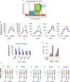
Figure 2. Nutlin reduces glycolysis and glycolytic genes in sensitive but not resistant cells
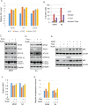
A. Graphic illustration of measurement of ECAR by Seahorse machine. B. The indicated cell lines were treated with vehicle (NT) or Nutlin (10 μM) for 16 hours and analyzed by Seahorse Extracellular Flux Analyzer for ECAR. C. Average glycolytic capacity from 6 replicates was presented as a graph with standard deviated indicated. There is significant difference between vehicle treated and Nutlin-treated MHM, S4, and SJSA1 cells (p < 0.01) but no difference for MCF7 and U2OS cells (p > 0.05). D. U2OS and MCF7 cells were treated with vehicle, Nutlin, 2-DG, or Nutlin plus 2-DG for three days and analyzed for apoptotic cells (Sub-G1). The average percentage of apoptotic cells from triplicate was presented as a graph with standard deviation indicated. D. The indicated cell lines were treated with vehicle (NT) or Nutlin (10 μM) for 24 hours. Relative mRNA of the indicated genes was quantitatively analyzed by Real-Time PCR and presented as graphs.
Figure 3
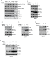
Figure 3. Nutlin inhibits autophagic flux in sensitive cells but promotes autophagic flux in resistant cells dependent on glycolysis
A. The indicated cell lines were treated with vehicle or Nutlin (10 μM) for 24 hours. Whole cell lysates were immunoblotted for phospho-AMPK (T172) and total AMPK, phospho-p70S6K or p70S6K, p53, LC3, and β-actin. MHM B., S4 C., SJSA1 D., and U2OS E. cells were treated with vehicle, Nutlin (10 μM), Bafilomycin A1 (BafA1, 10 μM), or Nutlin plus BafA1 for 24 hours. Whole cell lysates were immunoblotted for p62, LC3, p53, and β-actin. F. U2OS cells were treated with vehicle, Nutlin, 2-DG, or 2-DG plus Nutlin in the presence or absence of BafA1 for 24 hours. Whole cell lysates were immunoblotted for p62, LC3, and β-actin.
Figure 4
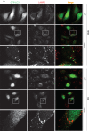
Figure 4. Nutlin affects formation of GFP-LC3-labeled autophagosomes differently in apoptosis sensitive and resistant cells
A. MHM and S4 cells were transiently transfected with GFP-LC3 for 24 hours and subsequently treated with vehicle (NT) or Nutlin (10 μM) for 24 hours. The cells were fixed and immunostained for LAMP2 and then scanned with a confocal microscope. The representative images for intracellular GFP-LC3 (green) and LAMP2 (red) were shown (the size of scale bar is 20 μM). B. U2OS cells were transiently transfected with GFP-LC3 for 24 hours and subsequently treated with vehicle (NT) Nutlin (10 μM), 2-DG (0.1 M), or Nutlin plus 2-DG for 24 hours. The cells were fixed and immunostained for LAMP2 and then scanned with a confocal microscope. The representative images for intracellular GFP-LC3 (green) and LAMP2 (red) were shown (the size of scale bar is 20 μM).
Figure 5
Figure 5. Nutlin increases autophagosomes in resistant cells dependent on glycolysis
Inhibition of autophagy sensitizes resistant cells to Nutlin-induced apoptosis. A. MHM, S4, and U2OS cells were transiently transfected with GFP-LC3 for 24 hours and subsequently treated with vehicle (NT) or Nutlin (10 μM) for 24 hours. The cells were scanned by a confocal microscope for GFP-LC-labeled autophagosomes. The percentage of the cells that showed punctate GFP-LC3 ( > 200 GFP-LC3 positive cells) were presented as a graph (representative of three independent experiments). B. MHM, S4, and U2OS cells were treated with vehicle (NT) or Nutlin (10 μM) for 24 hours. The cells were labelled with MDC and equal amount of cells were analyzed for MDC fluorescence. Average relative MDC fluorescence from triplicate was presented as a graph with standard deviation indicated. C. U2OS cells were transiently transfected with GFP-LC3 for 24 hours and subsequently treated with vehicle (NT) or Nutlin (10 μM) for 24 hours. The cells were scanned by a confocal microscope and percentage of the cells with formation of punctate GFP-LC3 in more than 200 GFP-LC3 positive cells were presented as a graph (representative of three independent experiments). D. U2OS cells were treated with vehicle (NT), Nutlin (10 μM), 2-DG (0.1 M), or Nutlin plus 2-DG for 24 hours. The cells were labelled with MDC and equal amount of cells were analyzed for MDC fluorescence. Average relative MDC fluorescence from triplicate was presented as a graph with standard deviation indicated. E. U2OS cells were treated with vehicle, Nutlin (10 μM), BafA1 (10 μM), Chloroquine (0.1 mM), or Nutlin plus BafA1 or Chloroquine for three days. The cells were analyzed for apoptosis (Sub-G1). Average percentage of apoptotic cells from triplicate was presented as a graph with standard deviation indicated.
Figure 6
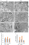
Figure 6. EM analysis of autophagic vacuoles
A. MHM, S4, and X2 cells were treated with vehicle (NT) or Nutlin (10 μM) for 24 hours. The cells were analyzed by a transmission microscope to visualize autophagic vacuoles (AVs). Representative images of early autophagic vacuoles (Avi, open arrow) and late autophagic vacuoles (Avd, filled arrow) were indicated. B. The autophagic vacuoles were quantified and average AVs per 100 square micrometer from whole grid scan was presented as a graph. C. The ratio of Avi vs. Avd is presented as a graph.
Figure 7
Figure 7. Nutlin downregulates ATG proteins in sensitive cells but not resistant cells
Inhibition of glycolysis also downregulates ATG proteins. A. MHM and S4 cells were treated with vehicle or Nutlin (10 μM) for 24 hours. Whole cell lysates were immunoblotted for ATG7, ATG10, and ATG5-12 conjugates. Note that the anti-ATG12 antibodies detected the 50 KD ATG5-12 conjugated proteins. Similar results were also shown using a ATG5 antibody (data not shown). B., SJSA1 cells were treated with vehicle or Nutlin (10 μM) in the presence or absence of BafA1 for 24 hours. Whole cell lysates were immunoblotted for ATG7, ATG10, and ATG5-12 conjugates. Note that the downregulation of ATG proteins is not blocked by BafA1 suggesting they are not degraded in lysosomes. C. U2OS cells were treated with vehicle or Nutlin (10 μM) in the presence or absence of 2-DG (0.1 M) for 24 hours. Whole cell lysates were immunoblotted for ATG7, ATG10, ATG5-12 conjugates, and p53. D. MHM and S4 cells were grow in media containing 10 mM glucose or 0.1 mM glucose (Glu deprivation) for 12 and 24 hours. Whole cell lysates were immunoblotted for ATG10 and ATG5-12 conjugates. E. MHM and U2OS were transfected with control siRNA or GAPDH siRNA for 48 hours. Whole cell lysates were immunoblotted for ATG10, ATG5-12 conjugates, and GAPDH with β-actin as control.
Figure 8
Figure 8. Nutlin induces accumulation of p62 and ubiquitinated proteins in insoluble fraction of lysates accompanied by activation of caspase-8 in sensitive cells
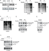
2-DG induces activation of caspase-8 in resistant cells. A. MHM, U2OS, and S4 cells were treated with vehicle or Nutlin for 24 hours. Equal number of cells were lysed in 1% Triton lysis buffer. The insoluble fraction (IF) and soluble fraction (SF) of the lysates were immunoblotted for p62. B. MHM and S4 cells were treated with vehicle or Nutlin for 24 hours. Equal amount of the SF and IF of the lysates were immunoblotted for ubiquitin. C. U2OS cells were treated with vehicle or Nutlin (10 μM) in the presence or absence of 2-DG (0.1 M) for 24 hours. Equal amount of the IF of the lysates were immunoblotted for ubiquitin. MHM and S4 cells D. and SJSA1 cells E. were treated with vehicle or Nutlin for 24 hours, U2OS cells F. were treated with vehicle or Nutlin (10 μM) in the presence or absence of 2-DG (0.1 M) for 24 hours. Whole cell lysates were immunoblotted for caspase-8 (upper part) and cleaved caspase-8 (lower part) with β-actin as loading control.
Figure 9
Figure 9. Inhibition of glycolysis induces O2-
The O2- scavenger Tiron promotes ATG protein expression, autophagy, and cell survival. A. MHM, S4, U2OS cells were treated with vehicle, Nutlin (10 μM), 2-DG (0.1 M), or Nutlin plus 2-DG for 24 hours. Intracellular O2- was measured with DHE and average relative O2- level from triplicate was presented as a graph with standard deviation indicated. B. MHM and S4 cells were treated with vehicle, Nutlin (10 μM), Tiron (5 mM), or Nutlin plus Tiron for 72 hours. The cells were analyzed for apoptosis (sub-G1). Average percentage of apoptotic cells from triplicate was presented with standard deviation indicated. MHM cells C. and S4 cells D. were treated with vehicle, Nutlin (10 μM), TIron (5 mM), or Nutlin plus Tiron for 24 hours. Whole cell lysates were immunoblotted for ATG3, 7, ATG10, ATG5-12 conjugates, LC3, and β-actin. E. MHM cells and S4 cells were treated with vehicle, Nutlin (10 μM), TIron (5 mM), or Nutlin plus Tiron in the presence or absence of BafA1 for 24 hours. Whole cell lysates were immunoblotted for p62 and β-actin. MHM and S4 cells were treated with vehicle, Nutlin (10 μM), Tiron (5 mM), or Nutlin plus Tiron for 24 hours, the cells were analyzed for MDC sequestration and autolysosomal volume. Average relative MDC fluorescence F. and autolysosomal volume G. from triplicate were presented as graphs with standard deviation indicated.
Figure 10
Figure 10. Proposed model
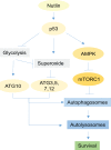
Activation of p53 by Nutlin leads to activation of AMPK and inhibition of mTORC1 which consequently initiate autophagy by activation of ULK1/2. Autophagy is a process consisting formation of autophagosomes and fusion with lysosomes to form autolysosomes. The early steps of formation of autophagosomes is regulated by ATG7 and ATG3, E1 and E2-like enzymes respectively that conjugate phosphatidylethanolamine to LC3 in order for attachment of LC3 to membrane. Maturation of autophagosomes to autolysosomes is regulated by conjugated ATG12-ATG5 proteins for which ATG7 and ATG10 are the respective E1 and E2 enzymes. Expression of ATG10 is dependent on glycolysis while expression of ATG3, 5, 7, 12 genes is suppressed by superoxide. In Nutlin-sensitive cells p53 inhibits glycolysis and induces superoxide which leads to downregulation of ATG3, 5, 7, 10, 12 genes and consequent disruption of autophagic process demonstrated as a decrease in autophagic flux. In contrast, in Nutlin-resistant cells p53 activates autophagy without affecting glycolysis and superoxide, leading to increased autophagic flux and completion of the autophagic process. Complete autophagy promotes survival by recycling essential nutrients and inhibition of caspase-8 activation.

References

    1. Schuler M, Green DR. Mechanisms of p53-dependent apoptosis. Biochemical Society transactions. 2001;29:684–688. - V体育官网入口 - PubMed
    1. Yu J, Zhang L. The transcriptional targets of p53 in apoptosis control. Biochemical and biophysical research communications. 2005;331:851–858. - VSports手机版 - PubMed
    1. Gartel AL, Serfas MS, Tyner AL. p21--negative regulator of the cell cycle. Proceedings of the Society for Experimental Biology and Medicine Society for Experimental Biology and Medicine. 1996;213:138–149. - PubMed
    1. Riley T, Sontag E, Chen P, Levine A. Transcriptional control of human p53-regulated genes. Nature reviews Molecular cell biology. 2008;9:402–412. - PubMed
    1. Madan E, Gogna R, Bhatt M, Pati U, Kuppusamy P, Mahdi AA. Regulation of glucose metabolism by p53: emerging new roles for the tumor suppressor. Oncotarget. 2011;2:948–957. - PMC - PubMed

Publication types

MeSH terms (VSports手机版)