Skip to main page content
U.S. flag

An official website of the United States government

Dot gov

The . gov means it’s official. Federal government websites often end in . gov or . mil. Before sharing sensitive information, make sure you’re on a federal government site. VSports app下载.

Https

The site is secure. The https:// ensures that you are connecting to the official website and that any information you provide is encrypted and transmitted securely. V体育官网.

. 2015 Oct;309(7):H1198-206.
doi: 10.1152/ajpheart.00393.2015. Epub 2015 Aug 14.

Vagus nerve stimulation mitigates intrinsic cardiac neuronal and adverse myocyte remodeling postmyocardial infarction

Affiliations

VSports app下载 - Vagus nerve stimulation mitigates intrinsic cardiac neuronal and adverse myocyte remodeling postmyocardial infarction

Eric Beaumont et al. Am J Physiol Heart Circ Physiol. 2015 Oct.

Abstract

This paper aims to determine whether chronic vagus nerve stimulation (VNS) mitigates myocardial infarction (MI)-induced remodeling of the intrinsic cardiac nervous system (ICNS), along with the cardiac tissue it regulates. Guinea pigs underwent VNS implantation on the right cervical vagus. Two weeks later, MI was produced by ligating the ventral descending coronary artery. VNS stimulation started 7 days post-MI (20 Hz, 0. 9 ± 0. 2 mA, 14 s on, 48 s off; VNS-MI, n = 7) and was compared with time-matched MI animals with sham VNS (MI n = 7) vs. untreated controls (n = 8). Echocardiograms were performed before and at 90 days post-MI. At termination, IC neuronal intracellular voltage recordings were obtained from whole-mount neuronal plexuses. MI increased left ventricular end systolic volume (LVESV) 30% (P = 0. 027) and reduced LV ejection fraction (LVEF) 6. 5% (P < 0. 001) at 90 days post-MI compared with baseline. In the VNS-MI group, LVESV and LVEF did not differ from baseline. IC neurons showed depolarization of resting membrane potentials and increased input resistance in MI compared with VNS-MI and sham controls (P < 0. 05). Neuronal excitability and sensitivity to norepinephrine increased in MI and VNS-MI groups compared with controls (P < 0. 05). Synaptic efficacy, as determined by evoked responses to stimulating input axons, was reduced in VNS-MI compared with MI or controls (P < 0. 05). VNS induced changes in myocytes, consistent with enhanced glycogenolysis, and blunted the MI-induced increase in the proapoptotic Bcl-2-associated X protein (P < 0 VSports手机版. 05). VNS mitigates MI-induced remodeling of the ICNS, correspondingly preserving ventricular function via both neural and cardiomyocyte-dependent actions. .

Keywords: autonomic regulation therapy; guinea pig; intrinsic cardiac nervous system; myocardial infarction; vagus nerve stimulation V体育安卓版. .


Figures

Fig. 1.
Fig. 1.
Evoked action potential (AP) frequencies with increasing intracellular stimulus intensities (0.1–0.6 nA, 500 ms) were determined by intracellular voltage recordings from intrinsic cardiac (IC) neurons in control preparations, in preparations at 90 days post-myocardial infarction (MI), and in preparations at 90 days post-MI that included 80 days of autonomic regulation therapy [vagus nerve stimulation (VNS)-MI], starting 10 days post-MI induction. A nonparametric Friedman test was used to evaluate difference among groups, followed by Wilcoxon signed-rank post hoc tests using a Bonferroni correction. Points represent the means ± SE from ∼60 cells for each condition. *P < 0.05, control vs. MI and VNS-MI.
Fig. 2.
Fig. 2.
Evoked action potential frequencies in response to increasing intracellular stimulus intensities were evaluated, concurrent with brief (1 s), local exposure to exogenous norepinephrine (NE) in IC soma derived from control animals and animals following MI, with and without chronic VNS. A nonparametric Friedman test was used to see differences among groups, followed by Wilcoxon signed-rank post hoc tests using a Bonferroni correction. Points represent the means ± SE from ∼60 cells for each condition. *P < 0.05, control vs. MI; #P < 0.05, control vs. VNS-MI.
Fig. 3.
Fig. 3.
Chronic VNS reduces synaptic efficacy of IC neurons. Nerve fibers synapsing with the IC neurons were stimulated via an extracellular concentric electrode (0.1–10 V, 2 ms) for 2 s at frequencies of 1, 2, 5, 10, and 20 Hz. A: representative examples of recordings derived from control, MI, and VNS-MI preparations when nerves were stimulated at 10 Hz. B: average data derived from ∼20 cells for each condition. An ANOVA analysis indicated significant differences among treatments and was followed by Newman-Keuls post hoc analysis. Points are the means ± SE. *P < 0.05, control and MI vs. VNS-MI neurons.
Fig. 4.
Fig. 4.
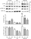
Phosphorylation status of GSK-3β and its substrate glycogen synthase (GS) in heart tissue derived from the MI (n = 5), VNS-MI (n = 4), and control (n = 3) animals. Shown are representative Western blots probed with antibodies specific for phosphorylated Ser641 of GS (p-GS), GS protein (GS), phosphorylated Ser9 of GSK-3β (p-GSK-3β), and GSK-3β protein (GSK-3β). Densitometry analysis of protein band intensity was performed for all Westerns. The graphs show the ratio of the p-GS/GS and p-GSK-3β/GSK-3β, where the protein bands were expressed in arbitrary densitometric units. ANOVA analysis indicating differences among the treatments was followed by Newman-Keuls post hoc analysis. *P < 0.05 vs. control central zone (CZ), intermediate zone (IZ), and distal zone (DZ); #P < 0.05 vs. MI-CZ, MI-IZ, and MI-DZ; and &P < 0.05 vs. VNS-MI-CZ.
Fig. 5.
Fig. 5.
The elevation of proapoptotic Bcl-2-associated X (BAX) in MI hearts is mitigated by VNS. A representative Western blot probed with antibodies specific for BAX protein is shown (30 μg total protein/lane). The experiment was repeated 4 times with all hearts. The graph shows the densitometry analysis of protein band intensity, which was performed for all Westerns for control (n = 3), MI (n = 5), and VNS-MI (n = 4). The blot stained with Ponceau S (Pon. S) is shown as protein-loading control. ANOVA analysis indicated significant differences among the treatments and was followed by Newman-Keuls post hoc analysis. *P < 0.05 MI vs. control and VNS-MI hearts.

References

    1. Ahonen A, Harkonen M, Juntunen J, Kormano M, Penttila A. Effects of myocardial infarction on adrenergic nerves of the rat heart muscle, a histochemical study. Acta Physiol Scand 93: 336–344, 1975. - PubMed
    1. Ajijola OA, Yagishita D, Patel KJ, Vaseghi M, Zhou W, Yamakawa K, So E, Lux RL, Mahajan A, Shivkumar K. Focal myocardial infarction induces global remodeling of cardiac sympathetic innervation: neural remodeling in a spatial context. Am J Physiol Heart Circ Physiol 305: H1031–H1040, 2013. - PMC - PubMed
    1. Antoni ML, Boden H, Hoogslag GE, Ewe SH, Auger D, Holman ER, van der Wall EE, Schalij MJ, Bax JJ, Delgado V. Prevalence of dyssynchrony and relation with long-term outcome in patients after acute myocardial infarction. Am J Cardiol 108: 1689–1696, 2011. - PubMed
    1. Ardell JL. Intrathoracic neuronal regulation of cardiac function In: Basic and Clinical Neurocardiology, edited by Armour JA, Ardell JL. New York: Oxford University Press, 2004, p. 118–152.
    1. Ardell JL, Cardinal R, Vermeulen M, Armour JA. Dorsal spinal cord stimulation obtunds the capacity of intrathoracic extracardiac neurons to transduce myocardial ischemia. Am J Physiol Regul Integr Comp Physiol 297: R470–R477, 2009. - PMC - PubMed

Publication types

"VSports" MeSH terms