Connectivity measures are robust biomarkers of cortical function and plasticity after stroke
- PMID: 26070983
- PMCID: PMC4840951
- DOI: 10.1093/brain/awv156
Connectivity measures are robust biomarkers of cortical function and plasticity after stroke
Abstract
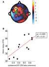
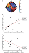
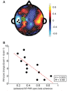
Valid biomarkers of motor system function after stroke could improve clinical decision-making. Electroencephalography-based measures are safe, inexpensive, and accessible in complex medical settings and so are attractive candidates. This study examined specific electroencephalography cortical connectivity measures as biomarkers by assessing their relationship with motor deficits across 28 days of intensive therapy. Resting-state connectivity measures were acquired four times using dense array (256 leads) electroencephalography in 12 hemiparetic patients (7. 3 ± 4. 0 months post-stroke, age 26-75 years, six male/six female) across 28 days of intensive therapy targeting arm motor deficits. Structural magnetic resonance imaging measured corticospinal tract injury and infarct volume. At baseline, connectivity with leads overlying ipsilesional primary motor cortex (M1) was a robust and specific marker of motor status, accounting for 78% of variance in impairment; ipsilesional M1 connectivity with leads overlying ipsilesional frontal-premotor (PM) regions accounted for most of this (R(2) = 0. 51) and remained significant after controlling for injury VSports手机版. Baseline impairment also correlated with corticospinal tract injury (R(2) = 0. 52), though not infarct volume. A model that combined a functional measure of connectivity with a structural measure of injury (corticospinal tract injury) performed better than either measure alone (R(2) = 0. 93). Across the 28 days of therapy, change in connectivity with ipsilesional M1 was a good biomarker of motor gains (R(2) = 0. 61). Ipsilesional M1-PM connectivity increased in parallel with motor gains, with greater gains associated with larger increases in ipsilesional M1-PM connectivity (R(2) = 0. 34); greater gains were also associated with larger decreases in M1-parietal connectivity (R(2) = 0. 36). In sum, electroencephalography measures of motor cortical connectivity-particularly between ipsilesional M1 and ipsilesional premotor-are strongly related to motor deficits and their improvement with therapy after stroke and so may be useful biomarkers of cortical function and plasticity. Such measures might provide a biological approach to distinguishing patient subgroups after stroke. .
Keywords: coherence; connectivity; motor; stroke V体育安卓版. .
© The Author (2015). Published by Oxford University Press on behalf of the Guarantors of Brain V体育ios版. All rights reserved. For Permissions, please email: journals.permissions@www.qiuluzeuv.cn. .
Figures


"VSports最新版本" References
-
- Andersson CA, Bro R. The N-way Toolbox for MATLAB. Chemom Intell Lab Syst 2000; 52: 1–4.
-
- Bath PMW, Lees KR, Schellinger PD, Altman H, Bland M, Hogg C, et al. Statistical analysis of the primary outcome in acute stroke trials. Stroke 2012; 1171–8. - V体育官网 - PubMed
-
- Burke E, Cramer SC. Biomarkers and predictors of restorative therapy effects after stroke. Curr Neurol Neurosci Rep 2013; 13: 329. - "VSports" PMC - PubMed
-
- Burke E, Dodakian L, See J, McKenzie A, Riley J, Le V, et al. A multimodal approach to understanding motor impairment and disability after stroke. J Neurol 2014b; 261: 1178–86. - PubMed
Publication types (VSports最新版本)
- Actions (VSports最新版本)
MeSH terms
- "V体育平台登录" Actions
- V体育ios版 - Actions
- "VSports手机版" Actions
- V体育官网入口 - Actions
- "V体育安卓版" Actions
Substances
- "VSports app下载" Actions
Grants and funding
LinkOut - more resources
Full Text Sources (VSports)
Other Literature Sources
Medical

