<area date-time="UjX2z"></area> Skip to main page content
U.S. flag

An official website of the United States government

Dot gov

The . gov means it’s official VSports app下载. Federal government websites often end in . gov or . mil. Before sharing sensitive information, make sure you’re on a federal government site. .

Https

The site is secure V体育官网. The https:// ensures that you are connecting to the official website and that any information you provide is encrypted and transmitted securely. .

. 2015 Jul 16;523(7560):352-6.
doi: 10.1038/nature14430. Epub 2015 May 25.

Improving survival by exploiting tumour dependence on stabilized mutant p53 for treatment

Affiliations

"VSports最新版本" Improving survival by exploiting tumour dependence on stabilized mutant p53 for treatment

E M Alexandrova et al. Nature. .

Erratum in (VSports在线直播)

Abstract

Missense mutations in p53 generate aberrant proteins with abrogated tumour suppressor functions that can also acquire oncogenic gain-of-function activities that promote malignant progression, invasion, metastasis and chemoresistance. Mutant p53 (mutp53) proteins undergo massive constitutive stabilization specifically in tumours, which is the key requisite for the acquisition of gain-of-functions activities. Although currently 11 million patients worldwide live with tumours expressing highly stabilized mutp53, it is unknown whether mutp53 is a therapeutic target in vivo. Here we use a novel mutp53 mouse model expressing an inactivatable R248Q hotspot mutation (floxQ) to show that tumours depend on sustained mutp53 expression. Upon tamoxifen-induced mutp53 ablation, allotransplanted and autochthonous tumours curb their growth, thus extending animal survival by 37%, and advanced tumours undergo apoptosis and tumour regression or stagnation. The HSP90/HDAC6 chaperone machinery, which is significantly upregulated in cancer compared with normal tissues, is a major determinant of mutp53 stabilization. We show that long-term HSP90 inhibition significantly extends the survival of mutp53 Q/- (R248Q allele) and H/H (R172H allele) mice by 59% and 48%, respectively, but not their corresponding p53(-/-) littermates. This mutp53-dependent drug effect occurs in H/H mice treated with 17DMAG+SAHA and in H/H and Q/- mice treated with the potent Hsp90 inhibitor ganetespib. Notably, drug activity correlates with induction of mutp53 degradation, tumour apoptosis and prevention of T-cell lymphomagenesis. These proof-of-principle data identify mutp53 as an actionable cancer-specific drug target. VSports手机版.


Figures
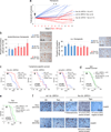
Extended Data Figure 1
Extended Data Figure 1. Generation and characterization of the conditionally inactivatable p53 flox R248Q allele
a–c, Generation of the conditionally inactivatable p53 R248Qflox allele. Mouse Exons 4–9 were replaced with human Exons 4–9 (called HUPKI, humanized p53 knockin) containing a p53 R248Q mutation in Exon 7 (marked by *). Exons 2–10 were flanked with loxP sites in Introns 1 and 10 (red arrows) to allow for Cre-targeted removal of the mutp53 allele upon addition of Tamoxifen/4OHT. A deletable Neo selection box was flanked by FRT sites (green). Knockin mice were mated with FLP mice to delete the Neo cassette in vivo, leaving behind the distal loxP site. The ‘floxQ’ allele thus has two loxP sites for subsequent Cre deletion. (b) For genotyping, the Neo-deleted floxQ knockin (KI) allele produces a 657 bp amplicon, in contrast to the 490 bp amplicon derived from the wtp53 allele. (c) Normal mouse embryo fibroblasts (MEFs) from floxQ/− embryos, which as non-malignant cells express non-stabilized mutp53, were adenovirally infected with empty vector (-Cre) or Cre-expressing vector (+Cre). Cre-mediated deletion of the mutp53 allele was confirmed by immunoblot analysis. d, The floxQ and constitutive Q (‘Q’) alleles behave identically in all aspects of gain-of-function including overall survival and tumor spectrum (not shown). Both floxQ and Q mice predominantly develop aggressive T-lymphomas, with some additional B-lymphomas and sarcomas. Also, the RosaCreERT2 allele has no discernable impact (data not shown). Kaplan-Meier analysis comparing overall survival of floxQ/− (red), Q/− (blue) and p53−/− (black) mice. Significance was assessed by Log Rank and Wilcoxon tests. e, Deletion of the mutp53 allele induces cell death in vitro. Viability of primary T- lymphoma cells freshly harvested from floxQ/−;ERT2/+ mice (n=4) and Q/−;ERT2/+ control mice (n=3), untreated or treated once with 4OHT or vehicle (EtOH) in short-term culture for 3–6 days. CTB assay, unpaired two-tailed Student’s t-test; mean ± s.e.m; n, number of independent T-lymphomas. Bottom, corresponding immunoblots of representative T-lymphomas at day 6. f, mutp53 deletion improves survival of host mice. Therapeutic protocol with primary floxQ/− T-lymphomas allotransplanted (black arrow on time axis) via subcutaneous injections into SCID mice. After visible tumors appeared, SCID mice were treated with daily intraperitoneal injections of oil or Tamoxifen (* on time axis). Mice were sacrificed when allowable endpoint size (1.5 cm3) was reached. Kaplan-Meier analysis, Log Rank test. Tamoxifen-induced allele deletion was strong but incomplete, shown by representative p53 immunofluorescence staining of tumors at endpoint (DAPI counterstain). g, Initial tumor volumes measured before treatment was started in the therapeutic protocol of the various groups shown in Fig. 1c. Unpaired two-tailed Student’s t-test; mean ± s.e.m; n, number of allografts. h, Control for Fig. 1d. Therapeutic treatment of nude mice allografted with p53−/−;ERT2/+ T-lymphoma cells and treated with Tam (150 mg/kg for 7 days) as indicated in the scheme in Fig. 1c (Endpoint 2). No response to Tam. Time-course, initial allograft volume and tumor mass at endpoint. Unpaired two-tailed Student’s t-test; mean ± s.e.m; n, number of allografts; NS, not significant.
Extended Data Figure 2
Extended Data Figure 2. Mutp53 ablation in floxQ/- mice causes autochthonous tumors regression or stagnation and extends T-lymphoma-specific survival
a, Time-course of imaged tumors, normalized to their initial tumor size (same as Fig. 2a but zoomed into the first 12 days of treatment). Stagnation or regression of floxQ/−;ERT2/+ tumors treated with Tam, in contrast to treated control tumors (floxQ/−;ERT2/+ on oil and Q/−;ERT2/+ on Tam) which grow robustly. #, sarcomas; all others are T-lymphomas. b, Similar mitotic index supports that cell cycle arrest and senescence are not significantly affected upon genetic mutp53 ablation in autochthonous T-lymphomas (from Fig. 2a–c) and s.c. allografts (from Fig. 1c, d). Instead, apoptosis is the main mechanism of tumor regression/stagnation. Quantification of phospho-S28 histone H3 (pH3) positive cells in individual autochthonous tumors or allografts. Five (left) or three (right) random 40× high power fields (with no or only minimal apoptosis) were counted for each tumor. Plotted are mean ± s.d. Representative pH3 immunostainings are shown. c–e, Kaplan-Meier analyses comparing Tamoxifen-treated T-lymphoma specific survival (c, e) and overall (d) survival of floxQ/−;ERT2/+ mice vs constitutive Q/−;ERT2/+ and p53−/−;ERT2/+ control mice from Fig. 2g. Animals were treated once (arrow) at 10 wks with oil or Tamoxifen by i.p injections for 5 consecutive days. f, At endpoint (death), like T-lymphomas (Fig. 2h), also most sarcomas in Tamoxifen-treated floxQ/−;ERT2/+ mice are again entirely composed of p53-positive cells. This indicates strong selective pressure for mutp53-positive tumor cells in that the small minority of non-recombined malignant cells outcompeted the vast majority of recombined mutp53-deleted cells and with time took over the tumor mass, supporting tumor addiction to high levels of mutp53. Out of 10 sarcomas, 9 (90%) stained positive for p53 and only 1 (10%) was negative for p53. p53 immunostainings of representative fibro- and angiosarcomas are shown. Note, the blue cells in oil-treated osteosarcoma are normal stromal cells.
Extended Data Figure 3
Extended Data Figure 3. Synergistic action of 17AAG+SAHA in subcutaneous xenografts of mutp53-harboring T47D (p53 L194F) human breast cancer cells
Representative images of nude mice and their dissected tumors with 1 Mio cells injected per site.
Extended Data Figure 4
Extended Data Figure 4. Ganetespib kills mutp53 human and mouse cells in a mutp53-dependent manner
a–c, On a molar basis, ganetespib is >50-fold more potent than 17AAG in degrading mutp53 and killing human mutp53 cancer cells. MDA468 (p53 R280K) (a) and T47D (p53 L194F) (c) breast cancer cells, as well as ES2 (p53 S241F) ovarian cancer cells (b) were seeded into 6-well plates and treated for 24–48h. After incubation, dead cells were washed off and total protein lysates from only live cells were immunoblotted as indicated. CTB assays on parallel cultures for cell viability show drug activity. (c) SkBr3, (p53 R175H) breast cancer cells; DU145, heterozygous (p53 P223L/V274F) prostate cancer cells; MDA231 (R280K) breast cancer cells. Mean ± s.e.m. of four (b) or three (c) technical replicas, unpaired two-tailed Student’s t-test. p-Akt and p-Erk are also Hsp90 clients; cleaved PARP indicates activated apoptosis. d–f, Ganetespib destabilizes mutp53 but not wtp53 in cultured human ovarian carcinoma cells: EFO21 (p53 C124R) and HOC7 (p53 C275F) (d), wtp53 COV434 and COLO704 (e), and in human non-small cell lung cancer xenografts H1975 (p53 R273H) (f). (f) Nude mice bearing tumor xenografts (each lane is an independent tumor) were treated with a single bolus of DMSO or ganetespib (50 mg/kg i.v.). Tumors were harvested at baseline (30 min), 48 hr and 72 hr. Cells were lysed and tumors homogenized and immunoblotted as indicated. Chk1 and CDK1 are other Hsp90 clients, cleaved PARP indicates activated apoptosis. g, h, Ganetespib decreases stabilized mutp53 levels in live Q/− T-lymphoma cells within 24 h, associated with induction of apoptosis. (g) Freshly isolated live Q/− T-lymphoma cells were treated with DMSO or 50 nM ganetespib for 24h, followed by immunoblots as indicated. Hsp70 indicates drug activity. Hsc70 is the loading control. (h) Death curves of freshly isolated Q/− and p53−/− T-lymphoma cells treated with DMSO or 50 nM ganetespib for the indicated times. CTB and trypan blue exclusion assays are plotted. All values are relative to DMSO treatment at the same time point. Mean ± s.e.m, unpaired two-tailed Student’s t-test, n = 4 independent isolates per genotype for every time point, * p<0.05, ** p<0.01. i, j, Ganetespib suppresses tumor growth. Subcutaneous allografts of Q/− B-lymphoma. (i) Prophylactic protocol, treatment days are indicated in red, tumor cell injection is marked by arrow. Time-course of allograft growth. n, number of allografts. (j) Therapeutic protocol (same as in Fig. 4b). Representative animals and tumor mass at endpoint are shown. Mean ± s.e.m; unpaired two-tailed Student’s t-test; n, number of allografts. k, Ganetespib yields synergistic anti-tumor effects in combination with cyclophosphamide. Subcutaneous allografts of Q/− and H/H T-lymphoma cells were treated once (arrow) with the indicated doses of ganetespib or cyclophosphamide alone or in combination. The mean (± s.e.m.) allograft size for Q/− and H/H at the start of treatment was 274 ± 36 mm3 and 323 ± 44 mm3, respectively. Unpaired two-tailed Student’s t-test; n, number of allografts. Each single drug is compared to the combination. * p<0.05 or ** p<0.01. l, Comparison of ganetespib treatment of floxQ/− vs corresponding p53−/− control mice from Fig. 4g. floxQ/− mice, which normally have a significantly shorter lifespan than p53-null littermates (median 139d vs 195d, respectively, see also Extended Fig. 1d) respond to ganetespib with significantly longer survival (right shift) and now resemble that of p53−/− mice. Kaplan-Meier analysis. m, n, Ganetespib monotherapy once a week improves overall survival more efficiently than either genetic mutp53 ablation or 17DMAG+SAHA given 5 times a week. (m) Comparison of Kaplan-Meier survival curves of Tam-treated floxQ/− animals from Fig. 2g and ganetespib-treated floxQ/− animals from Fig. 4g. Note, based on their phenotypic identity (see Extended Fig. 1d), floxQ/− mice were used in Fig. 4g in lieu of Q/− to ensure direct comparability with Tam treatment. (n) Comparison of Kaplan-Meier survival curves of 17DMAG+SAHA treated H/H animals from Fig. 3c and ganetespib-treated H/H animals from Fig. 4f.
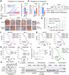
Figure 1
Figure 1. Genetic ablation of mutp53 curbs tumor growth in allografts
a–d, Various prophylactic (a, b) and therapeutic (c, d) protocols of primary floxQ/− vs Q/− and p53-null T-lymphomas allotransplanted (black arrows on time axes) via subcutaneous (a, c, d) or tail vein (b) injections into nude mice, treated with daily intraperitoneal injections of Tamoxifen or oil (* on time axes). (a) Experimental diagram, allograft mass, representative tissue immunostaining and immunoblot at endpoint. Unpaired two-tailed Student’s t-test; mean ± s.e.m; n, number of allografts. (b) mutp53 deletion improves survival of host mice. Kaplan-Meier analysis, Log Rank test; n, number of mice. (c) Two different therapeutic protocols (100 mg/kg Tam for 5 days vs 150 mg/kg for 7 days) show the dose-dependence of allograft growth on mutp53 depletion. Unpaired two-tailed Student’s t-test; mean ± s.e.m; n, number of allografts. (d) Allograft growth using therapeutic protocol (Fig. 1c, Endpoint 2) and mean fold changes at endpoint; n, number of allografts.
Figure 2
Figure 2. Mutp53 ablation in floxQ/− mice causes autochthonous tumor regression or stagnation and extends survival
(a) Fold growth over time until endpoint of clinically advanced tumors in floxQ/−;ERT2/+ mice imaged by ultrasound and treated with Tamoxifen (Tam) or oil (beginning at day 0, arrowhead); normalized to initial tumor size. (b) Daily growth rates of individual tumors during the first 5–12 days of Tam/oil treatment (left), and mean ± s.e.m. of all tumors (right). Unpaired two-tailed Student’s t-test. #, sarcomas; all others are T-lymphomas. (c) Representative examples of sagittal ultrasound images of T-lymphomas in Tam-treated floxQ/−;ERT2/+ and control mice. (d) Genetic ablation of mutp53 in autochthonous tumors induces apoptosis. Immunostaining for mutp53 and cleaved caspase 3 in representative control and mutp53-ablated T-lymphomas. (e) Lung metastasis in these mice by H&E and p53 immunostaining. (f) Organ-confined disease in young floxQ/− mice indicated by p53 immunostaining of malignant thymic cell clones. (g) Kaplan-Meier analysis comparing cancer-related overall survival of floxQ/−;ERT2/+ vs Q/−;ERT2/+ and p53−/−;ERT2/+ mice. Animals were treated once (arrow) at 10 wks with Tam or oil for 5 consecutive days. (h) p53 immunostaining at endpoint (death) of representative T-lymphomas (see also Extended Fig. 2f).
Figure 3
Figure 3. Pharmacological inhibition of the mutp53 stabilizing HSP90/HDAC6 axis with 17DMAG+SAHA prolongs survival of H/H mice in a mutp53-dependent manner
(a) 17AAG and SAHA synergize in degrading mutp53 and suppressing growth of subcutaneous xenografts of MDA231 cells expressing excess ectopic p53R280K. Mean ± s.e.m; n, number of xenografts, unpaired one-tailed Student’s t-test. (b) Organ-confined disease in young H/H mice, indicated by p53 immunostaining of malignant thymic cell clones. (c–e) cancer-related overall survival (c, d) and T-lymphoma specific survival (e) of 17DMAG+SAHA treated mutp53 H/H and p53-null mice, Kaplan-Meier analyses, Log Rank statistics. (f) Immunoblot of p53 and Hsp70 in thymic tissues at endpoint from mice shown in (e), each lane represents a different vehicle- or drug-treated mouse. (g) Real-time qRT-PCR analysis of NQO1 in representative responder and escaper H/H T-lymphomas from (f).
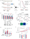
Figure 4
Figure 4. Treatment of H/H and Q/− mice with ganetespib suppresses tumor growth and extends survival in a mutp53-dependent manner
(a, b) Prophylactic (a) and therapeutic (b) treatment of subcutaneous Q/− allografts with ganetespib or vehicle (DMSO). (a) Arrowhead, tumor cell injection. (b) Initial allograft volume and mass at endpoint, mean ± s.e.m, unpaired two-tailed Student’s t-test; n, number of allografts. (c, d) Time-course of mutp53 levels and apoptosis (cleaved caspase 3) in ganetespib-treated (arrows) Q/− subcutaneous T-lymphoma allografts, analyzed by immunoblot (c) and immunostaining (d). Asterisks mark non-specific bands. Enlarged panels after the 1st and 2nd doses (d, far right) immunostained for p53 (arrows), dead cells stain blue. (e) Growth of subcutaneous Q/− T-lymphoma allografts treated once (arrow) with ganetespib or cyclophosphamide alone or in combination. Mean ± s.e.m, unpaired two-tailed Student’s t-test; n, number of allografts, * p<0.05, ** p<0.01 (single drugs vs combination). (f–h) Kaplan-Meier analysis of cancer-related overall survival (f, g) and T-lymphoma specific survival (h) of mutp53 H/H, Q/− and their respective p53−/− controls. (i) Fold growth over time of clinically advanced Q/− and p53−/− T-lymphomas imaged by ultrasound and treated with ganetespib; normalized to initial tumor size. Mean ± s.e.m, unpaired two-tailed Student’s t-test; n, number of tumors, * p<0.05. (j) Comparison of overall survival of ganetespib-treated mutp53 H/H and their respective p53−/− controls, shown in (f). (k, l) Immunoblot analysis of thymic tissues from same-litter siblings (k) and from Q/− mice from (h) at endpoint. Each lane represents individual DMSO or ganetespib-treated mice.

References

    1. Doyle B, et al. p53 mutation and loss have different effects on tumourigenesis in a novel mouse model of pleomorphic rhabdomyosarcoma. The Journal of pathology. 2010;222:129–137. - V体育平台登录 - PubMed
    1. Hanel W, et al. Two hot spot mutant p53 mouse models display differential gain of function in tumorigenesis. Cell death and differentiation. 2013;20:898–909. - "VSports" PMC - PubMed
    1. Lang GA, et al. Gain of function of a p53 hot spot mutation in a mouse model of Li-Fraumeni syndrome. Cell. 2004;119:861–872. - PubMed
    1. Morton JP, et al. Mutant p53 drives metastasis and overcomes growth arrest/senescence in pancreatic cancer. Proceedings of the National Academy of Sciences of the United States of America. 2010;107:246–251. - V体育平台登录 - PMC - PubMed
    1. Olive KP, et al. Mutant p53 gain of function in two mouse models of Li-Fraumeni syndrome. Cell. 2004;119:847–860. - PubMed

Supplementary References

    1. Ventura A, et al. Restoration of p53 function leads to tumour regression in vivo. Nature. 2007;445:661–665. - PubMed
    1. Dobbelstein M, Moll U. Targeting tumour-supportive cellular machineries in anticancer drug development. Nature Reviews. Drug Discovery. 2014;13:179–196. - PubMed
    1. Gorska M, et al. Geldanamycin and its derivatives as Hsp90 inhibitors. Frontiers in Bioscience. 2012;17:2269–2277. - PubMed
    1. Hrzenjak A, et al. Histone deacetylase inhibitor vorinostat suppresses the growth of uterine sarcomas in vitro and in vivo. Molecular Cancer. 2010;9:49. - PMC - PubMed
    1. Proia DA, et al. Multifaceted intervention by the Hsp90 inhibitor ganetespib (STA-9090) in cancer cells with activated JAK/STAT signaling. PloS One. 2011;6:e18552. - PMC - PubMed

"VSports app下载" Publication types

VSports - MeSH terms