V体育2025版 - Skip to main page content
U.S. flag

An official website of the United States government

Dot gov

The . gov means it’s official. Federal government websites often end in VSports app下载. gov or . mil. Before sharing sensitive information, make sure you’re on a federal government site. .

Https

The site is secure V体育官网. The https:// ensures that you are connecting to the official website and that any information you provide is encrypted and transmitted securely. .

. 2015 Apr 9;161(2):319-32.
doi: 10.1016/j.cell.2015.02.043. Epub 2015 Apr 2.

The BRAF pseudogene functions as a competitive endogenous RNA and induces lymphoma in vivo

Affiliations

The BRAF pseudogene functions as a competitive endogenous RNA and induces lymphoma in vivo

Florian A Karreth et al. Cell. .

Abstract

Research over the past decade has suggested important roles for pseudogenes in physiology and disease. In vitro experiments demonstrated that pseudogenes contribute to cell transformation through several mechanisms. However, in vivo evidence for a causal role of pseudogenes in cancer development is lacking VSports手机版. Here, we report that mice engineered to overexpress either the full-length murine B-Raf pseudogene Braf-rs1 or its pseudo "CDS" or "3' UTR" develop an aggressive malignancy resembling human diffuse large B cell lymphoma. We show that Braf-rs1 and its human ortholog, BRAFP1, elicit their oncogenic activity, at least in part, as competitive endogenous RNAs (ceRNAs) that elevate BRAF expression and MAPK activation in vitro and in vivo. Notably, we find that transcriptional or genomic aberrations of BRAFP1 occur frequently in multiple human cancers, including B cell lymphomas. Our engineered mouse models demonstrate the oncogenic potential of pseudogenes and indicate that ceRNA-mediated microRNA sequestration may contribute to the development of cancer. .


Figures

Figure 1:
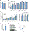
Figure 1:. The BRAF pseudogene regulates BRAF in a Dicer1-dependent manner.
(A) Western blot demonstrating increased BRAF and pERK expression upon ectopic BRAF pseudogene expression in mouse (NIH3T3, left panel) and human (PC9, right panel) cells. (B) Western blot of B-Raffl/fl fibroblasts overexpressing Braf-rs1 or control (yellow fluorescent protein, YFP) in the presence or absence of Adeno-Cre infection. (C) Increased proliferation of NIH3T3 fibroblasts upon ectopic Braf-rs1 expression. (D) Increased proliferation of PC9 cells upon ectopic BRAFP1 expression. (E, F) Western blot (E) and proliferation assay (F) of Dicer1fl/delta and Dicer1delta/delta murine sarcoma cells overexpressing Braf-rs1. (G, H) Western blot (G) and proliferation assay (H) of Dicer1WT and Dicer1Mut human HCT116 colon cancer cells overexpressing BRAFP1. Error bars represent mean±S.D; *, p≤0.05; **, p≤0.01; ***, p≤0.001. See also Figure S1.
Figure 2:
Figure 2:. The BRAF pseudogene functions as a miRNA sponge.
(A) BRAF 3’UTR-Luciferase reporter assay in Dicer1WT and Dicer1Mut HCT116 cells expressing BRAFP1 or control (YFP). (B) Luciferase reporter assay using the 3’UTRs of B-Raf and Braf-rs1 to analyze repression by the indicated miRNA mimics. miR141 serves as a negative control. (C) Braf-rs1 sequesters miRNAs to regulate MRE-Luc reporter activity. HEK293T cells were co-transfected with MRE-Luc reporter constructs, the respective miRNA mimics, and Braf-rs1-L277 or empty control L277 plasmids. The Luciferase activity relative to a Luc reporter without MRE is shown. (D) Luciferase activity measured in HEK293T cells co-expressing MRE-Luc reporters (Luc-653, Luc-134, or Luc-543) and wildtype, or MRE-mutant Braf-rs1 or empty vector. (E) qPCR showing tTA-induced Braf-rs1 expression in TRE-BPS MEFs. (F) Western blot for B-Raf and pERK in tTA-infected TRE-BPS MEFs. (G) Proliferation of TRE-BPS MEF1 shown in (F). Error bars represent mean±S.D; *, p≤0.05; **, p≤0.01; ***, p≤0.001. See also Figure S2.
Figure 3:
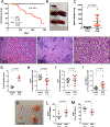
Figure 3:. Braf-rs1 expression in vivo results in a lymphoid malignancy.
BPS, TRE-BPS; CAG-rtTA3 mice on Dox; control, TRE-BPS or CAG-rtTA3 mice on Dox here and in all figures. (A) Survival of BPS and control mice. (B, C) Size (B) and weight (C) of BPS and control mouse spleens. (D, E) Photomicrograph of a spleen from a control (D) and BPS mouse (E). (F) Higher magnification photomicrograph showing tumor cells in a BPS spleen. White arrowheads denote plasma cells and black arrowhead highlights a mitotic figure. (G) Quantification of Ki-67 staining. (H-J) Flow cytometry-based quantification of splenic B220+ (H), CD3+ (I) and Gr-1+/Mac-1+ (J) populations. (K) Size of control and BPS mouse lymph nodes. (L, M) Flow cytometry-based quantification of B220+ (L) and CD3+ (M) populations in lymph nodes. Error bars represent mean±S.D. See also Figure S3.
Figure 4:
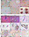
Figure 4:. Braf-rs1 induces diffuse large B-cell lymphoma.
(A) CD45R/B220 staining. Higher magnification inset shows staining of large lymphoma cells. (B) CD3 staining. Higher magnification inset shows positive staining of reactive T cells. (C) IgG staining. Arrowheads denote plasma cells. (D) Bcl6 staining. Lymphoma cells are negative and residual germinal center is positive. (E) Mum1 staining. Tumor cells are positive and residual germinal center is negative. (F) Photograph of control and BPS kidneys. Arrowheads denote tumor nodules. (G-I) H&E staining of kidney (G), liver (H), and lung (I) sections from BPS mice. (J-L) CD45R/B220 immunohistochemistry of kidney (J), liver (K), and lung (L) sections from BPS mice. (M-O) Mum1 immunohistochemistry of kidney (M), liver (N), and lung (O) sections from BPS mice.
Figure 5:
Figure 5:. Lymphomas are transplantable, addicted to Braf-rs1 expression, and activate the MAPK pathway.
(A) Transplanted lymphoma cells infiltrating the spleen, liver, kidney, and lungs of NSG recipient mice. (B) Spleen size measurements after Dox withdrawal. (C-F) H&E staining (C, D) and Mum1 immunohistochemistry (E, F) of BPS and control mouse spleens depicted in (B) after Dox withdrawal. (G) Immunohistochemical staining for B-Raf of lymphoma and adjacent normal white pulp in BPS spleen. (H) Immunohistochemical staining for pERK of lymphoma and adjacent normal white pulp in BPS spleen. (I) Percentage of liver infiltration of TRE-BPS; CAG-rtTA3; Pten+/− lymphoma cells transplanted into NSG mice in response to GSK1120212 treatment. Each symbol represents a liver section, and each recipient mouse is color-coded. Error bars represent mean±S.D; ***, p≤0.001. See also Figure S4.
Figure 6:
Figure 6:. Braf-rs1CDS and Braf-rs13’UTR possess oncogenic ceRNA activity similar to full length Braf-rs1.
(A, B) Weights of spleens (A) and inguinal lymph nodes (B) of the indicated mouse strains after 6 months on Dox. (C) Survival of TRE-BPS3’UTR and TREBPSCDS mice. (D) Table summarizing the penetrance, median survival, and disease onset of TRE-BPS, TRE-BPS3’UTR, and TRE-BPSCDS mice. (E) H&E staining of BPS3’UTR-induced lymphoma. White arrowheads indicate plasma cells, black arrowhead indicates mitotic figure. (F-J) immunohistochemical staining of BPS3’UTR-induced lymphoma for Ki-67 (F), CD45R/B220 (G), CD3 (H), Bcl6 (I), and Mum1 (J). Error bars represent mean±S.D. See also Figure S5.
Figure 7:
Figure 7:. BRAFP1 in human cancer.
(A) Percentage of primary human B-cells, primary human DLBCL, and human DLBCL cell lines expressing BRAFP1 as determined by qPCR analysis. (B, C) Positive correlation of BRAFP1 and BRAF expression in human DLBCL primary tumors (B) and cell lines (C). (D-G) Western blot for BRAF and pERK in OCILy18 (D), H1299 (E), PC9 (F), and OCI-Ly1 (G) cells in response to BRAFP1 silencing. (H-K) Proliferation of OCI-Ly18 (H), H1299 (I), PC9 (J), and OCI-Ly1 (K) cells in response to BRAFP1 silencing. (L) Western blot for BRAF and pERK in OCI-Ly1 cells overexpressing BRAFP1. (M) Proliferation of OCI-Ly1 cells. (N) Percentage of human CD19+ transplanted OCI-Ly1 cells in bone marrow of NSG recipients. (O) Model depicting the proposed oncogenic action of the BRAF pseudogene. Error bars represent mean±S.D; *, p≤0.05; **, p≤0.01. See also Figure S6.

References

    1. Ala U, Karreth FA, Bosia C, Pagnani A, Taulli R, Léopold V, Tay Y, Provero P, Zecchina R, and Pandolfi PP (2013). Integrated transcriptional and competitive endogenous RNA networks are cross-regulated in permissive molecular environments. Proc. Natl. Acad. Sci. U.S.a 110, 7154–7159. - PMC - PubMed
    1. Bea S, Zettl A, Wright G, Salaverria I, Jehn P, Moreno V, Burek C, Ott G, Puig X, Yang L, et al. (2005). Diffuse large B-cell lymphoma subgroups have distinct genetic profiles that influence tumor biology and improve gene-expression-based survival prediction. Blood 106, 3183–3190. - PMC - PubMed
    1. Beard C, Hochedlinger K, Plath K, Wutz A, and Jaenisch R. (2006). Efficient method to generate single-copy transgenic mice by site-specific integration in embryonic stem cells. Genesis 44, 23–28. - PubMed
    1. Bier A, Oviedo-Landaverde I, Zhao J, Mamane Y, Kandouz M, and Batist G. (2009). Connexin43 pseudogene in breast cancer cells offers a novel therapeutic target. Mol. Cancer Ther 8, 786–793. - PubMed (V体育ios版)
    1. Bosia C, Pagnani A, and Zecchina R. (2013). Modelling Competing Endogenous RNA Networks. PLoS ONE 8, e66609. - PMC - PubMed

V体育官网入口 - Publication types