Skip to main page content
U.S. flag

An official website of the United States government

Dot gov

The . gov means it’s official. Federal government websites often end in VSports app下载. gov or . mil. Before sharing sensitive information, make sure you’re on a federal government site. .

Https

The site is secure V体育官网. The https:// ensures that you are connecting to the official website and that any information you provide is encrypted and transmitted securely. .

. 2015 Mar 11;43(5):2575-89.
doi: 10.1093/nar/gkv101. Epub 2015 Feb 20.

"V体育2025版" Ovarian carcinoma CDK12 mutations misregulate expression of DNA repair genes via deficient formation and function of the Cdk12/CycK complex

Affiliations

Ovarian carcinoma CDK12 mutations misregulate expression of DNA repair genes via deficient formation and function of the Cdk12/CycK complex

Kingsley M Ekumi et al. Nucleic Acids Res. .

"VSports最新版本" Abstract

The Cdk12/CycK complex promotes expression of a subset of RNA polymerase II genes, including those of the DNA damage response. CDK12 is among only nine genes with recurrent somatic mutations in high-grade serous ovarian carcinoma. However, the influence of these mutations on the Cdk12/CycK complex and their link to cancerogenesis remain ill-defined. Here, we show that most mutations prevent formation of the Cdk12/CycK complex, rendering the kinase inactive. By examining the mutations within the Cdk12/CycK structure, we find that they likely provoke structural rearrangements detrimental to Cdk12 activation. Our mRNA expression analysis of the patient samples containing the CDK12 mutations reveals coordinated downregulation of genes critical to the homologous recombination DNA repair pathway. Moreover, we establish that the Cdk12/CycK complex occupies these genes and promotes phosphorylation of RNA polymerase II at Ser2 VSports手机版. Accordingly, we demonstrate that the mutant Cdk12 proteins fail to stimulate the faithful DNA double strand break repair via homologous recombination. Together, we provide the molecular basis of how mutated CDK12 ceases to function in ovarian carcinoma. We propose that CDK12 is a tumor suppressor of which the loss-of-function mutations may elicit defects in multiple DNA repair pathways, leading to genomic instability underlying the genesis of the cancer. .


Figures (VSports最新版本)

Figure 1.
Figure 1.
CDK12 mutations in HGS-OvCa abrogate the activity of Cdk12 predominantly by impairing the interaction between Cdk12 and CycK. (A) Schematic depiction of the wild-type and mutant Cdk12 proteins containing individual CDK12 mutations analyzed in this study. Highly structured kinase domain (KD; red), arginine/serine rich region (RS; green) and two regions with proline-rich motifs (PRM1 and PRM2; blue) are depicted. The ruler on top indicates the length of Cdk12 protein in amino acids. Vertical lines denote sites of individual missense and insertion mutations. Finally, ‘fs’ stands for a frame-shift mutation and the associated number indicates the number of altered amino acids at the C-terminus of mutant Cdk12 protein (dotted). (B) Overall structure of human Cdk12/CycK and positions of the mutations. Cdk12 is shown as cartoon representation in blue and CycK as surface representation in grey. CDK12 missense, insertion and internal deletion mutations are located in the C-terminal lobe of the KD. The mutated amino acid residues are highlighted in red. pT893 is highlighted in orange. (C) Effects of the CDK12 mutations on the interaction between Cdk12 and CycK. The indicated wild-type and mutant FLAG epitope-tagged Cdk12 proteins (Cdk12-F) were immuno-purified from whole cell extracts (WCEs) of the individual HEK 293 Flp-In T-Rex cell lines using FLAG-M2 agarose (FLAG IP) and examined for their interaction with endogenous CycK. Levels of Cdk12-F and CycK proteins in WCEs (INPUT, 5% of WCEs; top) and IPs (FLAG IP; bottom) were detected by Western blotting using FLAG and CycK antibodies. (D) CDK12 mutations abrogate the kinase activity of Cdk12. The indicated wild-type and mutant Cdk12-F proteins were immuno-purified (IP) as in panel C and the complexes were examined for their kinase activity by in vitro kinase assay (IVKA) toward the recombinant GST-CTD. Levels of Ser2-P GST-CTD isoforms and input GST-CTD (30%) were detected by Western blotting using Ser2-P-specific RNAPII and GST antibodies.
Figure 2.
Figure 2.
Structural analysis of the CDK12 mutations. (A) Schematic representation of secondary structure elements within the KD of Cdk12. Canonical α-helices and β-strands as well as boundaries of N- and C-terminal lobes are labeled. Positions of the mutated Cdk12 amino acids analyzed in this study are indicated on top of the schematic. (B) The hydroxyl group of the Y901 aromatic ring mediates a tight hydrogen bond (2.7 Å) to the carboxyl group of E928. Interestingly, the tyrosine is closely surrounded by two cysteines, C862 and C924, which could make the Y901C mutation unfavorable. Hydrophobic contacts of Y901 are formed to I925 and K861, with P934 stabilizing the conformation of E928. (C) L996 resides on helix αI. It is surrounded by T1014 and F1019 on helix αJ, by C922, G923 and L926 of helix αF, and M840 and M844 of helix αE. The replacement of L996 by the spacious aromatic ring of a phenylalanine might preclude the tight assembly of the four helical bundle. (D) R882 is part of the canonical arginine network in CDKs that stabilize the T-loop upon phosphorylation of the critical threonine. Together with R858 of the HRD motif, R882 mediates salt bridges to the phosphate group of pT893. Its mutation to leucine might prohibit the conformational arrangement of the T loop required for full activation of the kinase.
Figure 3.
Figure 3.
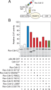
CDK12 mutations in HGS-OvCa decrease transcriptional activation by Cdk12. (A) Schematic depiction of the heterologous RNA tethering assay. Plasmid reporter pSLIIB-CAT contains modified HIV1-LTR promoter in which the apical region of transactivation response RNA element (TAR) was substituted with 29-nucleotide stem-loop IIB (SLIIB) subdomain of the HIV-1 Rev response element (RRE), the Rev binding RNA sequence. The interaction between Rev (pink oval) and SLIIB within the TAR/SLIIB stem-loop structure at the 5′ end of nascent RNA tethers the Rev-Cdk12 chimeric protein to RNAPII engaged in transcription, resulting in elevated transcription of CAT reporter gene. CTD of the biggest RNAPII subunit Rbp1 is represented as a tail of RNAPII (yellow), wherein white circle depicts Ser2 residue to be phosphorylated by Rev-Cdk12 (dashed arrow) and gold circles depict Ser5 and Ser7 residues in an already phosphorylated form. Arrow within HIV1-LTR indicates transcription start site. (B) CDK12 HGS-OvCa mutations compromise stimulation of transcription by Rev-Cdk12. HEK 293 cells were co-transfected with pSLIIB-CAT reporter gene and plasmids encoding the proteins indicated below the graph. Transcriptional activities of Cdk12-F (white bar 1), Rev (white bar 2), the mutant Rev-Cdk12 chimeras (red bars) and catalytically dead Rev-Cdk12 D887N chimera (green bar) are represented as CAT activities relative to the activity of wild-type Rev-Cdk12 chimera (blue bar), which was set to 100%. Results are presented as the mean ± SD. Levels of the Rev-Cdk12 chimeras and endogenous Cdk12 protein are shown below the graph and were detected by Western blotting using Cdk12 antibody. The top asterisk (*) indicates migration of the endogenous Cdk12 protein and the bottom one indicates the position of an unspecific band recognized by the Cdk12 antibody, which serves as a loading control.
Figure 4.
Figure 4.
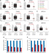
The crucial DDR genes are downregulated in HGS-OvCa patient samples with mutations in CDK12. (A, B and C) Graphs show comparisons of relative expression levels between the HGS-OvCa samples with the wild-type or mutated CDK12. The identity of genes is indicated on top of each graph. The data was generated using the following microarray probes: ATM (212672_at), ATR (209903_s_at), CHEK1 (205394_at), FANCI (213007_at), MDC1 (203062_s_at), RAD51D (209965_s_at), NEK9 (212299_at), ORCL3 (210028_s_at), TERF2 (203611_at), BRCA1 (211851_x_at), BRCA2 (208368_s_at). Whereas samples with the wild-type CDK12 are plotted as black triangles, those containing individual missense or nonsense/indel CDK12 mutations are depicted as colored circles or squares, respectively, as indicated by the legend in the top right corner. Results are presented as mean (red line) with standard error of the mean (SEM) (black whiskers). P-values are given next to red asterisks (*) and the number of asterisks indicates the degree of significance as follows: * = P ≤ 0.05; ** = P ≤ 0.01; *** = P ≤ 0.001. Panels A and C show genes related to the HR pathway, while other affected DDR genes are shown in panel B. (D and E) Depletion of Cdk12 decreases the mRNA levels of HR genes in Caov-3 cells. Relative mRNA levels of genes indicated below the bars were determined by RT-qPCR using total RNA samples isolated from Caov-3 cells treated with the control (blue bars) or Cdk12 #1 siRNA (red bars). The error bars represent the mean ± SD.
Figure 5.
Figure 5.
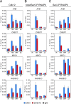
Cdk12/CycK is present at the novel HR genes to promote phosphorylation of the CTD of RNAPII at Ser2. (A, B and C) Control (blue bars) or Cdk12 knockdown HeLa DR-GFP cells (red bars) were subjected to ChIP-qPCR analysis to determine the levels of Cdk12 (panel A), total/Ser5-P RNAPII (panel B) and Ser2-P RNAPII (panel C) occupancy at RAD51D intergenic region (IR) and three gene-specific regions as indicated below the graphs. The identity of genes analyzed is indicated on top of each graph. Levels of IgG signals at IR and gene regions are presented as gray bars. Where these levels were significantly lower than those obtained with specific antibody, the error bars were omitted for simplicity reasons. Results are presented as percent of input DNA and plotted as the mean ± SD.
Figure 6.
Figure 6.
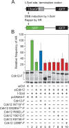
CDK12 mutations in HGS-OvCa abrogate the ability of Cdk12/CycK to stimulate the repair of DNA double-strand breaks by HR. (A) Schematic representation of the DR-GFP recombination substrate. The defective EGFP genes of the cassette, separated by 3.7 kb, are shown (top). The first one (SceGFP) contains an I-SceI endonuclease site and an in-frame termination codon (white vertical line), while the second one (iGFP) is an internal EGFP fragment, yielding GFP− cells. The homologous EGFP sequences are depicted as black rectangles and the non-repetitive EGFP sequence is depicted as gray rectangle. Induction of DSB by I-SceI triggers the repair of SceGFP by HR using the iGFP sequence as a donor DNA, resulting in wild-type EGFP (green rectangle) and GFP+ cells (bottom). (B) The mutant Cdk12 proteins fail to promote the repair of the DSB by HR. HeLa DR-GFP cells were treated with the control or Cdk12 #2 siRNA and transfected with the I-SceI expression plasmid together with plasmids encoding the wild-type (blue bar) or mutant (red bars) Cdk12-F proteins as indicated below the graph. The HR frequency for each experimental condition is represented as the frequency relative to the one reached by the I-SceI expression in the control siRNA-treated cells, which was set to 100% (green bar). Results are presented as the mean ± SD. Levels of the endogenous Cdk12 and Cdk12-F proteins are shown below the graph and were detected by Western blotting using Cdk12 antibody. The top asterisk (*) indicates migration of the endogenous Cdk12 protein and the bottom one indicates the position of an unspecific band recognized by the Cdk12 antibody, which serves as a loading control.

References

    1. Fuda N.J., Ardehali M.B., Lis J.T. Defining mechanisms that regulate RNA polymerase II transcription in vivo. Nature. 2009;461:186–192. - PMC - PubMed
    1. Eick D., Geyer M. The RNA polymerase II carboxy-terminal domain (CTD) code. Chem. Rev. 2013;113:8456–8490. - "VSports在线直播" PubMed
    1. Zhou Q., Li T., Price D.H. RNA polymerase II elongation control. Annu. Rev. Biochem. 2012;81:119–143. - PMC - PubMed
    1. Lenasi T., Barboric M. P-TEFb stimulates transcription elongation and pre-mRNA splicing through multilateral mechanisms. RNA Biol. 2010;7:145–150. - "VSports最新版本" PubMed
    1. Bartkowiak B., Liu P., Phatnani H.P., Fuda N.J., Cooper J.J., Price D.H., Adelman K., Lis J.T., Greenleaf A.L. CDK12 is a transcription elongation-associated CTD kinase, the metazoan ortholog of yeast Ctk1. Genes Dev. 2010;24:2303–2316. - VSports注册入口 - PMC - PubMed

Publication types

MeSH terms (VSports最新版本)