Skip to main page content
U.S. flag

An official website of the United States government

Dot gov

The . gov means it’s official. Federal government websites often end in . gov or . mil. Before sharing sensitive information, make sure you’re on a federal government site VSports app下载. .

Https

The site is secure. The https:// ensures that you are connecting to the official website and that any information you provide is encrypted and transmitted securely. V体育官网.

. 2014 Oct 16;10(10):e1004597.
doi: 10.1371/journal.pgen.1004597. eCollection 2014 Oct.

Oligoasthenoteratozoospermia and infertility in mice deficient for miR-34b/c and miR-449 loci

Affiliations

Oligoasthenoteratozoospermia and infertility in mice deficient for miR-34b/c and miR-449 loci

Stefano Comazzetto et al. PLoS Genet. .

Abstract

Male fertility requires the continuous production of high quality motile spermatozoa in abundance. Alterations in all three metrics cause oligoasthenoteratozoospermia, the leading cause of human sub/infertility. Post-mitotic spermatogenesis inclusive of several meiotic stages and spermiogenesis (terminal spermatozoa differentiation) are transcriptionally inert, indicating the potential importance for the post-transcriptional microRNA (miRNA) gene-silencing pathway therein. We found the expression of miRNA generating enzyme Dicer within spermatogenesis peaks in meiosis with critical functions in spermatogenesis. In an expression screen we identified two miRNA loci of the miR-34 family (miR-34b/c and miR-449) that are specifically and highly expressed in post-mitotic male germ cells. A reduction in several miRNAs inclusive of miR-34b/c in spermatozoa has been causally associated with reduced fertility in humans. We found that deletion of both miR34b/c and miR-449 loci resulted in oligoasthenoteratozoospermia in mice. MiR-34bc/449-deficiency impairs both meiosis and the final stages of spermatozoa maturation. Analysis of miR-34bc-/-;449-/- pachytene spermatocytes revealed a small cohort of genes deregulated that were highly enriched for miR-34 family target genes. Our results identify the miR-34 family as the first functionally important miRNAs for spermatogenesis whose deregulation is causal to oligoasthenoteratozoospermia and infertility VSports手机版. .


Conflict of interest statement (VSports在线直播)

The authors have declared that no competing interests exist.

Figures
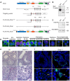
Figure 1
Figure 1. Expression and function of Dicer in adult spermatogenesis.
(A) Domain structure of the Dicer protein is shown. The organization of the 5′ portion of Dicer locus is depicted. The targeting vector used for introduction of FlagHA2 into the Dicer locus and the schematic map of the targeted Dicer gene before and after Cre mediated-recombination are shown. Triangles represent loxP sites as indicated. Rectangles indicate the position of Neomycin (Neo) and Diptheria toxin A (DTA) selection marker genes. The SacI restriction sites are indicated as well as the respective Southern fragments detected by the 3′probe. A schematic diagram of the resulting FlagHA2-Dicer protein is shown. (B) Southern blot of tail derived SacI-digested DNA from wild-type and Dcr+/FH-Neo mice is shown with the 3′ probe indicated in A. (C) Western blot using anti-HA and anti-SMC1 antibodies on extracts from adult wild type and Dcr+/FH testis is shown. (D) Immunofluorescence using anti-HA and anti-γH2AX antibodies on Dcr+/FH testis germ cells from adult testis sections is shown. Scale bar = 10 µm. (E) Hematoxylin and eosin stained testis section from adult DcrCtl and DcrC-KO mice with representative tubules shown. Scale bars = 50 µm and 20 µm in the upper and lower panel, respectively. (F) Increased apoptosis in DcrC-KO testis. A TUNEL assay counterstained with DAPI is shown on testis sections from adult DcrCtl and DcrC-KO mice. The apoptotic cells stain in green. Scale bars = 50 µm and 10 µm in the upper and lower panel, respectively. Abbreviations: P, pachytene and RS, round spermatid. Representative images are shown from at least 3 mice analyzed in panels D–F.
Figure 2
Figure 2. miR-34b/c and miR-449 are selectively expressed in post-mitotic spermatogenesis.
(A) Heat diagram summarizing the expression of miRNAs in mitotic in vitro cultured spermatogonial stem cell (SSC) lines, ex vivo isolated meiotic spermatocytes (sp.cytes) and spermiogenic round spermatids (RS). The average expression of biological replicates is shown. (B) The mature sequence of the murine miR-34 family miRNAs is shown with the seed sequence highlighted in red. The expression of the miR-34 family members is summarized from the array data. (C) The expression of miR-34 family was determined by Northern blotting of RNA derived from a broad panel of tissues. U6 snRNA and Let-7a was used as loading controls. Abbreviations: BM, bone marrow; Sp, spleen; Thy, Thymus; Lym, lymph node; Te, testis; Ov, ovary; Mu, muscle, He, heart; Li, liver; Lu, lung; Kid. Kidney and Pe, peritoneal cavity cells. (D) Expression of miR-34 family members was determined in the first wave of spermatogenesis by Northern blotting of total RNA from testis at the indicated day post partum (dpp), Ad indicates adult. U6 snRNA was used as a loading control. Representative data is shown from two independent experiments in panel C and D. (E) The onset of miR-34c expression in early pachytene spermatocytes during the first wave of spermatogenesis. The spatial expression of miR-34c (Green) is shown by in situ hybridization on sections of 14 dpp mouse testis, the section were counterstained with anti-γH2AX (Red) antibodies to precisely identify the meiotic stage. Scale bar = 30 µm. Abbreviations: L, leptotene; Z, zygotene; eP, early pachytene and P, pachytene. (F) The expression of miR-34c (Green) by in situ hybridization in the indicated adult spermatogenic populations is shown. Anti-γH2AX (Red) was used as in (E). Scale bar = 10 µm. Representative images from one of three independent experiments are shown for panel E and F.
Figure 3
Figure 3. Oligoasthenoteratozoospermia and infertility in miR-34bc−/−;449−/− mice.
(A) qRT-PCR of miR-34a, miR-34b, miR-34c and miR-449a from control (Ctl) and miR-34bc−/−;449−/− adult testis. (B) miR-34bc−/−;449−/− male mice are infertile. The number of pups born per plug from wild type and miR-34bc−/−;449−/− mice is shown. The number of animals tested and the mean ±s.e.m. are indicated. (C) Hematoxylin and eosin stained epididymis section from control (Ctl) and miR-34bc−/−;449−/− adult mice is shown. A representative image of 5 mice analyzed is shown. Scale bar = 50 µm. (D) Reduced sperm count in miR-34bc−/−;449−/− mice. Mean sperm count ±s.e.m. from control and miR-34bc−/−;449−/− adult mice is shown (n = 8). (E) Sperm morphology from the indicated genotypes is shown. Scale bar = 10 µm. *** and **** indicates a p value (unpaired t test with Welch correction) of <0.001 and <0.0001 respectively.
Figure 4
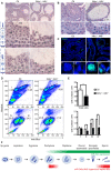
Figure 4. miR-34bc/449 are required for multiple stages of post-mitotic spermatogenesis.
(A) PAS stained testis section from adult control and miR-34bc−/−;449−/− mice is shown. Overview of several tubules is shown in upper panels. Magnified and staged tubules are presented in the lower panels, the schematic diagram summarizes the spermatogenic content of tubules in wild type mice. Abbreviations: Pl, preleptotene; Z, zygotene; P, pachytene; D, diplotene;; RS, round spermatid and ES, elongating spermatid. Scale bars = 50 µm and 10 µm in the upper and lower panels, respectively. (B) MiR-34bc−/−;449−/− testis sections and percentages of tubules with meiotic arrest at zygotene stage is shown. Scale bars = 30 µm and 20 µm in the upper and lower panel, respectively. Representative images are shown from 6 mice analysed is shown in panel A and B. (C) Increased apoptosis in miR-34bc−/−;449−/− testis. A TUNEL assay counterstained with DAPI (Blue) is shown on testis sections from adult control and miR-34bc−/−;449−/− mice (upper panel). The apoptotic cells stain in green. Apoptotic miR-34bc−/−;449−/− pachytene (P) and elongating spermatid (ES) are shown in the lower panel along with non-apoptotic control cells. Scale bars = 30 µm and 10 µm in the upper and lower panel, respectively. Representative images are shown from 3 mice analysed is shown. (D) FACS plot of adult testis shown, gated populations in upper panel I (lepto-zygotene) and II (pachytene-diplotene), in lower panel III (round spermatids) and IV (elongating spermatids). Numbers indicated the overall percentage of the respective populations. (E) Comparative enumeration of spermatogenic populations of control (Ctl) and miR-34bc/499−/− mice. Total testicular cell numbers (upper) are shown. Numbers plotted for the developmentally defined subpopulations indicated in (D) by roman numerals (lower panel). 8 animals per genotype were analyzed by FACS. Mean ±s.e.m. values are shown in the graph. (F) Schematic diagram indicating the expression and impact of loss of miR-34bc/449 expression. * and *** indicates a p value (unpaired t test with Welch correction) of <0.05 and <0.001 respectively.
Figure 5
Figure 5. miR-34bc/449 regulates a small cohort of genes in spermatocytes.
(A) Expression scatterplot showing relative average expression of affymetrix probes between control (x-axis) and miR-34bc−/−;449−/− (y-axis). Significantly deregulated (p = 0.05) genes with a log2 fold change of 0.5 (red) are shown. (B) The list of the 13 upregulated genes with predicted miR-34 seed binding sites is shown. Also indicated is the gene function as well as number of miR-34 binding sites. (C) qRT-PCR expression analysis of representative miR-34 family seed-containing deregulated genes identified. Normalized data are plotted as relative fold change in miR-34bc−/−;449−/− versus wild type pachytene spermatocytes. Standard error is shown and the asterisk indicates significantly upregulated expression (P<0.05). Other genes identified from the array that change in expression are also presented. The data in all panels are from biological quadruplicates of each genotype. (D) Sylamer enrichment landscape plot for all 876 7 nt words complementary to canonical mouse miRNA seed regions. The y-axis represents the sorted genelist of 21,560 genes from most up-regulated to most down-regulated in the miR-34bc−/−;449−/− pachytene spermatocytes. Each 7mer word was tested for significant enrichment across the 3′UTRs of genes in this list. The word corresponding to seed matching miR-34 family (Red) is enriched in the up-regulated genes.

"VSports最新版本" References

    1. Eddy EM (2002) Male germ cell gene expression. Recent Prog Horm Res 57: 103–128. - PubMed
    1. de Rooij DG, Russell LD (2000) All you wanted to know about spermatogonia but were afraid to ask. J Androl 21: 776–798. - PubMed
    1. Handel MA, Schimenti JC (2010) Genetics of mammalian meiosis: regulation, dynamics and impact on fertility. Nat Rev Genet 11: 124–136. - PubMed
    1. Monesi V (1964) Ribonucleic Acid Synthesis during Mitosis and Meiosis in the Mouse Testis. J Cell Biol 22: 521–532. - PMC - PubMed
    1. Paronetto MP, Messina V, Barchi M, Geremia R, Richard S, et al. (2011) Sam68 marks the transcriptionally active stages of spermatogenesis and modulates alternative splicing in male germ cells. Nucleic Acids Res 39: 4961–4974. - PMC - PubMed