"V体育平台登录" Epithelial-mesenchymal transition spectrum quantification and its efficacy in deciphering survival and drug responses of cancer patients
- PMID: 25214461
- PMCID: "V体育安卓版" PMC4287932
- DOI: V体育官网入口 - 10.15252/emmm.201404208
Epithelial-mesenchymal transition spectrum quantification and its efficacy in deciphering survival and drug responses of cancer patients
Abstract
Epithelial-mesenchymal transition (EMT) is a reversible and dynamic process hypothesized to be co-opted by carcinoma during invasion and metastasis. Yet, there is still no quantitative measure to assess the interplay between EMT and cancer progression. Here, we derived a method for universal EMT scoring from cancer-specific transcriptomic EMT signatures of ovarian, breast, bladder, lung, colorectal and gastric cancers VSports手机版. We show that EMT scoring exhibits good correlation with previously published, cancer-specific EMT signatures. This universal and quantitative EMT scoring was used to establish an EMT spectrum across various cancers, with good correlation noted between cell lines and tumours. We show correlations between EMT and poorer disease-free survival in ovarian and colorectal, but not breast, carcinomas, despite previous notions. Importantly, we found distinct responses between epithelial- and mesenchymal-like ovarian cancers to therapeutic regimes administered with or without paclitaxel in vivo and demonstrated that mesenchymal-like tumours do not always show resistance to chemotherapy. EMT scoring is thus a promising, versatile tool for the objective and systematic investigation of EMT roles and dynamics in cancer progression, treatment response and survival. .
Keywords: drug response; epithelial‐mesenchymal transition; gene expression signature; microarray; prognosis V体育安卓版. .
© 2014 The Authors V体育ios版. Published under the terms of the CC BY 4. 0 license. .
Figures

A six-step scheme illustrating the generation of a cancer-specific EMT signature. Note that tumours and cell lines have their own cancer-specific EMT signatures. (Top right panel) Red and green bars on sample enrichment score (ES) bar chart indicate, respectively, mesenchymal-like (Mes) and epithelial-like (Epi) samples selected for building the BinReg EMT signature. (Middle right panel) Heatmap of the EMT signature from Significance Analysis of Microarray (SAM)/Receiver Operating Characteristics (ROC) analysis. The colour bar shows the EMT phenotype probability of cell line or tumour samples, sorted from most Epi to most Mes VSports注册入口. Red and green bars indicate Mes and Epi samples selected for SAM/ROC analysis. (Bottom right panel) Plots of empirical cumulative distribution function of Mes (red) and Epi (green) gene sets.
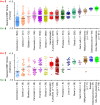
Dot plot of EMT score (mean ± SEM) for breast cancer cell lines (n = 34) with spindle- and non-spindle-like morphologies. Mann–WhitneyU-testP-value is shown V体育官网入口.
Immunohistochemistry staining heatmap of Oestrogen Receptor (ER), Progesterone Receptor (PR), and Epi (CDH1, ERBB2, CK19) as well as Mes (CK5, VIM, CDH2) markers (black = low, red = high, white = no data). Breast cancer cell lines (n = 39) are aligned from the most Epi to most Mes based on the EMT score, as shown by the bar chart. Dot plot is the-log10P-value of two-sample Kolmogorov–Smirnov test. Arbitrary threshold ofP < 0. 001 was used to define Epi, intermediate and Mes cell lines. Breast cancer cell line microarrays and subtype are from GSE16795 (Hollestelleet al, 2010). Subtype colour code: blue, Luminal; maroon, Basal VSports在线直播.

Circos plot illustrating the generic EMT signature: the overlap of ovarian (blue), breast (purple), lung (green), colorectal (yellow), bladder (red) and gastric (orange) cancer-specific EMT signatures is shown. Links indicate overlapping genes (red = mesenchymal, green = epithelial). Heatmap on the inner ring indicates weight computed based on Significance Analysis of Microarray (SAM) fold-change, false discovery rate, Receiver Operating Characteristics (ROC) and number of samples of a gene in each cancer-specific EMT signature (red = high, blue = low weight). On the outermost ring, genes are represented by ticks and aligned from the highest SAM fold-change to the lowest for each cancer type. Selected genes are labelled.
EMT score (mean ± SEM;y-axis) of breast cancer molecular subtypes as predicted using ssGSEA and signature from Pratet al (2010) in non-laser-capture micro-dissected (non-LCM) cohort (n = 3,992; upper panel) and LCM cohort (n = 417; lower panel). The Mann–WhitneyU-testP-value of binary comparison for each subtype is given. Colour code: maroon, Basal; yellow, Claudin-low; light blue, Luminal-A; dark blue, Luminal-B; orange, ERBB2+; green, Normal-like. N.A, not applicable. Note that noP-value is available for Claudin-low and Normal-like subtypes in lower panel becausen < 3.


Bar plots of breast (n = 270; left panel) and ovarian (n = 328; right panel) cancers stratified by EMT status and clinical response based on response evaluation criteria in solid tumours (RECIST). Regimen was neoadjuvant doxorubicin and cyclophosphamide for breast cancer, and platinum-based adjuvant/progression/recurrence chemotherapy for ovarian cancer. Percentage distribution of EMT status is given in each clinical response group. Abbreviation: CR, complete response; PR, partial response; SD, stable disease; PD, progressive disease. Green, epithelial-like (Epi); orange, intermediate; red, mesenchymal-like (Mes).
Volcano plot of EMT correlation with drug sensitivity regardless of cancer type.Rho ∈ [−1.0, +1.0] (x-axis) and-log10P-value (y-axis) were computed by Spearman's correlation coefficient test. Dashed line ofP-value = 0.1 is plotted. Red and green indicate higher drug resistance in Mes tumours (Rho ∈ [0, +1.0]) and Epi tumours (Rho ∈ [−1.0, 0]), respectively.
Kaplan–Meier analysis comparing overall survival (left panel) and disease-free survival (right panel) of Epi (green) and Mes (red) ovarian cancer patients who underwent a treatment regimen with (dark colour) or without (light colour) paclitaxel.P-value reported was computed by log-rank test. Abbreviation: HR = hazard ratio.
References
-
- Akalay I, Janji B, Hasmim M, Noman MZ, Andre F, De Cremoux P, Bertheau P, Badoual C, Vielh P, Larsen AK, et al. Epithelial-to-mesenchymal transition and autophagy induction in breast carcinoma promote escape from T-cell-mediated lysis. Cancer Res. 2013;73:2418–2427. - PubMed
-
- Baird RD, Tan DS, Kaye SB. Weekly paclitaxel in the treatment of recurrent ovarian cancer. Nat Rev Clin Oncol. 2010;7:575–582. - PubMed
-
- Bauer JA, Chakravarthy AB, Rosenbluth JM, Mi D, Seeley EH, De Matos Granja-Ingram N, Olivares MG, Kelley MC, Mayer IA, Meszoely IM, et al. Identification of markers of taxane sensitivity using proteomic and genomic analyses of breast tumors from patients receiving neoadjuvant paclitaxel and radiation. Clin Cancer Res. 2010;16:681–690. - PMC (VSports最新版本) - PubMed
Publication types
"V体育官网入口" MeSH terms
- Actions (V体育平台登录)
- V体育ios版 - Actions
- "V体育平台登录" Actions
- V体育平台登录 - Actions
- VSports注册入口 - Actions
- "VSports app下载" Actions
- "VSports在线直播" Actions
- Actions (V体育ios版)
- VSports在线直播 - Actions
- V体育官网 - Actions
- "V体育安卓版" Actions
VSports手机版 - Substances
- "VSports app下载" Actions
LinkOut - more resources
Full Text Sources
Other Literature Sources
Molecular Biology Databases (V体育ios版)

