Skip to main page content
U.S. flag

An official website of the United States government

Dot gov

The . gov means it’s official. Federal government websites often end in . gov or . mil. Before sharing sensitive information, make sure you’re on a federal government site VSports app下载. .

Https

The site is secure. The https:// ensures that you are connecting to the official website and that any information you provide is encrypted and transmitted securely V体育官网. .

. 2014 Sep 15;34(18):3421-34.
doi: 10.1128/MCB.00221-14. Epub 2014 Jul 7.

Nrf2- and ATF4-dependent upregulation of xCT modulates the sensitivity of T24 bladder carcinoma cells to proteasome inhibition

Affiliations

Nrf2- and ATF4-dependent upregulation of xCT modulates the sensitivity of T24 bladder carcinoma cells to proteasome inhibition

Peng Ye et al. Mol Cell Biol. .

Erratum in

"VSports在线直播" Abstract

The ubiquitin-proteasome pathway degrades ubiquitinated proteins to remove damaged or misfolded protein and thus plays an important role in the maintenance of many important cellular processes. Because the pathway is also crucial for tumor cell growth and survival, proteasome inhibition by specific inhibitors exhibits potent antitumor effects in many cancer cells. xCT, a subunit of the cystine antiporter system xc (-), plays an important role in cellular cysteine and glutathione homeostasis. Several recent reports have revealed that xCT is involved in cancer cell survival; however, it was unknown whether xCT affects the cytotoxic effects of proteasome inhibitors. In this study, we found that two stress-inducible transcription factors, Nrf2 and ATF4, were upregulated by proteasome inhibition and cooperatively enhance human xCT gene expression upon proteasome inhibition. In addition, we demonstrated that the knockdown of xCT by small interfering RNA (siRNA) or pharmacological inhibition of xCT by sulfasalazine (SASP) or (S)-4-carboxyphenylglycine (CPG) significantly increased the sensitivity of T24 cells to proteasome inhibition. These results suggest that the simultaneous inhibition of both the proteasome and xCT could have therapeutic benefits in the treatment of bladder tumors VSports手机版. .


Figures

FIG 1
FIG 1
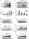
Proteasome inhibitors induce xCT in T24 cells. (A and B) T24 cells were treated with 20 to 100 nM BTZ or 50 nM EPO for 6 h, and then xCT mRNA expression was evaluated by RT-qPCR and normalized to cyclophilin A (CypA) expression levels. The data represent means and SEM from three independent experiments. Differences between groups were assessed with one-way ANOVA with a Bonferroni post hoc test (A) or Student's t test (B). **, P < 0.01. (C) T24 cells were treated with 20 nM BTZ for 3 to 24 h, and xCT mRNA expression was evaluated by RT-qPCR. (D) T24 cells were exposed to different amounts of BTZ or 10 nM EPO, as indicated, for 6 h, and then whole-cell lysates were subjected to immunoblot analysis. (E) T24 cells were treated with 20 nM BTZ for 3 to 24 h. xCT protein expression was evaluated by immunoblot analysis. Lamin B was used as the loading control.
FIG 2
FIG 2
Proteasome inhibitor induces xCT expression in an Nrf2- and ATF4-dependent manner. (A) T24 cells were exposed to 50 or 100 nM BTZ or EPO or 5 μM MG132 for 6 h, and Nrf2 and ATF4 protein expression levels were analyzed by immunoblot analysis. (B) T24 cells were exposed to 20 nM BTZ for 1 to 24 h, and eIF2α phosphorylation and ATF4 and Nrf2 protein expression levels were assessed by immunoblot analysis. (C) Nrf2 and ATF4 mRNA expression by 20 nM BTZ was evaluated by RT-qPCR analysis. Differences between groups (versus the 0-h control) were assessed by one-way ANOVA with a Bonferroni post hoc test. **, P < 0.01. (D) T24 cells were transfected with Nrf2 and/or ATF4 siRNA as described in Materials and Methods. At 24 h posttransfection, the T24 cells were treated with DMSO, 20 nM BTZ, or 100 nM bortezomib for another 6 h, and xCT mRNA expression was evaluated by RT-qPCR analysis. Differences between groups (versus control siRNA) were assessed with one-way ANOVA with a Bonferroni post hoc test. **, P < 0.01. (E and F) Immunoblot analysis of siRNA-transfected T24 cells. The arrowheads indicate Nrf2 or ATF4, and the asterisks indicate nonspecific bands. (G) HeLa cells were exposed to tBHQ or Tm, as indicated, for 24 h. Then, immunoblot analyses were performed to evaluate Nrf2 and ATF4 expression. (H) HeLa cells were treated with tBHQ and/or Tm, as indicated, for 24 h. xCT mRNA expression was analyzed by RT-qPCR and normalized with CypA expression. The data are presented as means and SEM from at least three independent experiments. Differences between groups were assessed by one-way ANOVA with a Bonferroni post hoc test. *, P < 0.05. (I and J) xCT protein induction by tBHQ and/or Tm treatment (24 h) in T24 cells was analyzed by immunoblot analysis.
FIG 3
FIG 3
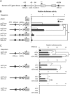
Reporter analysis of the human xCT gene promoter. (A) Schematic representation of the human xCT gene. The putative ARE (ARE-pro) and the two AAREs (AARE-F and AARE-R) are indicated by solid and open triangles, respectively. (B) T24 cells were transfected with either a wild-type reporter gene (pxCT pro WT-Luc) or mutated reporter genes and then incubated for 6 h in the presence of DMSO or 100 nM BTZ. Reporter activities were measured as described in Materials and Methods. (C) HeLa cells were cotransfected with each reporter plasmid in combination with the Nrf2 and/or ATF4 expression vector. After 24 h of incubation, the transfected cells were subjected to a luciferase assay. Luciferase activities were normalized with Renilla luciferase activities, and the bars represent the means and SEM from at least three independent experiments. Differences between groups were assessed by one-way ANOVA with a Bonferroni post hoc test. *, P < 0.05; **, P < 0.01; NS, not significant.
FIG 4
FIG 4
Reporter analysis of the human xCT gene intronic enhancer. (A) Reporter constructs containing a wild-type or mutated intronic ARE were transfected into T24 cells and analyzed in the same manner as for Fig. 3B. (B) Reporter constructs containing wild-type and a mutant intronic AREs were cotransfected with an Nrf2 or ATF4 expression vector as described for Fig. 3C. Luciferase activities were normalized with Renilla luciferase activities, and the bars represent the means and SEM from at least three independent experiments. Differences between groups were assessed by one-way ANOVA with a Bonferroni post hoc test. *, P < 0.05; NS, not significant.
FIG 5
FIG 5
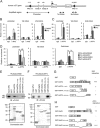
BTZ-induced Nrf2 and ATF4 recruitment to cis-regulatory elements of the human xCT gene. (A) Schematic representation of the human xCT gene locus and PCR-amplified regions used in the ChIP assay. (B and C) T24 cells were treated with DMSO or 100 nM BTZ for 6 h and subjected to a ChIP assay using anti-Nrf2 or anti-ATF4 antibody as described in Materials and Methods. The first intron region was used as a negative control. (D) T24 cells were transfected with control or Nrf2 siRNA. At 24 h posttransfection, the cells were treated with 100 nM BTZ for 6 h and subjected to a ChIP assay using anti-ATF4 antibody. The data are expressed relative to the corresponding values for the DMSO-treated and normal IgG-captured fragment, and the data represent the means and SEM from at least three independent experiments. Differences between groups were assessed by one-way ANOVA with a Bonferroni post hoc test. *, P < 0.05; **, P < 0.01; NS, not significant. (E) Interaction between bacterially expressed GST-ATF4 fusion proteins and FLAG-tagged Nrf2 expressed in 293T cells was analyzed by a GST pulldown experiment. GST-ATF4-bound FLAGx3-Nrf2 was detected by an anti-FLAG antibody (top), and GST-ATF4 fusion proteins were visualized by Coomassie staining (bottom). The arrowhead indicates FLAG-tagged Nrf2, and the asterisks indicate GST or GST-Nrf2 fusion proteins. (F) Interactions between bacterially expressed GST-Nrf2 fusion proteins and FLAG-tagged ATF4 expressed in 293T cells were assessed by GST pulldown experiments. GST-Nrf2-bound FLAGx3-ATF4 was detected by anti-FLAG antibody (top), and GST-Nrf2 fusion proteins were visualized by Coomassie staining (bottom). The arrowhead indicates FLAG-tagged ATF4, and the asterisks indicate GST or GST-ATF4 fusion proteins. (G and H) Schematic representations of GST-ATF4 fusion proteins and GST-Nrf2 fusion proteins used in pulldown experiments. The strength of interaction between FLAG-tagged protein and GST fusion proteins is represented as follows: +++, high; ++, medium; +, low; −, no interaction. TAD, transactivation domain; ODDD, oxygen-dependent degradation domain.
FIG 6
FIG 6
xCT knockdown (k/d) by siRNA and xCT inhibitors sensitizes T24 cells to BTZ. (A) T24 cells were transfected with either control or xCT siRNA. At 24 h posttransfection, the cells were reseeded on 96-well plates. After 24 h of incubation, the cells were exposed to different concentrations of BTZ for 48 h, and cell viability was analyzed using CCK-8. The data are the means ± SEM from at least three independent experiments. Differences between groups (versus the control) were assessed by one-way ANOVA with a Bonferroni post hoc test. *, P < 0.05. (B and C) T24 cells were pretreated with 0.3 mM SASP in the presence or absence of 66 μM 2-ME or 0.2 mM CPG for 6 h and then treated with 20 to 100 nM BTZ for another 48 h. Cell viability was measured by using CCK-8. The data are expressed as the means ± SEM from at least three independent experiments. Differences between groups (versus the respective controls) were assessed by one-way ANOVA with a Bonferroni post hoc test. *, P < 0.05; **, P < 0.01. (D) T24 cells were transfected with control or xCT siRNAs. After 24 h, the cells were treated with BTZ for 6 h and then subjected to immunoblot analysis. The arrowhead indicates xCT, and the asterisk indicates nonspecific bands. (E) The effect of SASP on BTZ-induced xCT protein expression was assessed by immunoblot analysis.
FIG 7
FIG 7
xCT knockdown or SASP treatment decreases intracellular cysteine and GSH levels. (A and B) Intracellular cysteine and GSH levels in control or xCT siRNA-transfected T24 cells. Twenty-four hours after siRNA transfection, the cells were exposed to DMSO or 20 nM BTZ for 18 h, and then cellular cysteine and GSH levels were measured as described in Materials and Methods. (C and D) T24 cells were pretreated with 0.3 mM SASP for 6 h and then incubated in the presence or absence of 20 nM BTZ for another 24 h. Intracellular cysteine and GSH levels were analyzed as described in Materials and Methods. (E) T24 cells were pretreated with 0.5 or 1 mM NAC for 30 min and then treated with 100 nM BTZ. After 48 h of incubation, cell viability was measured. The value for untreated cells was arbitrarily set as 100%, and the means of relative values are presented with SEM. Differences between groups were assessed by one-way ANOVA with a Bonferroni post hoc test. *, P < 0.05; **, P < 0.01; NS, not significant.
FIG 8
FIG 8
Effects of xCT inhibition on T24 cell sensitivity to other proteasome inhibitors. (A) T24 cells were transfected with xCT siRNA as described in the legend to Fig. 6A and then treated with 10 nM EPO. Cell viability was evaluated after 48 h by using CCK-8. (B) T24 cells were pretreated with 0.3 mM SASP in the presence or absence of 66 μM 2-ME for 30 min and then exposed to 20 to 60 nM EPO. After 48 h of incubation, cell viability was measured using CCK-8. (C) The effect of SASP on EPO-induced xCT expression was assessed by immunoblot analysis. The arrowhead indicates xCT. (D and E) T24 cells were pretreated with 0.3 mM SASP for 30 min and then treated with different concentrations of MG132 or CFZ. After 48 h of incubation, cell viability was measured as described for panels A and B. The data are expressed relative to the corresponding value for untreated cells. Differences between groups (versus the respective controls) were assessed by one-way ANOVA with a Bonferroni post hoc test. **, P < 0.01. The data are presented as means and SEM.
FIG 9
FIG 9
xCT knockdown increases BTZ sensitivity of other cancer cell lines. (A and B) Cell viability of xCT siRNA-transfected T98G and U373MG cells after 48-h BTZ treatment. The data represent means and SEM from three independent experiments. Differences between groups were assessed with Student's t test. *, P < 0.05. (C and D) T98G or U373MG cells were transfected with either control or xCT siRNA. After 24 h of transfection, the cells were exposed to BTZ for 6 h, and the whole-cell lysates were subjected to immunoblot analysis. (E) T24 cells were treated with 20 nM BTZ for 3 to 24 h, and then GCLC, GCLM, HSPA2, PSMB5, and BIRC5 mRNA expression was evaluated by RT-qPCR and normalized with cyclophilin A (CypA) expression levels. The data represent means and SEM from three independent experiments. Differences between groups (versus the respective 0-h controls) were assessed with one-way ANOVA and a Bonferroni post hoc test. **, P < 0.01.
FIG 10
FIG 10
Hypothetical model for xCT induction by proteasome inhibitors and its effect on proteasome inhibitor-induced cytotoxicity in T24 cells. Proteasome inhibitor-induced xCT protects the cells from the cytotoxic effects of the proteasome inhibitor. See the text for details.

References

    1. Hochstrasser M. 1995. Ubiquitin, proteasomes, and the regulation of intracellular protein degradation. Curr. Opin. Cell Biol. 7:215–223. 10.1016/0955-0674(95)80031-X - DOI - PubMed
    1. Jentsch S, Schlenker S. 1995. Selective protein degradation: a journey's end within the proteasome. Cell 82:881–884. 10.1016/0092-8674(95)90021-7 - DOI - PubMed
    1. Adams J. 2003. The proteasome: structure, function, and role in the cell. Cancer Treat. Rev. 29(Suppl. 1):3–9. 10.1016/S0305-7372(03)00081-1 - DOI - PubMed
    1. Voorhees PM, Dees EC, O'Neil B, Orlowski RZ. 2003. The proteasome as a target for cancer therapy. Clin. Cancer Res. 9:6316–6325 - PubMed
    1. Adams J. 2004. The development of proteasome inhibitors as anticancer drugs. Cancer Cell 5:417–421. 10.1016/S1535-6108(04)00120-5 - DOI (V体育ios版) - PubMed

Publication types

MeSH terms

Substances