Skip to main page content
U.S. flag

An official website of the United States government

Dot gov

The . gov means it’s official. Federal government websites often end in . gov or . mil VSports app下载. Before sharing sensitive information, make sure you’re on a federal government site. .

Https

The site is secure. The https:// ensures that you are connecting to the official website and that any information you provide is encrypted and transmitted securely V体育官网. .

. 2014 May 1;28(9):943-58.
doi: 10.1101/gad.239327.114. Epub 2014 Apr 14.

"VSports手机版" Rho-actin signaling to the MRTF coactivators dominates the immediate transcriptional response to serum in fibroblasts

Affiliations

Rho-actin signaling to the MRTF coactivators dominates the immediate transcriptional response to serum in fibroblasts

Cyril Esnault et al. Genes Dev. .

Abstract

The transcription factor SRF (serum response factor) recruits two families of coactivators, the MRTFs (myocardin-related transcription factors) and the TCFs (ternary complex factors), to couple gene transcription to growth factor signaling. Here we investigated the role of the SRF network in the immediate transcriptional response of fibroblasts to serum stimulation. SRF recruited its cofactors in a gene-specific manner, and virtually all MRTF binding was directed by SRF. Much of SRF DNA binding was serum-inducible, reflecting a requirement for MRTF-SRF complex formation in nucleosome displacement. We identified 960 serum-responsive SRF target genes, which were mostly MRTF-controlled, as assessed by MRTF chromatin immunoprecipitation (ChIP) combined with deep sequencing (ChIP-seq) and/or sensitivity to MRTF-linked signals. MRTF activation facilitates RNA polymerase II (Pol II) recruitment or promoter escape according to gene context. MRTF targets encode regulators of the cytoskeleton, transcription, and cell growth, underpinning the role of SRF in cytoskeletal dynamics and mechanosensing VSports手机版. Finally, we show that specific activation of either MRTFs or TCFs can reset the circadian clock. .

Keywords: MRTF; Rho; SRF; TCF; chromatin; signal transduction. V体育安卓版.


Figures
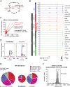
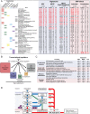
Figure 1.
Figure 1.
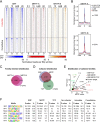
Much of SRF binding is controlled by growth factor signaling. (A) Signaling pathways, activators, and inhibitors affecting the SRF network. (B) Representative SRF, MRTF, and TCF cofactor-binding profiles as normalized reads per base pair. Cell culture conditions were resting cells (0.3% FCS), cells stimulated for 30 min (15% FCS), stimulated in presence of LatB (LatB), and stimulated in presence of U0126 (U0126). (C) Serum stimulation induces SRF binding. Scatter plot comparing ChIP-seq read counts in stimulated and resting cells. (Dotted line) Linear regression plot for all sites (Spearman r, 0.3; fold-inducibility, 1.7 ± 0.03). Note that division into inducible (>1.5-fold increase; red) and constitutive (<1.5-fold increase; black) populations greatly improves rank order correlations, respectively. Solid lines show linear regression plots for the two populations. (D) Metaprofile of SRF binding at constitutive and inducible sites. (E) SRF ChIP-seq peaks are associated with protein-coding genes (within ±2 kb of the TSS, P < 10−999; within a gene feature, P = 1.9 × 10−106; basic χ2 test). (Red) 5′ flanking sequences (−2 kb to the TSS); (pink) other gene features (5′ untranslated region [UTR], 338; introns, 740; coding exons, 39; 3′ UTR, 11); (blue) intergenic, <70 kb from TSS or pA site; (gray) intergenic, >70 kb from TSS or pA site. (F) Distribution of SRF sites around the TSS, shown as sites per 40-base-pair (bp) bin.
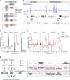
Figure 2.
Figure 2.
Gene-specific MRTF and TCF recruitment. (A) Heat maps showing correlation of SRF and MRTF binding at inducible and constitutive SRF sites across a 2-kb region centered on the SRF peak summit. Color intensity represents normalized reads per 8-bp window. See Supplemental Figure S2, D and E. (B) Metaprofiles of MRTF binding, centered on SRF peak summits. (C) Most MRTF-A sites detected in ChIP-seq bind MRTF-B. See Supplemental Figure S2F. (D) Venn diagram showing relationships between sites bound by SRF, either MRTF or any TCF. See Supplemental Figure S2, G and H. (E) Scatter plot showing relative binding of MRTFs and TCFs by binding score (see the Supplemental Material). Among the 123 genes binding both families, more than twofold difference in score is associated with preferential response to ERK or Rho signaling (Gineitis and Treisman 2001). (F) SRF-associated sequence motifs within 100 bp of SRF peak summits, classified by cofactor-binding and SRF-binding inducibility. For motifs associated with LatB-sensitive and LatB-insensitive “no-cofactor” SRF sites, see Supplemental Figure S2I.
Figure 3.
Figure 3.
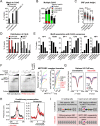
Characterization of MRTF- and TCF-associated SRF sites. (A) Match to the SRF CArG consensus (CCW6GG) within 100 bp of the SRF peak summit. Expected values are number of matches expected by chance with randomly selected sequences. (B) Multiple matches to the CArG consensus exist within 100 bp of each SRF peak summit. Peaks are grouped according to the best CArG match. (C) SRF peak height increases with matches to the CArG consensus. P < 0.0001, Mann-Whitney test. (D) Relationships between CArG consensus match and SRF-binding inducibility (left) or cofactor specificity (right). (E) SRF-associated sequence motif frequency plotted as function of match to the CArG consensus. (F) MRTF–SRF complex-binding properties at constitutive and inducible sites are similar in EMSA. (Left) EMSA analysis performed with whole-cell extract from NIH3T3 cells transfected with SRF expression plasmid (SRF) or vector alone (control) together with recombinant MRTF-A123-1A (non-actin-binding mutant) (Vartiainen et al. 2007). An asterisk marks the binding conditions used for antibody supershift assays (αSRF and αMRTF). (Right) Yield of the total MRTF–SRF complex quantified relative to SRF alone, taken as 1 (left plot) or percentage of maximum (right panel). Inducible and constitutive SRF sites are coded red and black, respectively. (G) H3 ChIP-seq metaprofiles at constitutive and inducible SRF sites under different assay conditions. (H) Evolutionary conservation across SRF-binding sites, determined by the Phastcons algorithm. (Left) All sites. (Right) Promoter-associated sites (transcription at the right). (I) Indirect cooperativity model for inducible SRF binding. Constitutive SRF binding and low nucleosome occupancy are facilitated by low nucleosome affinity and/or binding of other transcription factors nearby, and MRTF activation therefore has no effect. Inducible SRF binding is associated with high nucleosome affinity and/or the absence of other transcription factor-binding events; at these sites, SRF binding alone is insufficient for nucleosome displacement, which requires formation of the MRTF/SRF complex.
Figure 4.
Figure 4.
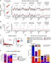
The majority of serum-inducible genes are MRTF-controlled. (A, left) Scatter plot display of total (top) and intronic (bottom) RNA-seq read counts before and after serum stimulation. Serum-stimulated genes (FDR = 0.2) are highlighted in red. (Right) Definition of serum-inducible genes sensitive to SRF-linked signal pathways (FDR = 0.08). (Red bars) Median. See Supplemental Table S2. (B) Signaling to ncRNA genes, as in A. See Supplemental Table S3. (C) SRF sites are overrepresented within 70 kb of transcriptionally active genes. (Left) Frequencies of SRF sites relative to active and inactive genes (per 10-kb bin relative to TSS or pA site). Zero indicates sites within 2 kb of the TSS or within a gene feature. (Asterisks) Significant at P < 0.05, multiple t-test with Holmes-Sidak correction. (Right) SRF-binding sites are significantly closer to active genes. (Asterisks) Significant at P < 0.0001, Mann-Whitney test. See Supplemental Table S4. (D) Relationship between SRF peak properties and activity of their closest associated gene. The left bars indicate peaks classified according to activity of the closest gene, gray bars at the side indicate the fraction of these peaks associated with regulatory events at genes up to 70-kb distant, the center bars represent classification of peaks by SRF-binding inducibility, and the right bars represent classification of peaks by cofactor association. (Direct) Sites within 2 kb of 5′ flanking sequences or within a gene feature; (near) sites within 70 kb of the TSS or pA site. See Supplemental Table S4. (E) Candidate MRTF serum-inducible genes (defined by MRTF binding, sensitivity to LatB or CD, or both) categorized by distance from the nearest SRF sites, as in D. Many inducible genes whose closest SRF site lies within 70 kb share that site with a second gene. See Supplemental Table S4A.
Figure 5.
Figure 5.
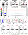
MRTF acts at Pol II recruitment and post-recruitment steps. (A) Serum-induced genes exhibit increased Pol II loading. RNA Pol II ChIP-seq analysis with antibodies as follows: 8WG16 (Pol II CTD un-P), total reads from −2 kb to +1 kb from TSS; H14 (Pol II CTD S5P), total reads from −2 kb to the gene 3′ end or +70 kb, the limit of Pol II progress after 30 min of stimulation; and H5 (Pol II CTD S2P), total reads from +1 to the gene 3′ end or +70 kb. Statistical significance, Mann-Whitney test, (**) P < 0.01; (****) P < 0.0001. (B) Representative MRTF-B and Pol II ChIP-seq tracks on Acta2 and Klf7. (C) LatB inhibits serum induction of Klf7 and Acta2, assessed by qRT–PCR. SEM of three independent experiments. (D) MRTF is required for Pol II recruitment on a subset of target genes. Four-hundred-eighty-three genes were analyzed whose serum-induced activation was LatB-sensitive in the absence of U0126. (Left) Scatter plot summary of the Pol II ChIP-seq signal from −2 to +1 kb around the TSS. Genes exhibiting >30% reduction (group I) are shown in black, and those exhibiting <30% reduction (group II) are in red. (Right) Summary of LatB’s effect on the two groups. Statistical significance, Mann-Whitney test, (***) P < 0.001. (E) LatB inhibits group I and group II gene RNA synthesis to a similar extent in RNA-seq. (F) Metaprofiles of Pol II ChIP-seq for group I and group II genes. (Top) 8WG16, H14, and H5 normalized ChIP-seq read counts are shown across gene loci, standardized to 20 kb, and flanking 5 kb. (Bottom) Read counts from −1 kb to +1 kb from the TSS.
Figure 6.
Figure 6.
GO analysis of SRF targets. (A) SRF- and MRTF-linked gene signatures were analyzed using DAVID. (SRF) Serum-inducible genes with an SRF-binding site within 70 kb; (MRTF) serum-inducible genes with MRTF–SRF binding within 70 kb or LatB and/or CD sensitivity; (stringent MRTF) MRTF–SRF binding within 70 kb and sensitive to LatB or CD. The signatures are compared with SRF-linked serum-inducible or constitutively transcribed “direct” target genes (i.e., with SRF sites within 2 kb of a 5′ flanking sequence or within a gene feature). See also Supplemental Tables S6 and S7. (F.E.) Fold enrichment. (B) SRF-controlled genes involved in transcriptional regulation are subdivided into functional categories. (C) Relationship between the inducible SRF, MRTF, and TCF gene signatures and previously defined sets of genes up-regulated between two experimental conditions; statistical significance by two-tailed Fisher test. See Supplemental Table S8. (D) MRTF–SRF signaling is a nuclear component of integrin-mediated “inside-out” signaling. Classes of SRF target genes involved in adhesion signaling and mechanosensation are shown. Engagement with the ECM induces changes in actin dynamics and actomyosin contractility, promoting focal adhesion assembly (blue arrows). MRTF–SRF target gene expression provides an additional long-term “inside-out” signaling mechanism (red arrows). The green dashed line indicates direct physical coupling between actomyosin, focal adhesions, and the nucleus.
Figure 7.
Figure 7.
SRF and MRTF can both reset the circadian clock. (A) SRF targets among circadian clock circuits (for discussion of regulatory loops, see Koike et al. 2012). (B) SRF, MRTF, and TCF ChIP-seq peaks are shown below in blue, red, and green, respectively, aligned with binding sites in the liver for the clock genes Per1 and Per2 (core loop components) and Nr1d1, Nr1d2, Dbp, and Nfil3 (interlocking loop components) (Koike et al. 2012), shown in gray. (C) Clock resetting by MRTF activation. NIH3T3 cells were treated with 2 µM CD for 2 h followed by washout; transcripts were quantified by qRT–PCR over 36 h. For serum stimulation kinetics, see Supplemental Figure S7C. (D) Clock resetting by TCF activation. Wild-type and SAP-1−/− Elk1−/− Net∂/∂ MEFs were treated with TPA, and transcripts were quantified by qRT–PCR. (E) Circadian clock synchronization by MRTF- and TCF-linked SRF signaling.

Comment in

References

    1. Akashi M, Nishida E 2000. Involvement of the MAP kinase cascade in resetting of the mammalian circadian clock. Genes Dev 14: 645–649 - PMC - PubMed
    1. Anders S, Huber W 2010. Differential expression analysis for sequence count data. Genome Biol 11: R106. - PMC - PubMed
    1. Balamotis MA, Pennella MA, Stevens JL, Wasylyk B, Belmont AS, Berk AJ 2009. Complexity in transcription control at the activation domain-mediator interface. Sci Signal 2: ra20. - PMC (V体育安卓版) - PubMed
    1. Balsalobre A, Damiola F, Schibler U 1998. A serum shock induces circadian gene expression in mammalian tissue culture cells. Cell 93: 929–937 - PubMed
    1. Barker HE, Cox TR, Erler JT 2012. The rationale for targeting the LOX family in cancer. Nat Rev Cancer 12: 540–552 - PubMed

Publication types

MeSH terms

Associated data

"V体育ios版" Grants and funding