Skip to main page content
U.S. flag

An official website of the United States government

Dot gov

The . gov means it’s official. Federal government websites often end in VSports app下载. gov or . mil. Before sharing sensitive information, make sure you’re on a federal government site. .

Https

The site is secure. The https:// ensures that you are connecting to the official website and that any information you provide is encrypted and transmitted securely. V体育官网.

. 2014 Apr 1;127(Pt 7):1585-94.
doi: 10.1242/jcs.141069. Epub 2014 Jan 24.

Modulation of hypoxia-signaling pathways by extracellular linc-RoR

Affiliations

Modulation of hypoxia-signaling pathways by extracellular linc-RoR

Kenji Takahashi et al. J Cell Sci. .

Abstract

Resistance to adverse environmental conditions, such as hypoxia, contributes to the reduced efficacy of anticancer therapies and tumor progression. Although deregulated expression of many long noncoding RNA (lncRNA) occurs in human cancers, the contribution of such RNA to tumor responses to hypoxia are unknown. RNA expression profiling identified several hypoxia-responsive lncRNAs, including the long intergenic noncoding RNA, regulator of reprogramming (linc-RoR), which is also increased in expression in malignant liver cancer cells. Linc-RoR expression was increased in hypoxic regions within tumor cell xenografts in vivo. Tumor cell viability during hypoxia was reduced by small interfering RNA (siRNA) to linc-RoR. Compared with controls, siRNA to linc-RoR decreased phosphorylation of p70S6K1 (RPS6KB1), PDK1 and HIF-1α protein expression and increased expression of the linc-RoR target microRNA-145 (miR-145) VSports手机版. Linc-RoR was highly expressed in extracellular RNA released by hepatocellular cancer (HCC) cells during hypoxia. Incubation with extracellular vesicle preparations containing extracellular RNA increased linc-RoR, HIF-1α expression and cell survival in recipient cells. These studies show that linc-RoR is a hypoxia-responsive lncRNA that is functionally linked to hypoxia signaling in HCC through a miR-145-HIF-1α signaling module. Furthermore, this work identifies a mechanistic role for the extracellular transfer of linc-RoR in intercellular signaling to promote cell survival during hypoxic stress. .

Keywords: Cell stress; Exosome; Extracellular vesicle; Gene expression; Hypoxia; Liver cancer; linc-RoR; microRNA. V体育安卓版.


Figures

Fig. 1.
Fig. 1.
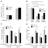
Expression of lncRNA in HepG2 cells. (A) LncRNA expression was assessed by RT-PCR in malignant HepG2 cells and non-malignant human hepatocytes. Each analysis was performed on three independent samples for each lncRNA. Each bar represents the relative expression for an individual lncRNA. Deregulated lncRNA expression, with a >2-fold change in malignant cells compared to non-malignant cells, was identified for 39 lncRNAs. (B) LncRNA expression was examined in HepG2 HCC cells under conditions of hypoxia or normoxia. 89 lncRNAs were identified in HepG2 cells, of which 20 lncRNA were increased by a >2-fold change under hypoxia conditions. Seven of these lncRNAs, including linc-RoR, were also increased in expression in malignant cells. The Venn diagram depicts the number of individual lncRNAs that were increased in HepG2 cells compared to non-malignant hepatocytes, or in response to hypoxia compared to normoxia.
Fig. 2.
Fig. 2.
Expression of linc-RoR expression in human HCC cells during hypoxia. RNA was extracted and RT-PCR performed, as described in the Materials and Methods section. (A) Basal expression level of linc-RoR in non-malignant human hepatocytes (HHs) and HCC cell lines. Expression of linc-RoR was normalized to expression of RNU6B and expressed relative to expression in HHs. (B) Linc-RoR expression was assessed in malignant liver cancer cell lines and HH cells incubated under hypoxia or normoxia for 24 hours. Expression of linc-RoR was normalized to RNU6B expression and expressed relative to the value in normoxia. Ct values of RNU6B were similar across samples and not altered during hypoxia. Bars represent the mean±s.e.m. of three separate studies. *P<0.05.
Fig. 3.
Fig. 3.
Linc-RoR is increased in hypoxic areas in vivo. HCC tumor cell xenografts were established in athymic mice to examine the expression of linc-RoR in vivo. Intratumoral hypoxic areas were identified by immunohistochemistry for Hypoxyprobe-1 (A). Scale bar: 500 µm. Immunohistochemistry for Hypoxyprobe-1 (B), in situ hybridization for linc-RoR (C) or in situ hybridization for negative control probe (D) in representative high-power fields from adjacent sections. Scale bars: 50 µm. The arrows show Hypoxyprobe-1- or linc-RoR-positive cells. (E) The number of linc-RoR-positive cells was quantified in hypoxic or nonhypoxic areas of tumor tissues. Data represents mean±s.e.m. of the number of positive cells with detectable linc-RoR in ten high-power fields. *P<0.05.
Fig. 4.
Fig. 4.
Knockdown of linc-RoR decreases tumor cell viability during hypoxia. (A) HepG2 cells were seeded (1×10⁴/well) into 96-well collagen-coated plates and cultured under conditions of normoxia or hypoxia, and cell viability was assessed after 24 or 48 hours. (B) HepG2 cells or PLC-PRF-5 cells were transfected with two different siRNAs to linc-RoR (1 or 2) or nontargeting control siRNA. After 48 hours, RNA was isolated and qPCR for linc-RoR was performed. (C,D) HepG2 cells were transfected with siRNA 1 or 2 to linc-RoR (C and D, respectively) or nontargeting control. After 24 hours, cells were plated (1×10⁴/well) in 96-well plates and cultured under normoxia or hypoxia conditions. Cell viability was assessed after 24 or 48 hours and is expressed relative to controls.
Fig. 5.
Fig. 5.
Knockdown of linc-RoR decreases HIF-1α and PDK1 expression. HepG2 cells were transfected using siRNA 1 to linc-RoR or ‘ontargeting’ control. After 48 hours, cells were plated (1×10⁴/well) on 96-well amine-coated plates and incubated under normoxia or hypoxia conditions. After 48 hours of incubation, quantitative immunocytochemistry for (A) HIF-1α and (B) PDK1 were performed using a HIF-1α + PDK1 Hypoxia Human In-Cell ELISA kit (Abcam) and imaged using a LI-COR Odyssey system. Expression of HIF-1α and PDK1 were normalized to the corresponding Janus Green fluorescence for each well.
Fig. 6.
Fig. 6.
Linc-RoR modulates expression of miR-145 and downstream signaling. (A–C) HepG2 cells were transfected with two different siRNAs to linc-RoR (1 or 2) or nontargeting control as indicated, and cultured under hypoxia (A,B) or normoxia (C). After 48 hours, RNA was isolated and RT-PCR for (A,C) HIF-1α or (B,C) miR-145 was performed. (D,E) HepG2 cells were transfected with siRNA 1 to linc-RoR, or nontargeting control. After 48 hours, cells were lysed and immunoblot analysis was performed using specific antibodies against p70S6K1 and phospho-p70S6K1 (pp70S6K1). A representative immunoblot (D) and quantitative densitometric data (E) of the ratio of phosphorylated to total p70S6K1, from three independent experiments, are shown. (F) HepG2 cells were transfected with either HA-HIF-1α-pcDNA3 or pcDNA3 vector and, 24 hours later, cells were transfected with siRNA no. 1 to linc-RoR, or nontargeting control. After 24 hours, cells were seeded (1×10⁴/well) into 96-well plates and cultured under hypoxia conditions; cell viability was assessed after 72 hours.
Fig. 7.
Fig. 7.
Effect of linc-RoR knockdown in tumor xenografts in vivo. Xenograft tumors were established following ex vivo transfection of PLC-PRF-5 cells with siRNA 1 to linc-RoR or control siRNA as described in the Materials and Methods section. (A) Tumor volume was estimated at the indicated time-points. Data represent the average of estimated tumor volume from three separate xenografts. (B) Tumors were excised at 6 weeks after implantation. The bars represent average and standard deviation of xenograft tumor weight. Scale bar: 10 mm. (C–E) RNA was isolated from xenograft tumors and PCR was performed for (C) linc-RoR, (D) HIF-1α mRNA or (E) miR-145. (F) Immunoblot analysis from tumor lysates was performed using specific antibodies against p70S6K1 or phospho-p70S6K1. A representative immunoblot and quantitative densitometric data showing the ratio of phosphorylated to total p70S6K1 from three separate tumors is shown.
Fig. 8.
Fig. 8.
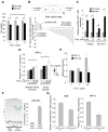
Extracellular linc-RoR during tumor cell responses to hypoxia. (A) HepG2 cells were transfected with siRNA 1 to linc-RoR. After 48 hours, cells were plated (1×10⁴/well) into 96-well plates in vesicle-depleted medium and incubated with varying concentrations of EVs under conditions of normoxia or hypoxia. Cell viability was assessed after 48 hours. (B) The expression of lncRNA, within EV preparations, released by HepG2 tumor cells from three independent replicates was assessed using LncProfilerTM qPCR Array Kit. Each bar represents the relative expression of extracellular RNA and donor-cell RNA for an individual lncRNA. Nine lncRNAs, including linc-RoR, were predominantly expressed in extracellular-RNA isolations compared to their donor cells. (C) Tumor cells were incubated under hypoxia or normoxia conditions, and extracellular RNA released by these cells was obtained after 48 or 72 hours. qRT-PCR for linc-RoR was then performed. (D) HepG2 cells were plated (1×10⁴/well) on 96-well amine-coated plates in vesicle-depleted medium and incubated with varying concentrations of EVs that had been derived from HepG2 cells under normoxia or hypoxia conditions. Recipient cells were then cultured under hypoxia conditions for 48 hours. Quantitative immunocytochemistry for HIF-1α was performed in recipient cells using an in-cell ELISA assay. (E) EVs were isolated from HepG2 cells under normoxia and added to recipient HepG2 cells. After 48 hours incubation under normoxia with those EVs, recipient-cell RNA was isolated and qRT-PCR for HIF-1α or miR-145 was performed. (F,G) HepG2 cells were transfected with siRNA 1 to linc-RoR, or nontargeting control, and cultured under normoxia. (F) After 72 hours, extracellular RNA from HepG2 cells was isolated and droplet digital PCR for miR-145 was performed. The number of positive droplets and concentration of miR-145 from three independent experiments are shown. (G) After 72 hours, EVs were collected from each group, and 10 µg/ml of EV was added to recipient HepG2 cells. After 48 hours incubation under normoxia, recipient-cell RNA was isolated and RT-PCR for linc-RoR or HIF-1α was performed.

References

    1. Ariel I., Miao H. Q., Ji X. R., Schneider T., Roll D., de Groot N., Hochberg A., Ayesh S. (1998). Imprinted H19 oncofetal RNA is a candidate tumour marker for hepatocellular carcinoma. Mol. Pathol. 51, 21–25 10.1136/mp.51.1.21 - DOI - PMC - PubMed
    1. Batagov A. O., Kuznetsov V. A., Kurochkin I. V. (2011). Identification of nucleotide patterns enriched in secreted RNAs as putative cis-acting elements targeting them to exosome nano-vesicles. BMC Genomics 12, Suppl. 3S18 10.1186/1471--2164--12--S3--S18 - DOI - PMC - PubMed
    1. Boyer L. A., Plath K., Zeitlinger J., Brambrink T., Medeiros L. A., Lee T. I., Levine S. S., Wernig M., Tajonar A., Ray M. K. et al. (2006). Polycomb complexes repress developmental regulators in murine embryonic stem cells. Nature 441, 349–353 10.1038/nature04733 - DOI - PubMed
    1. Braconi C., Valeri N., Kogure T., Gasparini P., Huang N., Nuovo G. J., Terracciano L., Croce C. M., Patel T. (2011a). Expression and functional role of a transcribed noncoding RNA with an ultraconserved element in hepatocellular carcinoma. Proc. Natl. Acad. Sci. USA 108, 786–791 10.1073/pnas.1011098108 - DOI (V体育ios版) - PMC - PubMed
    1. Braconi C., Kogure T., Valeri N., Huang N., Nuovo G., Costinean S., Negrini M., Miotto E., Croce C. M., Patel T. (2011b). microRNA-29 can regulate expression of the long non-coding RNA gene MEG3 in hepatocellular cancer. Oncogene 30, 4750–4756 10.1038/onc.2011.193 - DOI - PMC - PubMed

V体育平台登录 - Publication types

MeSH terms (V体育2025版)

Substances

LinkOut - more resources