Skip to main page content
U.S. flag

An official website of the United States government

Dot gov

The . gov means it’s official. Federal government websites often end in . gov or VSports app下载. mil. Before sharing sensitive information, make sure you’re on a federal government site. .

Https

The site is secure. The https:// ensures that you are connecting to the official website and that any information you provide is encrypted and transmitted securely V体育官网. .

. 2014 Apr;59(4):1435-47.
doi: 10.1002/hep.26790. Epub 2014 Feb 18.

"V体育2025版" Differential effects of sorafenib on liver versus tumor fibrosis mediated by stromal-derived factor 1 alpha/C-X-C receptor type 4 axis and myeloid differentiation antigen-positive myeloid cell infiltration in mice

Affiliations

Differential effects of sorafenib on liver versus tumor fibrosis mediated by stromal-derived factor 1 alpha/C-X-C receptor type 4 axis and myeloid differentiation antigen-positive myeloid cell infiltration in mice

Yunching Chen et al. Hepatology. 2014 Apr.

Abstract

Sorafenib--a broad kinase inhibitor--is a standard therapy for advanced hepatocellular carcinoma (HCC) and has been shown to exert antifibrotic effects in liver cirrhosis, a precursor of HCC. However, the effects of sorafenib on tumor desmoplasia--and its consequences on treatment resistance--remain unknown. We demonstrate that sorafenib has differential effects on tumor fibrosis versus liver fibrosis in orthotopic models of HCC in mice VSports手机版. Sorafenib intensifies tumor hypoxia, which increases stromal-derived factor 1 alpha (SDF-1α) expression in cancer and stromal cells and, subsequently, myeloid differentiation antigen-positive (Gr-1(+)) myeloid cell infiltration. The SDF-1α/C-X-C receptor type 4 (CXCR4) pathway directly promotes hepatic stellate cell (HSC) differentiation and activation through the mitogen-activated protein kinase pathway. This is consistent with the association between SDF-1α expression with fibrotic septa in cirrhotic liver tissues as well as with desmoplastic regions of human HCC samples. We demonstrate that after treatment with sorafenib, SDF-1α increased the survival of HSCs and their alpha-smooth muscle actin and collagen I expression, thus increasing tumor fibrosis. Finally, we show that Gr-1(+) myeloid cells mediate HSC differentiation and activation in a paracrine manner. CXCR4 inhibition, using AMD3100 in combination with sorafenib treatment, prevents the increase in tumor fibrosis--despite persistently elevated hypoxia--in part by reducing Gr-1(+) myeloid cell infiltration and inhibits HCC growth. Similarly, antibody blockade of Gr-1 reduces tumor fibrosis and inhibits HCC growth when combined with sorafenib treatment. .

Conclusion: Blocking SDF-1α/CXCR4 or Gr-1(+) myeloid cell infiltration may reduce hypoxia-mediated HCC desmoplasia and increase the efficacy of sorafenib treatment. V体育安卓版.


Figures

Figure 1
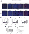
Figure 1. Tumor hypoxia and SDF1α expression are increased after sorafenib treatment in orthotopic HCC
A, Representative immunofluorescence staining of CAIX and SDF1α in HCA-1 tumor implanted in mice with liver fibrosis. Images are 636μm across. B,C, The hypoxic tumor tissue fraction (B) and SDF1α expression (C) increased significantly after sorafenib treatment in orthotopic HCA-1 tumors in mice (n=5–9). D, Immunofluorescence imaging analysis showed that SDF1α expression is increased after 14 days of sorafenib treatment in spontaneous HCCs in Mst1−/− Mst2F/− transgenic mice. The increased hypoxia and SDF1α expression persisted when sorafenib treatment was combined with AMD3100 (n=5–9). E,F, Expression of CAIX (E) and SDF1α (F) in the liver was not significantly changed after sorafenib, AMD3100 or combination treatment (n=4–7). The number of random regions of interest used for quantification is shown in parentheses. Data are presented as mean ± SEM; *P<0.05, **P<0.01.
Figure 2
Figure 2. Immunostaining for SDF1α in liver tissue samples from HCC patients
Paraffin embedded tissue sections were stained for SDF1α by immunohistochemistry. A,B SDF1α expression in non-malignant liver tissue (A) was consistently lower than in malignant HCC tumor area (B). Both in cirrhotic liver tissue (C) and in HCC nodules (D), SDF1α expression was colocalized with ECM components/fibrous septa supporting the potential pro-fibrotic role of SDF1α in human hepatic fibrogenesis and HCC desmoplasia. E, Specimens of tumors that re-occurred after transcatheter arterial chemoembolization and radiofrequency ablation – both of which are known to cause severe hypoxia – consistently showed high expression of SDF1α, which was also associated with more pronounced desmoplasia in HCC area. Error bars represent score ± SEM (n=10 regions of interest per sample).
Figure 3
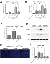
Figure 3. Sorafenib treatment selectively increases tumor-associated fibrosis in HCC in an SDF1α/CXCR4 pathway-dependent manner
A,B, Sorafenib significantly increased the collagen I content (A) and the number of α-SMA+ myofibroblasts (B) in orthotopic HCA-1 tumors implanted in mice with fibrotic livers. The analysis was performed using immunofluorescence in tumor tissue, and maker-positive area was normalized to the area of DAPI (nuclear stain) (N=5–7). C–D, Sorafenib treatment significantly increased the collagen I content (C and D) and the expression of α-SMA (D) (detected by Western blotting) in spontaneously arising HCCs (N=7–8). E, Representative images of immunofluorescence of Collagen I staining in spontaneous HCCs. Images are 636μm across. Addition of AMD3100 to sorafenib treatment prevented the increase in collagen I content and the number of α-SMA+ myofibroblasts in HCCs (A–E). F, In tumor-bearing mice, sorafenib significantly reduced the collagen I content in the surrounding liver tissue in mice with underlying liver fibrosis to levels comparable to those seen in livers from non-CCl4-treated mice (N=4–8). The number of random regions of interest used for quantification is shown in parentheses. *p<0.05; **p<0.01; Data are shown as mean ± SEM.
Figure 4
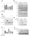
Figure 4. SDF1α/CXCR4 axis promotes HSC to myofibroblast differentiation in the face of PDGFR blockade by sorafenib
A, Exposure to recombinant PDGF-B increased HSC proliferation, while treatment with sorafenib reduced the viability of HSCs. Exposure to recombinant SDF1α increased the viability of HSCs despite PDGFR inhibition using sorafenib treatment, in a dose-dependent manner. HSC viability was measured by MTT assay (N=6 experimental repeats). B, Exposure to recombinant SDF1α increased the expression of α-SMA and collagen I and reduced cleaved caspase-3 expression (evaluated by Western blotting) despite sorafenib treatment, in a dose-dependent manner. C, Exposure to recombinant SDF1α increased viability of HSCs despite sorafenib treatment (N=3–5 experimental repeats). D, Exposure to recombinant SDF1α upregulated collagen I and α-SMA expression levels as well as ERK and AKT activation in HSCs, consistent with their myofibroblast differentiation. Inhibition of CXCR4 with AMD3100 (D) or using siRNA (E) prevented the effects of SDF1α. F, ERK inhibition with FR-180204 (2μM) decreased α-SMA expression in HSCs treated with recombinant SDF1α. Data are presented as mean ± SEM.
Figure 5
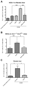
Figure 5. Intratumoral infiltration of Gr1+ myeloid cells is increased after sorafenib (SOR) treatment, and is prevented by CXCR4 inhibition in HCC
A–B, After SOR treatment, the number of 7AAD–CD11b+Gr1+ monocytes (measured by flow cytometry) significantly increased in HCA-1 tumors growing C3H mice with liver fibrosis (A) of as well as in spontaneous HCCs in Mst1−/− Mst2F/− transgenic mice (B). Treatment with the CXCR4 inhibitor AMD3100 prevented this effect (A–B). C, The number of 7AAD–CD11b+Gr1+ myeloid cells was significantly reduced in the fibrotic liver tissues from sorafenib-treated mice. Data are shown as percentages of the total number of cells evaluated in enzymatically digested tissue. Data are presented as mean ± SEM (N=5–14 mice per group). **P<0.01.
Figure 6
Figure 6. Gr-1+ myeloid cell infiltration increases fibrosis in HCC after sorafenib in an SDF1α/CXCR4 pathway-dependent manner
A–C, Depletion of Gr-1+ myeloid cells using systemic therapy with anti-Gr-1 blocking antibodies prevents the shift towards a pro-fibrotic environment after sorafenib treatment. Sorafenib significantly increased the number of 7AAD–CD11b+Gr1+ monocytes (A) (N=3–7 mice per group), the collagen I content (B) and the number of α-SMA+ myofibroblasts (C) in orthotopic HCA-1 tumors growing in mice with liver fibrosis, evaluated by immunohistochemistry. Combining anti-Gr-1 antibodies with sorafenib treatment prevented these effects. Quantification was performed in 5–12 confocal microscopy images per mouse. D–F, Tumor-infiltrating Gr-1+ myeloid cells directly mediate HSC differentiation to myofibroblasts via SDF1α/CXCR4 axis. Co-culture with tumor-tissue isolated Gr1+ myeloid cells (obtained using magnetic beads) increased collagen I and α-SMA expression levels as well as ERK activation in HSCs (D). Co-culture with Gr1+ myeloid cells increased collagen I and α-SMA expression in HSCs despite sorafenib treatment, in a dose-dependent manner (E). Recombinant PDGF-B and sorafenib alone were used as positive and negative control, respectively, for HSC differentiation (E). Inhibition of CXCR4 with AMD3100 prevented the increase in collagen I, α-SMA, and p-ERK expression in HSCs co-cultured with Gr-1+ myeloid cells (F). Data are presented as mean ± SEM. * P<0.05.
Figure 7
Figure 7. Sorafenib induces a modest growth delay in HCC, and CXCR4 inhibition or blockade of Gr-1 synergize with sorafenib treatment by increasing cell apoptosis
A, Sorafenib (SOR) treatment induces a minor but significant delay in orthotopic HCC models after 14 days of treatment. HCA-1 tumor growth was delayed by sorafenib in syngeneic C3H mice with underlying liver fibrosis (N=6–14 mice per group). Whereas treatment with the CXCR4 inhibitor AMD3100 (AMD) had no effect as monotherapy, AMD3100 with sorafenib induces a more significant tumor growth delay. B, Representative images of immunofluorescence TUNEL in HCA-1 tumors. Images are 636μm across. C, Cell apoptosis, measured by TUNEL, increased after sorafenib treatment in HCA-1 tumors (N=6–11). D, Representative images of immunofluorescence of TUNEL in spontaneous HCCs. Images are 636μm across. E, Cell apoptosis, measured by TUNEL, increased after sorafenib treatment in spontaneously arising HCCs in Mst1−/− Mst2F/− transgenic mice (N=6–11). Cell apoptosis was significantly increased when sorafenib treatment was combined with AMD3100 in both tumor models. F, Combining anti-Gr-1 antibodies with sorafenib treatment induced a significant tumor growth delay of orthotopic HCA-1 tumors growing in mice with liver fibrosis (N=7–13 mice per group). Data are presented as mean ± SEM; *P<0.05, **P<0.01.
Figure 8
Figure 8. Differential impact of sorafenib on liver versus tumor-associated fibrosis mediated by SDF1α/CXCR4 axis and Gr-1+ cells in HCC
The differential effects of sorafenib are the result of increased intratumoral hypoxia, leading to elevated SDF1α expression and Gr-1+ myeloid cell infiltration. Blocking CXCR4 prevents Gr-1+ myeloid cell infiltration and hepatic stellate cells differentiation and activation, and synergizes with the anti-tumor effects of sorafenib.

"VSports在线直播" References

    1. Hernandez-Gea V, Toffanin S, Friedman SL, Llovet JM. Role of the microenvironment in the pathogenesis and treatment of hepatocellular carcinoma. Gastroenterology. 2013;144:512–527. - PMC - PubMed
    1. Hui CK, Leung N, Shek TW, Yao H, Lee WK, Lai JY, Lai ST, et al. Sustained disease remission after spontaneous HBeAg seroconversion is associated with reduction in fibrosis progression in chronic hepatitis B Chinese patients. Hepatology. 2007;46:690–698. - PubMed
    1. Luedde T, Schwabe RF. NF-kappaB in the liver--linking injury, fibrosis and hepatocellular carcinoma. Nat Rev Gastroenterol Hepatol. 2011;8:108–118. - V体育平台登录 - PMC - PubMed
    1. Friedman SL. Evolving challenges in hepatic fibrosis. Nat Rev Gastroenterol Hepatol. 2010;7:425–436. - PubMed
    1. Wilhelm SM, Carter C, Tang L, Wilkie D, McNabola A, Rong H, Chen C, et al. BAY 43-9006 exhibits broad spectrum oral antitumor activity and targets the RAF/MEK/ERK pathway and receptor tyrosine kinases involved in tumor progression and angiogenesis. Cancer Res. 2004;64:7099–7109. - PubMed (V体育官网入口)

Publication types

VSports注册入口 - MeSH terms