Skip to main page content
U.S. flag

An official website of the United States government

Dot gov

The . gov means it’s official. Federal government websites often end in VSports app下载. gov or . mil. Before sharing sensitive information, make sure you’re on a federal government site. .

Https

The site is secure V体育官网. The https:// ensures that you are connecting to the official website and that any information you provide is encrypted and transmitted securely. .

Clinical Trial
. 2013 Nov 22;288(47):33542-33558.
doi: 10.1074/jbc.M113.511170. Epub 2013 Oct 2.

"VSports app下载" Targeting aberrant glutathione metabolism to eradicate human acute myelogenous leukemia cells

Affiliations
Clinical Trial

Targeting aberrant glutathione metabolism to eradicate human acute myelogenous leukemia cells

Shanshan Pei et al. J Biol Chem. .

Abstract

The development of strategies to eradicate primary human acute myelogenous leukemia (AML) cells is a major challenge to the leukemia research field. In particular, primitive leukemia cells, often termed leukemia stem cells, are typically refractory to many forms of therapy. To investigate improved strategies for targeting of human AML cells we compared the molecular mechanisms regulating oxidative state in primitive (CD34(+)) leukemic versus normal specimens. Our data indicate that CD34(+) AML cells have elevated expression of multiple glutathione pathway regulatory proteins, presumably as a mechanism to compensate for increased oxidative stress in leukemic cells. Consistent with this observation, CD34(+) AML cells have lower levels of reduced glutathione and increased levels of oxidized glutathione compared with normal CD34(+) cells. These findings led us to hypothesize that AML cells will be hypersensitive to inhibition of glutathione metabolism. To test this premise, we identified compounds such as parthenolide (PTL) or piperlongumine that induce almost complete glutathione depletion and severe cell death in CD34(+) AML cells VSports手机版. Importantly, these compounds only induce limited and transient glutathione depletion as well as significantly less toxicity in normal CD34(+) cells. We further determined that PTL perturbs glutathione homeostasis by a multifactorial mechanism, which includes inhibiting key glutathione metabolic enzymes (GCLC and GPX1), as well as direct depletion of glutathione. These findings demonstrate that primitive leukemia cells are uniquely sensitive to agents that target aberrant glutathione metabolism, an intrinsic property of primary human AML cells. .

Keywords: Anticancer Drug; CD34+; Cancer Stem Cells; Glutathione; Human; Leukemia; Parthenolide; Redox Regulation; Tumor Metabolism. V体育安卓版.


Figures
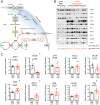
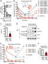
FIGURE 1.
FIGURE 1.
Primitive primary human AML cells differentially express genes required for control of redox state. A, schematic diagram showing major antioxidant machineries required for control of the redox state. B, expression of major antioxidant proteins in freshly isolated primary human CD34+ NBM (n = 4) and CD34+ AML (n = 9) specimens. Lysates from an equal number of cells (100,000) were loaded in each lane. The total amount of protein was quantified and presented as micrograms of protein per lane. C, relative mRNA expression of major antioxidant genes in freshly isolated primary human CD34+ NBM (n = 4) and CD34+ AML (n = 9) specimens. Mean expression of HPRT1, GUSB, and TBP was used as reference to internally normalize the expression of each gene within each specimen. Average expression of each gene in CD34+ NBM (n = 4) cells was set to 1, and the relative expression of each gene in each specimen was calculated accordingly and presented as dot plot. Mean ± S.E. of each group is presented. * indicates a significant difference.
FIGURE 2.
FIGURE 2.
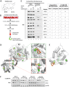
Primary human AML cells have aberrant glutathione metabolism. Amount of reduced (GSH) (A), oxidized (GSSG) (B), total glutathione (sum of GSH + GSSG) (C), and GSH to GSSG ratio (D) in each specimen. Measurement of glutathione turnover rate (E). Time-dependent increase of newly synthesized glutathione ([13C,15N]glutathione) as percentage of total glutathione in CD34+ AML cells (n = 5) (F) and CD34+ NBM cells (n = 3) (G). H, glutathione turnover at 8 h post-culturing CD34+ NBM (n = 3) or CD34+ AML (n = 5) cells in media with [13C,15N]cystine. In A–D and H, each dot represents the value of each specimen. Median ± IQR (Inter-Quartile Range) of each group is presented as error bar. * indicates a significant difference.
FIGURE 3.
FIGURE 3.
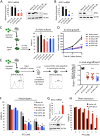
Primitive human AML cells are differentially sensitive to agents that deplete glutathione. A, PTL-induced dose-dependent glutathione depletion in primary human AML cells (n = 3). B, structure of PTL and PTL-induced cellular glutathione level change as a function of time. Red lines indicate changes in CD34+ AML cells (n = 5) and AML cell line M9-ENL cells. Black lines indicate changes in CD34+ NBM cells (n = 5). C, percentage of viable cells after being cultured with 7.5 μm PTL for 24 h. D, Western blot showing 7.5 μm PTL induced protein expression change of oxidative stress and apoptosis markers. E, structure of PLM and PLM-induced cellular glutathione level change as a function of time. Red lines indicate changes in CD34+ AML cells (n = 3) and AML cell line M9-ENL cells. Black lines indicate changes in CD34+ NBM cells (n = 2). F, percentage of viable cells after being cultured with 10 μm PLM for 24 h. In C and F, mean ± S.D. of each group is presented as bar graph. ***, p < 0.0005; *, p < 0.05.
FIGURE 4.
FIGURE 4.
Parthenolide directly binds to and interferes with multiple glutathione pathway components. A, chemical structure of MMB-biotin. Dashed box outlines intact structure of MMB, a stereoisomer of PTL. B, competitive binding assay. C, antioxidant proteins identified as PTL binding targets via competitive binding assay. Input and pull-down products from AML cells treated with biotin (lanes 1 and 4), MMB-biotin (lanes 2 and 5), or PTL followed by MMB-biotin (lanes 3 and 6). Pull-down products from MMB-biotin-treated AML cells were also subjected to LC-MS/MS analysis, and the results were presented in the chart for comparison. + and / signs indicate positive and negative results, respectively. Numbers of peptides recognized by LC-MS/MS were listed as well. D and E, covalent docking results of PTL to GCLC/GCLM holoenzyme complex (D) and GPX1 (E). The l-glutamate binding site of GCLC is enlarged in green box, and the heterodimer interface of GCLC-GCLM complex is in red box. The cysteines covalently attached with PTL are shown as surface representations, whereas the PTL structure is shown as thick sticks. F, Western blots showing time-dependent GCLC and GPX1 protein expression in primary human AML cells (n = 3) and M9-ENL cells treated with 7.5 μm PTL.
FIGURE 5.
FIGURE 5.
Targeting glutathione pathway is important for the anti-leukemia activity of PTL. Knockdown of GCLC (A) and GPX1 (B) in M9-ENL cells. C, percentage of viable cells after 48 h culture in vitro. D, number of cells at 24 and 48 h culture in vitro. E, procedure and results of in vivo competitive engraftment assay. F, percentage of viable cells treated with the indicated doses of PTL for 24 h. G, overexpression of GCLC in M9-ENL cells. H, percentage of viable cells treated with indicated doses of PTL for 24 h. Error bars represent mean ± S.D. of triplicates. C indicates control used for statistical comparison, *, p < 0.05; ***, p < 0.0005.
FIGURE 6.
FIGURE 6.
Parthenolide and piperlongumine represent a novel class of anti-leukemic agents. Viability of CD34+ AML cells (n = 9) treated 24 h with increasing doses of Ara-C (A), IDA (B), PTL (C), or PLM (D). E, representative graph showing the cellular glutathione level change over time in CD34+ AML cells treated with Ara-C, IDA, PTL, or PLM. F, viability of M9-ENL cells treated 24 h with Ara-C, IDA, PTL, or PLM at the indicated doses. G, cellular glutathione level change over time in M9-ENL cells treated with Ara-C, IDA, PTL, or PLM. In A–D and F, mean ± S.D. of triplicates is presented for each data point. In F, ***, p < 0.0005.
FIGURE 7.
FIGURE 7.
Toxicity of PTL in combination with conventional anti-leukemic agents. Viability of CD34+ AML cells treated with PTL + Ara-C (A) or PTL + IDA (B) at various dose combinations. Mean of triplicates is presented. Numbers indicate mean viability. In A, viability of cells treated with 5 μm PTL alone, 5 μm Ara-C alone, and 5 μm PTL + 5 μm Ara-C in combination are highlighted as red bars. In B, viability of cells treated with 5 μm PTL alone, 60 nm IDA alone, and 5 μm PTL + 60 nm IDA in combination are highlighted as red bars as well. C, heat map showing the degree of synergy for each drug combination at fixed dose combos in each AML specimen. Synergism is determined by the CI calculated using Calcusyn software. Color key for each category is presented at the top of graph: synergism (0.4 < CI < 0.6), moderate synergism (0.6 < CI < 0.8), slight synergism (0.8 < CI < 0.9), additive or none (CI > 0.9).

References

    1. Couzin J. (2002) Cancer drugs. Smart weapons prove tough to design. Science 298, 522–525 - PubMed
    1. Patel J. P., Gönen M., Figueroa M. E., Fernandez H., Sun Z., Racevskis J., Van Vlierberghe P., Dolgalev I., Thomas S., Aminova O., Huberman K., Cheng J., Viale A., Socci N. D., Heguy A., Cherry A., Vance G., Higgins R. R., Ketterling R. P., Gallagher R. E., Litzow M., van den Brink M. R., Lazarus H. M., Rowe J. M., Luger S., Ferrando A., Paietta E., Tallman M. S., Melnick A., Abdel-Wahab O., Levine R. L. (2012) Prognostic relevance of integrated genetic profiling in acute myeloid leukemia. N. Engl. J. Med. 366, 1079–1089 - PMC (VSports最新版本) - PubMed
    1. Skrtić M., Sriskanthadevan S., Jhas B., Gebbia M., Wang X., Wang Z., Hurren R., Jitkova Y., Gronda M., Maclean N., Lai C. K., Eberhard Y., Bartoszko J., Spagnuolo P., Rutledge A. C., Datti A., Ketela T., Moffat J., Robinson B. H., Cameron J. H., Wrana J., Eaves C. J., Minden M. D., Wang J. C., Dick J. E., Humphries K., Nislow C., Giaever G., Schimmer A. D. (2011) Inhibition of mitochondrial translation as a therapeutic strategy for human acute myeloid leukemia. Cancer Cell 20, 674–688 - PMC - PubMed
    1. Trachootham D., Alexandre J., Huang P. (2009) Targeting cancer cells by ROS-mediated mechanisms. A radical therapeutic approach? Nat. Rev. Drug Discov. 8, 579–591 - PubMed
    1. Conklin K. A. (2004) Chemotherapy-associated oxidative stress. Impact on chemotherapeutic effectiveness. Integr. Cancer Ther. 3, 294–300 - PubMed

Publication types (VSports)

"V体育平台登录" MeSH terms