"VSports注册入口" Skip to main page content
U.S. flag

An official website of the United States government

Dot gov

The . gov means it’s official. Federal government websites often end in VSports app下载. gov or . mil. Before sharing sensitive information, make sure you’re on a federal government site. .

Https

The site is secure V体育官网. The https:// ensures that you are connecting to the official website and that any information you provide is encrypted and transmitted securely. .

. 2014 Jul 10;33(28):3696-706.
doi: 10.1038/onc.2013.336. Epub 2013 Aug 19.

"V体育2025版" A peptide that inhibits function of Myristoylated Alanine-Rich C Kinase Substrate (MARCKS) reduces lung cancer metastasis

Affiliations

A peptide that inhibits function of Myristoylated Alanine-Rich C Kinase Substrate (MARCKS) reduces lung cancer metastasis

C-H Chen et al. Oncogene. .

Abstract

Myristoylated Alanine-Rich C Kinase Substrate (MARCKS), a substrate of protein kinase C, is a key regulatory molecule controlling mucus granule secretion by airway epithelial cells as well as directed migration of leukocytes, stem cells and fibroblasts VSports手机版. Phosphorylation of MARKCS may be involved in these responses. However, the functionality of MARCKS and its related phosphorylation in lung cancer malignancy have not been characterized. This study demonstrated elevated levels of MARCKS and phospho-MARCKS in highly invasive lung cancer cell lines and lung cancer specimens from non-small-cell lung cancer patients. siRNA knockdown of MARCKS expression in these highly invasive lung cancer cell lines reduced cell migration and suppressed PI3K (phosphatidylinositol 3'-kinase)/Akt phosphorylation and Slug level. Interestingly, treatment with a peptide identical to the MARCKS N-terminus sequence (the MANS peptide) impaired cell migration in vitro and also the metastatic potential of invasive lung cancer cells in vivo. Mechanistically, MANS peptide treatment resulted in a coordination of increase of E-cadherin expression, suppression of MARCKS phosphorylation and AKT/Slug signalling pathway but not the expression of total MARCKS. These results indicate a crucial role for MARCKS, specifically its phosphorylated form, in potentiating lung cancer cell migration/metastasis and suggest a potential use of MARCKS-related peptides in the treatment of lung cancer metastasis. .


Conflict of interest statement

CONFLICT OF INTEREST

The authors declare no conflict of interest.

Figures
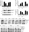
Figure 1
Figure 1
Expression of MARCKS and its phosphorylated molecule are involved in lung cancer cell migration. (a) MARCKS expression in various lung cancer cell lines. Top, cells from near-confluent cultures were harvested for RNA isolation and the level of expression was quantified with real-time RT-PCR and normalized with the β-actin level. Bottom, MARCKS protein and its Ser159/163 phosphorylation were confirmed by western blotting and expressed as fold change relative to CL1-0 cells. (b) The invasion (top) and migratory (bottom) abilities of CL1-0, CL1-0/F3, CL1-5, NCI-H292, A549 and PC9 cells as analyzed by transwell assays with or without matrigel, respectively. (c) Western blot analyses of MARCKS and its Ser159/163 phosphorylated molecule in cells of various lung cancer cell lines with or without treatment with TPA. Cultures were treated with 100 ng/ml TPA or control vehicle for 20 min and harvested for protein lysates. (d) Determination of the major kinase that led to high levels of MARCKS phosphorylation in these malignant lung cancer cell lines. The cell lysates were prepared from CL1-5, A549 and PC9 cultures that were pre-treated with a ROCK inhibitor (Y27632; 10 µm) and/or a PKC inhibitor (Calphostin C; 250 nm) for 2 h. Western blot analysis was carried out to assess MARCKS and its Ser159/163 phosphorylated molecule in these cell lysates.
Figure 2
Figure 2
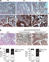
High levels of MARCKS phosphorylation are found in lung cancer specimens. (a) Higher IHC staining of Ser159/163 phosphorylated MARCKS in tumor (T) vs adjacent non-tumor areas (N) in 14/18 patients. P3, P5 and P6 are three representative stainings from these 14 patients. (b) Representative images of negative control (secondary antibody only) and positive staining of MARCKS phosphorylation at Ser159/163 by using IHC staining in tumor specimens from patients with NSCLC. The low expression (Score = 1) and high expression (Score = 2 or 3) of MARCKS phoshporylation by scoring system as described in Materials and methods. Bar = 20 µm. (c) Percentage of patients with high and low MARCKS phoshporylation with respect to adenocarcinoma (Adeno) vs non-adenocarcinoma (Non-Adeno) (Left), and poorly differentiated vs well differentiated (Right). Numbers in bars represent the percentage of patients for each condition.
Figure 3
Figure 3
MARCKS expression is crucial for lung cancer cell migration and invasion. (a–c) siRNA knockdown of MARCKS decreases migration capability of CL1-0/F3 (a), CL1-5 (b) and PC9 (c) cells. Cells were transfected with MARCKS-specific siRNAs or control non-specific siRNA (Native control) as indicated. After 48 h of transfection, cells were subjected to scratching/wound-healing assay, and the numbers of cells migrated to the wound area were quantified at 12 h post scratching. (left; n = 3, *P < 0.05 vs Native control). Cell lysates from these transfectants were prepared and subjected to western blotting (right). (d) Transwell migration (top) and matrigel invasion (bottom) assays confirmed the importance of MARCKS expression in cell motility and invasiveness after silencing MARCKS expression of PC9 cells by MARCKS-specific or non-specific siRNA transfeciton (n = 3, *P < 0.05 vs Native control). (e) siRNA knockdown of MARCKS also diminishes pAKT/Slug pathway. PC9 cells were transfected with MARCKS-specific or non-specific siRNA (Native control), and cell lysates were prepared 72 h after transfection. Western blot analyses were carried out with specific antibodies as shown in the left panel of figure. Right, the mean results for densitometric scans of three blots from multiple experiments were expressed as fold relative to that of parental PC9 cells.
Figure 4
Figure 4
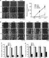
MANS peptide treatment impairs migration and invasiveness of malignant lung cancer cells. (a) Scratch/wound-healing assay for evaluating the inhibitory effects of MANS peptide on cell migration. Confluent cultures of CL1-0/F3 cells were scratched and wound-healing repair was monitored microscopically examined at 12 and 24 h after the scratch and the addition of RNS or MANS peptide (100 µm). Left, phase-contrast pictures. Right, quantification of the migration distance in cultures after the scratch. Data are representative of three independent experiments, *P < 0.05 compared with untreated culture (Con). (b) MANS peptide (100 µm), but not RNS peptide, also inhibits CL1-5 (Left) and PC9 (Right) cell migration in a scratch/wound-healing assay. These results are representative of three independent experiments. (c) Transwell migration assay also demonstrated the inhibitory effects of MANS peptide on invasive cell migration. Dissociated cells of CL1-0/F3, CL1-5, PC9 and A549 cultures were plated on transwells with or without RNS or MANS peptide (100 µm); 12 h later, cells that migrated to the lower chamber were fixed, stained and counted using light microscopy. Quantification of migrated cells to the lower chamber. Data expressed as mean ± s.d. (n = 4), *P < 0.05 compared with Con (untreated cells). (d) Invasion ability of cells with or without MANS peptide (100 µm) as determined by matrigel invasion assays (n = 3, *P < 0.05 vs Con).
Figure 5
Figure 5
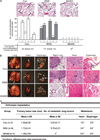
The suppressive effect of MANS peptide on cancer metastasis in vivo. (a) The nude mice bearing subcutaneous tumors were treated with intratumoral injections of PBS (Con), RNS or MANS peptide at the dosage of 50 nmol every 2 days. Top, representative hematoxylin and eosin (H&E)-stained sections of the lungs from nude mice (n = 6) with subcutaneous tumors. The black arrow indicates the micrometastasis of subcutaneous PC9 cells to the lung. Bottom, total numbers of lung micrometastatic colonies in individual mice were counted under the dissection scope (*P < 0.05 vs Con). (b, c) Dissociated PC9 cells were orthotopically injected to the left lobe of the mouse lung as described in Materials and methods. After a week, mice were injected intraperitoneally with 500 µl of PBS or with PBS containing either the RNS or MANS peptide (50 nmol) once every 3 days thereafter for a total of six injections up to day 25. At day 25, mice were killed, and the organs were removed and examined. (b), Gross (left) and H&E-stained (right) pictures of various organs removed from mice. The arrows indicate tumor nodules in the organ. Bar = 20 µm. (c) Quantification of the average pulmonary metastasis nodules from mice with injected cancer cells and treated with RNS or MANS peptide as described (*P < 0.05 vs Con).
Figure 6
Figure 6
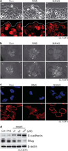
Enhancing cell–cell contact with increase of E-cadherin expression in cancer cells after MANS peptide treatment. Experiments were carried out with CL1-0/F3 cultures. (a) MANS peptide decreased cell spreading and increased cell–cell contacts at wound margins. Top, phase-contrast pictures 16 h after scratch and MANS peptide (100 µm) treatment; bottom, fluorescent microscopy pictures on scratch/wound-healing cultures stained with the F-actin-specific fluorescent dye, phalloidin. A white dotted line represents the wound margin. Bar = 10 µm. (b) Effects of the MANS peptide on inhibition of cell spreading. Passage CL1-0/F3 cells were seeded at low cell density (1000 cells per well of six-well plate) and allowed to grow as cell colonies. After 24 h of serum-free starvation, cells were stimulated with complete medium with RNS or MANS peptide (100 µm). Representative phase-contrast pictures were taken at 0 and 16 h after the treatment. Bar = 20 µm. (c) These cells as described in panel (b) were stained for E-cadherin and F-actin. The fluorescence of FITC-conjugated E-cadherin (green), TRITC-conjugated phalloidin (F-actin stained: red) and DAPI (nucleus counter-stained: blue) was visualized under a confocal laser-scanning microscope. Bar = 10 µm (d) Western blot analysis of E-cadherin expression in CL1-0/F3 cells after RNS or MANS peptide (10–100 µm) treatment. Sixteen hours after the treatment, cell lysates were prepared for western blot analysis as indicated.
Figure 7
Figure 7
MANS peptide coordinately suppressed MARCKS phosphorylation and AKT/Slug pathway in lung cancer cells. (a) The effect of MANS peptide on MARCKS phosphorylation in various lung cancer cell lines. Cells were incubated in a medium containing 100 µm RNS or MANS peptide for 16 h and harvested. Cell lysates were analyzed by western blotting for MARCKS and phospho-MARCKS. (b) MANS peptide suppressed TPA-induced MARCKS phosphorylation of NHBE cells. NHBE cells were treated with or without TPA (10 nm) and also with RNS or MANS (100 µm) at the same time. After co-treatment for 16 h, NHBE cells were harvested and cell lysates prepared for western blot analyses with appropriate antibodies. (c) Western blot analysis of the repression of pAKT/Slug pathway in PC9 cells after MANS peptide (100 µm) treatment. The mean results for densitometric scans of three blots from three separate experiments are shown in right panel. (d) Proposed hypothetical models for MARKS protein-mediated lung cancer cell invasion/migration, under un-treated condition (i), and also the mechanism of MANS peptide-mediated coordinative suppression of MARCKS phosphorylation and pAKT/Slug pathway (ii).

References

    1. Valastyan S, Weinberg RA. Tumor metastasis: molecular insights and evolving paradigms. Cell. 2011;147:275–292. - PMC - PubMed
    1. Teicher BA. Protein kinase C as a therapeutic target. Clin Cancer Res. 2006;12:5336–5345. - PubMed
    1. Roffey J, Rosse C, Linch M, Hibbert A, McDonald NQ, Parker PJ. Protein kinase C intervention: the state of play. Curr Opin Cell Biol. 2009;21:268–279. - "VSports" PubMed
    1. Harlan DM, Graff JM, Stumpo DJ, Eddy RL, Jr, Shows TB, Boyle JM, et al. The human myristoylated alanine-rich C kinase substrate (MARCKS) gene (MACS). Analysis of its gene product, promoter, and chromosomal localization. J Biol Chem. 1991;266:14399–14405. - PubMed
    1. Thelen M, Rosen A, Nairn AC, Aderem A. Regulation by phosphorylation of reversible association of a myristoylated protein kinase C substrate with the plasma membrane. Nature. 1991;351:320–322. - PubMed

Publication types

MeSH terms