Skip to main page content
U.S. flag

An official website of the United States government

Dot gov

The . gov means it’s official. Federal government websites often end in . gov or . mil. Before sharing sensitive information, make sure you’re on a federal government site VSports app下载. .

Https

The site is secure. The https:// ensures that you are connecting to the official website and that any information you provide is encrypted and transmitted securely V体育官网. .

. 2013 Jul 1;27(13):1447-61.
doi: 10.1101/gad.219642.113.

Autophagy suppresses progression of K-ras-induced lung tumors to oncocytomas and maintains lipid homeostasis

Affiliations

"V体育官网入口" Autophagy suppresses progression of K-ras-induced lung tumors to oncocytomas and maintains lipid homeostasis

Jessie Yanxiang Guo et al. Genes Dev. .

Abstract

Macroautophagy (autophagy hereafter) degrades and recycles proteins and organelles to support metabolism and survival in starvation. Oncogenic Ras up-regulates autophagy, and Ras-transformed cell lines require autophagy for mitochondrial function, stress survival, and engrafted tumor growth. Here, the essential autophagy gene autophagy-related-7 (atg7) was deleted concurrently with K-ras(G12D) activation in mouse models for non-small-cell lung cancer (NSCLC). atg7-deficient tumors accumulated dysfunctional mitochondria and prematurely induced p53 and proliferative arrest, which reduced tumor burden that was partly relieved by p53 deletion. atg7 loss altered tumor fate from adenomas and carcinomas to oncocytomas-rare, predominantly benign tumors characterized by the accumulation of defective mitochondria. Surprisingly, lipid accumulation occurred in atg7-deficient tumors only when p53 was deleted VSports手机版. atg7- and p53-deficient tumor-derived cell lines (TDCLs) had compromised starvation survival and formed lipidic cysts instead of tumors, suggesting defective utilization of lipid stores. atg7 deficiency reduced fatty acid oxidation (FAO) and increased sensitivity to FAO inhibition, indicating that with p53 loss, Ras-driven tumors require autophagy for mitochondrial function and lipid catabolism. Thus, autophagy is required for carcinoma fate, and autophagy defects may be a molecular basis for the occurrence of oncocytomas. Moreover, cancers require autophagy for distinct roles in metabolism that are oncogene- and tumor suppressor gene-specific. .

Keywords: K-ras; NSCLC; autophagy; fatty acid oxidation; metabolism; mitochondria; oncocytoma; p53 V体育安卓版. .


Figures (VSports)

Figure 1.
Figure 1.
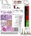
Atg7 deletion in K-ras-driven lung tumors blocks autophagy and reduces lung tumor size. (A) Representative IHC staining for LC3 and p62 in 14-wk-old tumors. Red arrows in K-rasG12D/+;atg7+/+ tissue point to autophagosomes, and black arrows in K-rasG12D/+;atg7−/− tissue point to LC3 or p62 aggregates. (B) Western blot of LC3-I-to-LC3-II processing and p62 accumulation in tumors at 18 wk after tumor initiation. Numbers identify individual mice. (C) Representative gross lung pathology (n = 5 mice for each time point in K-rasG12D/+ mice). (D) Graph of wet lung weight from C. The error bars represent the SEM; (*) P < 0.05 (t-test). (E) Representative histology (H&E) of lungs and tumor burden at the indicated times (n = 3 mice for each time point in K-rasG12D/+ mice). Full scans of all lobes are shown in Supplemental Figure S3. (F) Quantification of tumor burden from E. The error bar represents the SEM; (*) P < 0.05; (**) P < 0.01 (two-way ANOVA with Bonferroni post-test). (G) K-rasG12D/+;atg7+/+ and K-rasG12D/+;atg7−/− tumor-bearing mice (n = 3 for each time and genotype) were scanned by micro-CT at the indicated times to monitor lung tumor progression. K-ras+/+;atg7+/+ mice provided a normal lung control. The number in each panel represents the mouse identification number. The top panels show three-dimensional reconstruction of mouse lungs. Blue indicates lung airspace. The bottom panels show transverse section of the chest, with increased white areas indicating tumor. (H) Quantification of normal lung volume from G. Each spot represents an individual mouse. Normal lung volume decreased faster in mice with K-rasG12D/+;atg7+/+ tumors compared with those with atg7 deletion. (I) Quantification of Ki67-positive tumor cells. The error bar represents the SEM; (*) P < 0.05; (**) P < 0.01 (two-way ANOVA with Bonferroni post-test). Representative images of IHC are shown in Supplemental Figure S5C.
Figure 2.
Figure 2.
Atg7 deficiency converts adenomas/carcinomas to oncocytomas and causes accumulation of dysfunctional mitochondrial in K-ras-driven tumors. (A) Histology of lung tumors shows progression of K-rasG12D/+;atg7+/+ tumors to adenocarcinomas and of K-rasG12D/+;atg7−/− tumors to oncocytomas. Note the development over time of expansive cytoplasm in oncocytomas and necrotic cells (arrows) (n = 3 mice for each time point and genotype). (B) Representative electron microscope images of tumors at 18 wk. (N) Nuclear; (M) mitochondria; (Au-P) autophagosome; (Au-L) autolysosome; (L) lamellar body; (ER) endoplasmic reticulum. (C) Representative IHC of Tom20 shows accumulation of mitochondria in atg7-deficient tumors. (D) Cytochrome c oxidase activity in tumors at 25 wk after tumor induction.
Figure 3.
Figure 3.
Inflammation is the major cause of death of mice bearing K-rasG12D/+;atg7/ tumors. (A) Kaplan-Meier survival curve of mice with K-rasG12D/+ tumors that were atg7+/+, atg7+/−, and atg7−/− (P > 0.05, log-rank test). (B) Representative gross lung pathology at 42 wk after tumor initiation (n = 4 mice for K-rasG12D/+;atg7+/+; n = 5 mice for K-rasG12D/+;atg7−/−). (C) Quantification of wet lung weight from B. (*) P < 0.05 (t-test). (D) Quantification of tumor and crystalline macrophage burden in the lungs at 42 wk (n = 4 mice for K-rasG12D/+;atg7+/+; n = 5 mice for K-rasG12D/+;atg7−/−) (t-test). (E) Tumor and lung pathology at 42 wk. The top panels show representative lung lobes with K-rasG12D/+;atg7+/+ tumors or with inflammation in K-rasG12D/+;atg7−/− tumor-bearing lungs. Full scans of all lobes are shown in Supplemental Figure S3. The histology below shows typical adenocarcinoma (atg7+/+) or oncocytoma (atg7−/−) (arrow indicates necrotic cell). Below are representative IHC stainings for the macrophage marker CD68. The bottom panels show representative tumor with adjacent air sacs in mice with K-rasG12D/+;atg7+/+ tumors or inflammation in K-rasG12D/+;atg7−/− tumor-bearing lungs. The inset panels at right show representative histology and electron microscopy of crystalline macrophages in the lungs of mice with K-rasG12D/+;atg7−/− tumors. (F) The table shows the percentage of desquamative interstitial pneumonia in K-rasG12D/+;atg7+/+ and K-rasG12D/+;atg7−/− mice at the indicated times. (G) Western blot of YM1 in normal lung tissue and tumors. (H) Comparison analysis of the gene expression data showing pathways and molecular functions most significantly enriched in K-rasG12D/+ lung tumors compared with normal lung tissue (P = 0.05) (yellow line) by ingenuity pathway analysis. (I) Heat map diagram of differential gene expression in atg7+/+ and atg7−/− tumors compared with normal lung tissue. The bar represents log2-transformed fold change in signal intensities. (J) Quantification of cytokine expression levels in mouse lungs. Mouse cytokine array panels are shown in Supplemental Figure S9. (**) P < 0.01; (***) P < 0.001 (t-test).
Figure 4.
Figure 4.
atg7 deficiency suppresses p53−/−-K-rasG12D-driven NSCLC. (A) Representative IHC for Atg7, LC3, and p62 in tumors at 14 wk after tumor initiation. Red arrows are autophagosomes, and black arrows are LC3 or p62 aggregates. (B) Western blot of LC3-I-to-LC3-II processing and p62 accumulation in tumors at 14 wk after tumor initiation. (C) Representative lung images from tumor-bearing mice (n = 3 mice for each time point and genotype) by micro-CT scanning. A wild-type (p53+/+;K-ras+/+;atg7+/+) mouse provided a normal lung reference. The top panels show three-dimensional reconstruction of mouse lungs, with blue indicating airspace. The bottom panels show transverse section of the chest, with increased white areas indicating tumor. (D) Quantification of normal lung volume from micro-CT-scanned mice from C. Each spot represents an individual mouse. The error bar represents the SEM; (*) P < 0.05 (t-test). (E) Representative histology (H&E) of lungs and tumor burden at the indicated times (n = 3 mice for each time and genotype). Full scans of all lobes are shown in Supplemental Figure S10. (F) Quantification of tumor burden from E. The error bar represents the SEM; (*) P < 0.05; (**) P < 0.01 (two-way ANOVA with Bonferroni post-test). (G) Histology of lung tumors shows a representative adenocarcinoma in p53−/−;K-rasG12D/+;atg7+/+ tumors and oncocytoma in p53−/−;K-rasG12D/+;atg7−/− tumors. IHC of Tom20 shows mitochondrial accumulation in atg7-deficient tumors compared with wild type at 16.5 wk after tumor initiation (n = 3 mice for each genotype). The complete time course is shown in Supplemental Figure S11. (H) Representative IHC for Ki67 in tumors at 18 wk after tumor induction, with quantification below. (I) Kaplan-Meier survival curve of mice with p53−/−;K-rasG12D/+ tumors that were atg7+/+ or atg7−/− (P < 0.01, log-rank [Mantel-Cox] test).
Figure 5.
Figure 5.
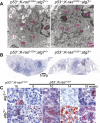
atg7 deficiency promotes tumor lipid accumulation. (A) Representative electron microscope images of tumors showing lipid droplet accumulation with atg7 deletion at 18 wk after tumor initiation. (Nu) Nucleus; (M) mitochondria; (L) lamellar body; (G) glycogen; (LD) lipid droplet. (B) Representative low-magnification images of Oil red O staining of representative lung lobes (frozen sections) at 18 wk after tumor induction (n = 2 mice for each genotype). (C) Representative high-magnification images of Oil red O staining of tumor tissue at the indicated times (n = 2 mice for each genotype).
Figure 6.
Figure 6.
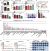
atg7-deficient TDCLs accumulate lipids and are sensitized to starvation-induced death. (A) Western blot for Atg7 and LC3 in TDCLs. (B) Western blot for LC3, p62, and active caspase-3 during starvation (HBSS) of TDCLs at the indicated times. (C) Clonogenic survival assay of TDCLs following 3 d of starvation (HBSS) and 4 d of recovery in normal medium (RPMI). (D) Growth of TDCL tumors in nude mice (n = 6 tumors for each TDCL). (E) Representative gross pathology of tumors derived from TDCLs from D. (F, left) Histology (H&E) of representative tumors from atg7 wild-type and atg7-deficient TDCL tumors showing hollow centers with atg7 deletion. (Right) Quantification of hollow tumor formation. (G) Oil red O staining of TDCL tumors (n = 4 tumors for each genotype). (H) Quantitation of cholesterol esters by thin-layer chromatography (TLC) (Supplemental Fig. S16A). (I) The graph shows the increased total pool size of FAs in atg7-deficient TDCLs compared with wild-type TDCLs under steady status. The error bar represents ±SD; P < 0.05 (t-test).
Figure 7.
Figure 7.
Autophagy is required for FAO. (A) Total MM (top) and MMP (bottom) in TDCLs under normal conditions (atg7+/+: n = 10; atg7−/−: n = 9 independent clones). (**) P < 0.01; (***) P < 0.001 (t-test). (B) OCR of TDCLs (two independent clones for each genotype) under normal conditions without (left) or with (right) FCCP challenge. The error bar represents ±SD; P > 0.05 (t-test). (C) OCR of TDCLs (two independent clones from each genotype) under normal or starvation (HBSS for 4 h) following addition of FCCP (0.3 μM) to establish maximum respiratory capacity. The error bar represents ±SD; (*) P < 0.05 (t-test). (D) Clonogenic survival assay of TDCLs with glutamine addition (1 mM) under 3 d of starvation (HBSS) and 3 d of recovery in normal medium (RPMI). (E, top) OCR trace of TDCLs in response to palmitate (400 μM) showing relative percentage change upon palmitate addition. (Bottom) OCR of TDCLs from the top panel at 71 min. The error bar represents ±SD; (*) P < 0.05 (t-test). (F) The graph shows autophagy-dependent changes in intracellular metabolite levels (52 metabolites; log2-transformed ratios of ion signals) in two atg7-deficient compared with two atg7 wild-type TDCLs in normal conditions. (G) The graphs show representative increased amino acid levels in two atg7-deficient compared with two atg7 wild-type TDCLs at each indicated starvation (HBSS) time. Additional amino acids are shown in Supplemental Figure S19. (H) Clonogenic survival assay for TDCLs following starvation (HBSS for 15 h and 25 h) in the presence of the CPT1 inhibitor etomoxir (50 μm) with and without pyruvate (1 mM). The complete assay is shown in Supplemental Figure S20.

References

    1. Amaravadi RK, Lippincott-Schwartz J, Yin XM, Weiss WA, Takebe N, Timmer W, DiPaola RS, Lotze MT, White E 2011. Principles and current strategies for targeting autophagy for cancer treatment. Clin Cancer Res 17: 654–666 - PMC - PubMed
    1. Cadwell K, Liu JY, Brown SL, Miyoshi H, Loh J, Lennerz JK, Kishi C, Kc W, Carrero JA, Hunt S, et al. 2008. A key role for autophagy and the autophagy gene Atg16l1 in mouse and human intestinal Paneth cells. Nature 456: 259–263 - PMC - PubMed
    1. Chesney J, Telang S 2013. Regulation of glycolytic and mitochondrial metabolism by Ras. Curr Pharm Biotechnol 14: 251–260 - "V体育ios版" PubMed
    1. Cheung EC, Vousden KH 2010. The role of p53 in glucose metabolism. Curr Opin Cell Biol 22: 186–191 - PubMed
    1. Degenhardt K, Mathew R, Beaudoin B, Bray K, Anderson D, Chen G, Mukherjee C, Shi Y, Gelinas C, Fan Y, et al. 2006. Autophagy promotes tumor cell survival and restricts necrosis, inflammation, and tumorigenesis. Cancer Cell 10: 51–64 - "VSports最新版本" PMC - PubMed

Publication types

MeSH terms (VSports在线直播)