<time lang="Wq3ffgG"></time><small id="25e9P"></small> VSports在线直播 - Skip to main page content
U.S. flag

An official website of the United States government

Dot gov

The . gov means it’s official. Federal government websites often end in . gov or . mil. Before sharing sensitive information, make sure you’re on a federal government site. VSports app下载.

Https

The site is secure. The https:// ensures that you are connecting to the official website and that any information you provide is encrypted and transmitted securely. V体育官网.

. 2013 Jul;65(7):1736-46.
doi: 10.1002/art.37981.

"V体育平台登录" Evidence that CXCL16 is a potent mediator of angiogenesis and is involved in endothelial progenitor cell chemotaxis : studies in mice with K/BxN serum-induced arthritis

Affiliations

Evidence that CXCL16 is a potent mediator of angiogenesis and is involved in endothelial progenitor cell chemotaxis : studies in mice with K/BxN serum-induced arthritis

Takeo Isozaki et al. Arthritis Rheum. 2013 Jul.

"VSports注册入口" Abstract

Objective: To examine the possibility that CXCL16 recruits endothelial cells (ECs) to developing neovasculature in rheumatoid arthritis (RA) synovium VSports手机版. .

Methods: We utilized the RA synovial tissue SCID mouse chimera system to examine human microvascular EC (HMVEC) and human endothelial progenitor cell (EPC) recruitment into engrafted human synovium that was injected intragraft with CXCL16-immunodepleted RA synovial fluid (SF). CXCR6-deficient and wild-type (WT) C57BL/6 mice were primed to develop K/BxN serum-induced arthritis and evaluated for angiogenesis V体育安卓版. HMVECs and EPCs from human cord blood were also examined for CXCR6 expression, by immunofluorescence and assessment of CXCL16 signaling activity. .

Results: CXCR6 was prominently expressed on human EPCs and HMVECs, and its expression on HMVECs could be up-regulated by interleukin-1β. SCID mice injected with CXCL16-depleted RA SF exhibited a significant reduction in EPC recruitment. In experiments using the K/BxN serum-induced inflammatory arthritis model, CXCR6(-/-) mice showed profound reductions in hemoglobin levels, which correlated with reductions in monocyte and T cell recruitment to arthritic joint tissue compared to that observed in WT mice. Additionally, HMVECs and EPCs responded to CXCL16 stimulation, but exhibited unique signal transduction pathways and homing properties. V体育ios版.

Conclusion: These results indicate that CXCL16 and its receptor CXCR6 may be a central ligand/receptor pair that is closely associated with EPC recruitment and blood vessel formation in the RA joint. VSports最新版本.


Conflict of interest statement

Conflicts of interest: none

Figures

Figure 1
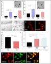
Figure 1. HMVECs express CXCR6 and are chemotactic for CXCL16
We show by Western blotting that HMVECs express CXCR6 and can be upregulated by IL-1β (figures 1A and B). C) HMVEC expression of CXCR6 is shown by red fluorescence staining. D) Control IgG staining for HMVECs is also shown. E) Intense HMVEC CXCR6 staining in response to rhuIL-1β stimulation is clearly seen, showing that CXCR6 is inducible in ECs (see arrow). F) Control IgG staining in response to rhIL-1β stimulation does not show EC staining for CXCR6. G) HMVEC vWF staining in response to rhIL-1β stimulation is apparent. H) Control IgG staining for IL-1β induced vWF (all images 40X). I) HMVECs migrate towards CXCL16 in a dose-dependent manner. HMVECs are chemotactic for CXCL16 at 1µM, indicating CXCL16 functions as an EC chemotactic factor.
Figure 2
Figure 2. CXCL16 forms tubes in Matrigel and in the human ST SCID mouse chimera
A, B and C) Capillary tube formation was determined blindly by evaluating circular tube networks and nodular contacts between at least 3 endothelial cell tubes. D) The left panel shows a representative NL ST section with scattered dye-tagged HMVECs migrating in response to PBS (see arrow in D). In the right panel, clearly organized migration of dye-tagged HMVECs is shown in response to intragraft administration of CXCL16 (40X). E) Fewer scattered HMVECs are observed in the RA ST SCID mouse chimera to CXCL16, likely due to HMVEC clumping. F) Intragraft injections of CXCL16 or TNF-α induced expression of CXCL16 mRNA in engrafted RA ST (n=number of mice). G) The far left panel shows a serial section of PKH26 dye-tagged HMVECs migrating into engrafted human ST. The middle panel shows a serial section taken from the same tissue stained with FITC labeled rabbit anti-human/mouse vWF (factor VIII). The right panel is a merger of panels. The green color shows mouse endothelium (arrow pointed at “M”), whereas the yellow color identifies human endothelium (arrow pointed at “H”) demonstrating the formation of a true chimera (40X).
Figure 3
Figure 3. EPCs express CXCR6 and migrate to CXCL16 in vivo
A) EPC expression of CXCR6 is shown by yellow/orange fluorescence staining (see beige arrow, 40X). B) EPCs express CD31 and can also be identified and sorted by CD34 expression, and found that >95% of these cells are AC133+ cells. The isolated cell population also expressed CXCR4, the receptor for SDF-1α/CXCL12, even without any external stimuli (e.g. cytokines, LPS, PMA, etc.). C and D) CXCR6 can be shown by Western blotting to be strongly expressed on EPCs, but not upregulated by IL-1β. E) SCID mice were engrafted with NL human synovium. Mice receiving intragraft injections of CXCL16 had approximately a 3 fold increase in EPC infiltration to engrafted tissue compared to mice receiving intragraft injections of PBS (n = no. of sections counted from 3 separate mice per group; 10X). F and G) NL synovium was engrafted into SCID mice and allowed several weeks to take. Mice injected with sham immunoneutralized RA SF showed robust recruitment of fluorescently dye-tagged EPCs, compared to mice injected with RA SF treated with anti-CXCL16 (see arrows; n = no. of sections evaluated from 3 different mice per group; 10X).
Figure 4
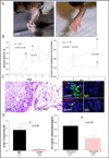
Figure 4. CXCR6 deficiency reduces joint inflammation and vascularity in K/BxN serum induced arthritis
A) Representative clinical images of inflamed joints revealed significant edema and inflammation in Wt, but not CXCR6−/− mice following arthritis induction with K/BxN serum. B) Mice lacking CXCR6 have significantly lower AI scores compared to Wt mice on days 5 and 7 of K/BxN serum induced arthritis [4 mice (16 joints) per group ± SEM]. Absolute increases in joint circumference after arthritis induction showed significantly less joint swelling in day 5 CXCR6−/− compared to Wt mice (p<0.05). C) In the left panel, joint histology of K/BxN serum induced day 9 Wt mice showed increased cellular infiltrate and severe tissue damage compared to CXCR6−/− mice (hematoxylin and eosin stain; 5µm, 40X). In the right panel, reduced vascular staining is also clearly seen in CXCR6−/− compared to Wt mice by immunofluorescence staining. D) The left graph shows the reduction in the number of blood vessels in CXCR6−/− joint tissue from the same mice (n corresponds to the number of tissue sections counted). Joint homogenates show significantly less Hb (normalized to total protein concentrations) in day 9 K/BxN serum induced CXCR6−/− compared to similarly treated Wt mice (right graph).
Figure 5
Figure 5. Signaling data shows HMVECs and EPCs are activated by CXCL16
Blots A, B, C and D show the signaling of CXCL16 in HMVECs. Blots E and F show signaling in EPCs. The upper band for each respective blot show the phosphorylated signaling molecule only. The lower band shows the total (T) amount of the signaling molecule containing phosphorylated (*p) and unphosphoylated forms of each molecule. Therefore, the upper blot is an indication of the amount of phosphorylated protein contained in the lower band for each signaling molecule. As shown, HMVEC *pP38, *pErk½ and *pJnk are activated by CXCL16, whereas *pSrc is downregulated. With respect to EPCs, *pP38 (E) and *pSrc (F) are activated in response to CXCL16. The table (G) lists all the signaling data for HMVECs and EPCs to CXCL16. Most interesting is the finding that both cell types signal through *pP38, indicating that CXCL16 angiogenic and/or vasculogenic activity can be targeted by *pP38 inhibition. All *p blots (upper panel of bands) were normalized to blots of total P38, Erk½, Jnk, or Src (lower panel of bands) for all respective blots; representative blots from n=3 separate blots are shown above graphs).
Figure 6
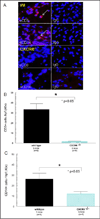
Figure 6. CXCR6−/− deficiency results in reduced leukocyte recruitment to inflamed joint tissue
Joints from day 9 CXCR6−/− and Wt mice induced with K/BxN serum were harvested and stained for CD3+ and CD14+ expression by immunofluorescence histology. A) Upper panel (Wt) shows prominent CD3+ and CD14+ expression in Wt K/BxN serum induced mice with the negative IgG control staining shown. In the lower panel (CXCR6−/−) of figure 6A, we found profound reductions in both CD3+ and CD14+ expressing cells in K/BxN serum induced mice lacking CXCR6 with the negative IgG control staining shown. The number of CD3+ (figure 6B) and CD14+ (figure 6C) expressing cells was significantly reduced in joints of arthritic mice deficient in CXCR6 (n = number of fields counted per mouse tissue section; taken from at least 3 different mice). Leukocyte recruitment was quantified by counting the number of fluorescing cells in a 40X field.

References

    1. Firestein GS. Evolving concepts of rheumatoid arthritis. Nature. 2003;423(6937):356–361. - PubMed
    1. Corrigall VM, Panayi GS. Autoantigens and immune pathways in rheumatoid arthritis. Crit Rev Immunol. 2002;22(4):281–293. - "VSports在线直播" PubMed
    1. Paleolog EM. Angiogenesis in rheumatoid arthritis. Arthritis Res. 2002;4(Suppl 3):S81–S90. - PMC (V体育ios版) - PubMed
    1. Folkman J, Haudenschild C. Angiogenesis by capillary endothelial cells in culture. Trans Ophthalmol Soc U K. 1980;100(3):346–353. - PubMed
    1. Masuda H, Asahara T. Post-natal endothelial progenitor cells for neovascularization in tissue regeneration. Cardiovasc Res. 2003;58(2):390–398. - PubMed

Publication types

MeSH terms