Skip to main page content
U.S. flag

An official website of the United States government

Dot gov

The . gov means it’s official. Federal government websites often end in . gov or . mil VSports app下载. Before sharing sensitive information, make sure you’re on a federal government site. .

Https

The site is secure. The https:// ensures that you are connecting to the official website and that any information you provide is encrypted and transmitted securely. V体育官网.

. 2013 May;15(5):533-43.
doi: 10.1038/ncb2730. Epub 2013 Apr 28.

Quantitative imaging of haematopoietic stem and progenitor cell localization and hypoxic status in the bone marrow microenvironment

Affiliations

Quantitative imaging of haematopoietic stem and progenitor cell localization and hypoxic status in the bone marrow microenvironment

César Nombela-Arrieta et al. Nat Cell Biol. 2013 May.

"VSports" Erratum in

  • Nat Cell Biol. 2013 Aug;15(8):1016

V体育2025版 - Abstract

The existence of a haematopoietic stem cell niche as a spatially confined regulatory entity relies on the notion that haematopoietic stem and progenitor cells (HSPCs) are strategically positioned in unique bone marrow microenvironments with defined anatomical and functional features. Here, we employ a powerful imaging cytometry platform to perform a comprehensive quantitative analysis of HSPC distribution in bone marrow cavities of femoral bones. We find that HSPCs preferentially localize in endosteal zones, where most closely interact with sinusoidal and non-sinusoidal bone marrow microvessels, which form a distinctive circulatory system VSports手机版. In situ tissue analysis reveals that HSPCs exhibit a hypoxic profile, defined by strong retention of pimonidazole and expression of HIF-1α, regardless of localization throughout the bone marrow, adjacency to vascular structures or cell-cycle status. These studies argue that the characteristic hypoxic state of HSPCs is not solely the result of a minimally oxygenated niche but may be partially regulated by cell-specific mechanisms. .


"VSports在线直播" Conflict of interest statement

The authors declare no competing financial interests.

V体育ios版 - Figures

Figure 1
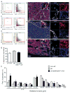
Figure 1. Quantitative image analysis of the spatial distribution of c-kit+ cells in whole longitudinal femoral sections
(a) Low magnification image of a longitudinal femoral BM cryosection immunostained for DAPI (blue) and c-kit (green) (PM=proximal metaphysis, DIA=diaphysis, DM=distal metaphysis). (b) Representative LSC scattergrams of isotype control and c-kit stained BM sections used to quantify percentages of c-kit+ cells in different regions of the BM in d and e. Intensity in the DAPI channel is shown in the X axis. BM cells contained in the c-kit+ gate in (b) can be automatically identified and visualized in high-resolution field images (c). Representative image of a BM stained for DAPI (blue) and c-kit (green) (top panel). Cells gated as positive based on fluorescence intensity in the c-kit channel, are automatically marked by the LSC analysis software (green rectangles, bottom panel). (d) Representative tissue maps and frequencies of c-kit+ hematopoietic progenitors in the metaphysis (MET) and diaphysis (DIA) of femoral BM (Data are represented as Mean±SEM calculated from n= 9 mice, a total of 17 sections were analyzed). (e) Frequencies of c-kit+ progenitors in longitudinal gates, spanning the width of the diaphysis of femoral sections (representative gates in left panel). ER=endosteal regions, CMR= central medullary region (*p< 0,05 Mean±SEM calculated from n= 7 mice, a total of 11 sections). (f) Image of the diaphysis of a BM section co-stained for c-kit and Laminin. See also Supplementary Video 1. (g) LSC analysis of perivascular cells in BM sections. Laminin fluorescent signal is thresholded and an integration contour in which the original vascular structure is expanded by 20 pixels (10 μm in the original 40x magnification) is defined to generate spatial gates containing only perivascular spaces (green outline right panel). (h) Representative LSC dot plots of total BM, perivascular and non-perivascular cell populations used to quantify cells in (i) Frequency of c-kit+ cells in perivascular and non-perivascular fractions (dashed line frequency of total BM) (**p< 0,01 Mean±SEM calculated from n=7 mice, a total of 10 sections analyzed).
Figure 2
Figure 2. The vast majority of HSPCs lie in direct contact with BM microvessels
Mapping of three subsets of committed progenitors and HSPCs in BM cavities via LSC. Representative dot plots of BM femoral sections stained for lineage markers and c-kit (a) Sca-1 and c-kit (c), or lineage, CD41, CD48 and c-kit (e). Isotype control stained sections were used to determine specific gates. Individual cells falling in the target gates were systematically visualized and confirmed. Representative images of examples of Linc-kit+, SK, and LinCD48CD41lo/−c-kit+ cells are shown in b, d and f, respectively. (g) Frequency of perivascular (<10μm from nearest blood vessel) Linc-kit+, SK and LinCD48CD41lo/−c-kit+ cells. (h) Histogram showing the distribution of the distances of Linc-kit+ (Mean±SEM calculated from n=3 mice, a total of 3 sections and 1915 individually validated cells) SK (n= 8 mice, 8 sections and 1589 individually validated cells) and LinCD48CD41lo/−c-kit+ HSPC (n=4 mice, 8 sections and 312 individually validated cells) populations to nearest bone surface, * p< 0,05.
Figure 3
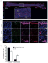
Figure 3. Three-dimensional study of BM microvasculature heterogeneity
(a) Representative 3D reconstruction of the femoral diaphysis stained with Laminin (green) and Sca-1 (red). Sca-1+ arteries run centrally along the diaphysis, emitting branches of smaller arterial vessels, which gradually narrow as they run laterally towards endosteal surfaces. Within endosteal regions LamininhiSca-1+ endosteal vessels give rise to Sca-1 sinusoids (in b and c), which return towards the central area of the diaphysis and coalesce in the draining central sinus (shown in c). a=arteries, s=sinusoid, cs=central sinus. Bone surfaces are shown in blue as revealed by second harmonic generation signals. (d) 3D overview of arterial and sinusoidal networks in the femoral metaphysis. (e) Transition from arterial to sinusoidal vessels (white arrow) in the endosteal surface of trabecular bone areas. See also Supplementary Videos 3, 4, 5, 6 and 7.
Figure 4
Figure 4. HSPCs directly interact with structurally and phenotypically diverse types of BM microvessels
(a) Low magnification immunofluorescent image of a whole femoral section stained for Laminin (red), DAPI (blue), c-kit (green) and Sca-1 (magenta). Lower panel shows the enlarged image of a section of the diaphysis in which central arteries (Sca-1+Laminin+), sinusoids (Laminin+/loSca-1−/lo) and endosteal vessels (Sca-1+Laminin+) can be visualized. (b) High-resolution images enlarged from boxed areas in (a) depicting examples of SK progenitors interacting with arteries, sinusoids and endosteal vessels. (c) Quantification of the percentages of perivascular SK progenitors and LinCD48CD41lo/−c-kit+ cells in contact with sinusoidal and non-sinusoidal endothelium (Mean±SEM calculated from n= 8 mice, a total of 8 sections and 1589 cells and n=4 mice, a total of 8 sections and 312 individually validated cells, respectively).
Figure 5
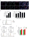
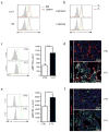
Figure 5. c-kit+ progenitors display a hypoxic status regardless of their perivascular localization and distribution in the BM cavity
(a) Representative flow cytometry histograms of Pimo incorporation (green) of total BM cells, B220+cells, LSK (Linc-kit+Sca-1) progenitors, ST-HSCs (LSKCD34+) and LT-HSCs (LSKCD34). Grey histograms depict background levels of Pimo staining in PBS-injected control mice. (b) Quantification of the shift in the mean fluorescence intensity of the different BM populations in the Pimo channel (ΔMFI, in fluorescence arbitrary units) from mice injected with Pimo compared to non-injected Pimo controls (n= 7 mice ** p< 0,01). (c) Images of femoral sections of control PBS-injected mice and Pimo-injected mice stained with anti-Pimo. (d) Representative images showing multiple examples of perivascular c-kit+ cells brightly stained for Pimo (white arrowheads) in both endosteal and central marrow regions. Pimohic-kit+ cells are found in many cases adjacent to B220+Pimo cells (orange arrowheads). (e) LSC analysis of sections stained for Pimo. Representative histograms used to quantify the mean fluorescence intensity (MFI) in the Pimo-specific channel of total BM cells, B220+, and ckit+ cells in total BM sections. (f) MFI of the Pimo fluorescent signal of total BM cells, B220+ cells and c-kit+ cells localized in the diaphysis (DIA) and the proximal and distal metaphysis (PM and DM respectively) (Mean±SEM calculated from n= 5 mice, a total of 9 sections * p< 0,05 ** p< 0,01). (g) Quantification of Pimo signal (MFI) of c-kit+ cells present in consecutive longitudinal regions of the diaphysis as shown in Fig. 1e. ER= endosteal regions, CMR= central medullary region (Mean±SEM calculated from n= 6 mice, a total of 9 sections). (h) Representative histograms of the fluorescence intensity (left) and quantification of the MFI (right) in the Pimo channel of non-perivascular and perivascular c-kit+ cells (Mean±SEM calculated from n= 7 mice, a total of 14 sections).
Figure 6
Figure 6. The hypoxic status of HSPCs is independent of their localization in BM regions and their cycling status
(a) Representative images of perivascular SK cells that strongly stained for Pimo. (b) LSC-quantified Pimo specific signal for total BM cells, c-kit+ progenitors and SK cells (Mean±SEM calculated from n=3 mice, a total of 3 sections* p< 0,05) (c) Quantification of Pimo fluorescent signal of SK cells located at various distances from nearest bone surface (Mean±SEM calculated from n=3 mice, a total of 3 sections 742 cells individually validated SK cells, *p< 0,05). (d) Pimo incorporation of LSK HSPCs present in BM, circulating blood and spleen after mobilization with AMD3100. (e and f) Flow cytometric analysis of Pimo incorporation of LSKCD34+ and LSKCD34 at different stages of the cell cycle. (e) Bottom panels show representative histograms of Pimo specific fluorescence for fractions of each population at G0 and G1. (f) Quantification of Pimo intensity for LSKCD34+ and LSKCD34 HSPCs in G0 and G1 (n=6 mice). For source data for graphs presented in panels 6b and 6c, see SI table 3.
Figure 7
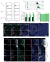
Figure 7. Stable HSPC expression of HIF-1α is not dictated by localization in the BM
(a) Flow cytometric analysis of intracellular HIF-1α expression in subsets of hematopoietic progenitors and HSPCs. (b) Representative LSC-obtained histograms depicting HIF-1α expression of total BM cells and c-kit+ progenitors in immunostained BM femoral sections. (c) MFI of the Pimo fluorescent signal of total BM cells, and c-kit+ cells localized in the diaphysis (DIA) and the proximal and distal metaphysis (PM and DM respectively) (Mean±SEM calculated from n= 5 mice, a total of 10 sections *** p< 0,005) (d) Quantification of HIF-1α expression (MFI) of c-kit+ cells present along consecutive longitudinal regions of the diaphysis (Mean±SEM calculated from n= 5 mice, a total of 10 sections). (e) Representative images depicting multiple examples of HIF-1α+c-kit+ cells (white arrowheads) in direct contact with isolectin B4+ vessels in the context of a large area of a BM femoral section. (f) Images of perivascular HIF-1α-expressing, Sca-1+c-kit+ cells in central and endosteal areas of the BM (marked by asterisks). Distances to the closest endosteal surface are indicated.
Figure 8
Figure 8. HIF-1α expression on cycling and actively proliferating HSPCs
(a) Expression of HIF-1α in HSPCs and hematopoietic progenitors from BM and spleen. (b) Flow cytometric analysis of HIF-1α expression in HSPCs in different stages of cell cycle. (c and e) Representative histograms and quantification of Pimo incorporation (c) and HIF-1α expression (e) of LSK cells in control and 5-FU-treated mice (7–9 days post-treatment). (c) n= 8 mice *** p<0,005, and (e) n= 5 mice * p<0,05. (d and f) Representative images of BM sections from control and 5FU-treated mice stained for Pimo, HIF-1α and vascular markers. Pimo+ and HIF-1α+ cells localize in close contact with disrupted vascular structures during the recovery phase post 5-FU treatment.

References

    1. Orkin SH, Zon LI. Hematopoiesis: an evolving paradigm for stem cell biology. Cell. 2008;132:631–644. - PMC - PubMed
    1. Wang LD, Wagers AJ. Dynamic niches in the origination and differentiation of haematopoietic stem cells. Nat Rev Mol Cell Biol. 12:643–655. - PMC - PubMed
    1. Adams GB, Scadden DT. The hematopoietic stem cell in its place. Nat Immunol. 2006;7:333–337. - PubMed
    1. Wilson A, et al. c-Myc controls the balance between hematopoietic stem cell self-renewal and differentiation. Genes Dev. 2004;18:2747–2763. - PMC - PubMed
    1. Kiel MJ, Morrison SJ. Uncertainty in the niches that maintain haematopoietic stem cells. Nat Rev Immunol. 2008;8:290–301. - PubMed

Publication types

MeSH terms