Skip to main page content
U.S. flag

An official website of the United States government

Dot gov

The . gov means it’s official. Federal government websites often end in . gov or . mil. Before sharing sensitive information, make sure you’re on a federal government site. VSports app下载.

Https

The site is secure. The https:// ensures that you are connecting to the official website and that any information you provide is encrypted and transmitted securely. V体育官网.

. 2013 Mar 19;108(5):1079-91.
doi: 10.1038/bjc.2012.484. Epub 2013 Feb 28.

"V体育ios版" Marked improvement of cytotoxic effects induced by docetaxel on highly metastatic and androgen-independent prostate cancer cells by downregulating macrophage inhibitory cytokine-1

Affiliations

V体育ios版 - Marked improvement of cytotoxic effects induced by docetaxel on highly metastatic and androgen-independent prostate cancer cells by downregulating macrophage inhibitory cytokine-1

M Mimeault et al. Br J Cancer. .

Abstract

Background: Overexpression of macrophage inhibitory cytokine-1 (MIC-1) frequently occurs during the progression of prostate cancer (PC) to androgen-independent (AI) and metastatic disease states and is associated with a poor outcome of patients VSports手机版. .

Methods: The gain- and loss-of-function analyses of MIC-1 were performed to establish its implications for aggressive and chemoresistant phenotypes of metastatic and AI PC cells and the benefit of its downregulation for reversing docetaxel resistance. V体育安卓版.

Results: The results have indicated that an enhanced level of secreted MIC-1 protein in PC3 cells is associated with their acquisition of epithelial-mesenchymal transition features and higher invasive capacity and docetaxel resistance. Importantly, the downregulation of MIC-1 in LNCaP-LN3 and PC3M-LN4 cells significantly decreased their invasive capacity and promoted the antiproliferative, anti-invasive and mitochrondrial- and caspase-dependent apoptotic effects induced by docetaxel V体育ios版. The downregulation of MIC-1 in PC3M-LN4 cells was also effective in promoting the cytotoxic effects induced by docetaxel on the side population (SP) endowed with stem cell-like properties and the non-SP cell fraction from PC3M-LN4 cells. .

Conclusion: These data suggest that the downregulation of MIC-1 may constitute a potential therapeutic strategy for improving the efficacy of current docetaxel-based chemotherapies, eradicating the total mass of PC cells and thereby preventing disease relapse and the death of PC patients. VSports最新版本.


"VSports app下载" Figures
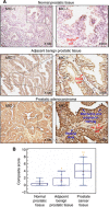
Figure 1
Figure 1
Immunohistochemical analyses of expression levels of the MIC-1 protein in nonmalignant and malignant prostatic tissues. (A) Representative pictures of immunohistochemical analyses of the expression level of MIC-1 in normal prostatic tissues from biopsies and adjacent benign and prostatic adenocarcinoma tissues from same PC patients are shown at original magnifications of × 100 and × 400. (B) Comparison of the composite scores of expression levels of MIC-1 in nonmalignant and malignant tissues from PC patients. Box plots showing the expression levels of MIC-1 in normal prostatic tissues from biopsies and prostatic adenocarcinoma specimens and their adjacent benign prostatic tissues. *P<0.005 and 0.0001 indicates a significant increase between the means of composite scores obtained for adjacent benign prostatic tissues and prostatic adenocarcinoma tissues relative to mean of composite scores obtained for normal prostatic tissue specimens. **P<0.0001 indicates a significant increase between the mean of composite scores obtained for prostatic adenocarcinoma tissues compared with mean of composite scores obtained for adjacent benign prostatic tissue specimens.
Figure 2
Figure 2
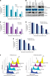
Effect of the MIC-1 protein on the antiproliferative effect induced by docetaxel on metastatic and AI PC cell lines. The PC cells were untreated (control) or treated with indicated docetaxel (Doc) concentrations for 2 days and the cell proliferation was evaluated by MTT assays and FACS analyses. (A) Data obtained by MTT assay for parental PC3 cells expressing a low level of MIC-1 untreated or treated with exogenous rhMIC-1 protein. (B) Immunoblot analyses of the expression levels of MIC-1 protein in culture supernatant and lysates from tested PC cell lines. Data obtained by MTT assays for (C) PC3-vect expressing low level of endogenous MIC-1 vs PC3-MIC-1 engineered for overexpressing MIC-1 and (D) LNCaP-LN3-Con and (E) PC3M-LN4-Con cells overexpressing MIC-1 vs MIC-1-silenced LNCaP-LN3-siMIC-1 and PC3M-LN4- siMIC-1 cells, respectively. (F) FACS analyses of number of PC cells in the cell cycle phase for PC3M-LN4-Con vs PC3M-LN4-siMIC-1 cells untreated or treated with indicated docetaxel concentrations for 2 days.
Figure 3
Figure 3
Effect of the MIC-1 protein on the invasive ability of PC cells and the anti-invasive effect induced by docetaxel treatment on PC cell lines expressing low and high levels of the MIC-1 protein. The PC cells were untreated (control) or treated with the indicated agents, plated on matrigel-coated membrane for invasion assays and incubated for 24 h. Data of the invasive ability obtained for (A) parental PC3 cells after a treatment with 1 ng ml−1 rhMIC-1 protein in the absence or presence of 10 μℳ SB431542 or 10 nℳ docetaxel. The results from comparative analyses by (B) confocal microscopy and (C) western blot of the effect induced by a treatment of starved PC3 cells with 1 ng ml−1 rhMIC-1 in the absence or presence of 10 μℳ SB431542 on expression levels and intracellular localisation of different gene products associated with the EMT process. Data of the invasive ability obtained for (D) PC3-Vect cells expressing a low level of MIC-1 vs PC3-MIC-1 cells engineered for overexpressing MIC-1 protein, and (E) scrambled LNCaP-LN3-Con and (F) PC3M-LN4-Con cells overexpressing high levels of endogenous MIC-1 vs MIC-1-silenced LNCaP-LN3-siMIC-1 and PC3M-LN4-siMIC-1 cells, respectively.
Figure 4
Figure 4
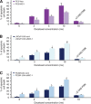
Fluorescence-activated cell sorting (FACS) analyses of the apoptotic effect induced by docetaxel on PC cell lines expressing low and high levels of the MIC-1 protein. Plots showing the percentages of apoptotic cell death induced after 4 days of treatment of PC cells with indicated docetaxel concentrations in the absence or presence of broad caspase inhibitor, Z-VAD-FMK, at 50 mM, which was estimated by the number of apoptotic cells detected in the sub-G1 phase by FACS analyses. Quantitative data obtained for (A) PC3-Vect cells expressing a low level of MIC-1 vs PC3-MIC-1 cells engineered for overexpressing MIC-1 protein, and (B and C) scrambled LNCaP-LN3-Con and PC3M-LN4-Con cells overexpressing high levels of endogenous MIC-1 vs MIC-1-silenced LNCaP-LN3-siMIC-1 and PC3M-LN4-siMIC-1 cells, respectively.
Figure 5
Figure 5
Stimulatory effect induced by docetaxel on mitochondrial membrane depolarisation, cytosolic cytochrome c releasing, caspase pathway activation and DNA fragmentation in PC3M-LN4-Con and MIC-1-silenced PC3M-LN4-siMIC-1 cells. (A) Representative profiles of effect induced by docetaxel on MMP in PC cells are shown. (B) Plots showing the percentage of depolarised PC cells induced after treatment with different docetaxel concentrations. (C) Western blot analyses of the amounts of cytochrome c released into cytosol, cleaved fragments of caspase-9 and caspase-3 and PARP, and (D) DNA fragmentation detected by agarose gel electrophoresis in PC cells untreated (control) or treated with 10 nℳ docetaxel for 4 days.
Figure 6
Figure 6
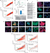
Characterisation of phenotypic and functional features of SP and non-SP PC3M-LN4 cell fractions and the implication of MIC-1 in their sensibility to the cytotoxic effect induced by docetaxel. (A) Hoechst dye efflux profiles obtained for parental PC3M-LN4 cells stained with fluorescent Hoechst dye in the absence or presence of 50 μℳ verapamil showing SP cells (green) and the non-SP fraction (blue) and (B) FACS profiles obtained after staining of PC3M-LN4 cells with phycoerythrin-labelled anti-CD133 antibody. (C) Clone formation efficacy of SP and non-SP PC3M-LN4 fractions and (D) representative pictures of dense prostaspheres formed by SP PC3M-LN4 cells as compared with small aggregates formed by non-SP PC3M-LN4 cells. (E–G) Comparative western blot and immunoconfocal analyses of expression levels of different markers in SP and non-SP PC3M-LN4 cell fractions. (H) Hoechst dye efflux profiles obtained for PC3M-LN4-Con cells overexpressing endogenous MIC-1 and MIC-1-silenced PC3M-LN4-siMIC-1 cells untreated or treated with 10 nℳ docetaxel for 4 days.

References

    1. Ago T, Kuroda J, Pain J, Fu C, Li H, Sadoshima J. Upregulation of Nox4 by hypertrophic stimuli promotes apoptosis and mitochondrial dysfunction in cardiac myocytes. Circ Res. 2010;106:1253–1264. - PMC - PubMed
    1. Bauskin AR, Brown DA, Junankar S, Rasiah KK, Eggleton S, Hunter M, Liu T, Smith D, Kuffner T, Pankhurst GJ, Johnen H, Russell PJ, Barret W, Stricker PD, Grygiel JJ, Kench JG, Henshall SM, Sutherland RL, Breit SN. The propeptide mediates formation of stromal stores of PROMIC-1: role in determining prostate cancer outcome. Cancer Res. 2005;65:2330–2336. - PubMed
    1. Birnie R, Bryce SD, Roome C, Dussupt V, Droop A, Lang SH, Berry PA, Hyde CF, Lewis JL, Stower MJ, Maitland NJ, Collins AT. Gene expression profiling of human prostate cancer stem cells reveals a pro-inflammatory phenotype and the importance of extracellular matrix interactions. Genome Biol. 2008;9:R83. - PMC - PubMed
    1. Boyle GM, Pedley J, Martyn AC, Banducci KJ, Strutton GM, Brown DA, Breit SN, Parsons PG. Macrophage inhibitory cytokine-1 is overexpressed in malignant melanoma and is associated with tumorigenicity. J Invest Dermatol. 2009;129:383–391. - VSports在线直播 - PubMed
    1. Brown DA, Lindmark F, Stattin P, Balter K, Adami HO, Zheng SL, Xu J, Isaacs WB, Gronberg H, Breit SN, Wiklund FE. Macrophage inhibitory cytokine 1: a new prognostic marker in prostate cancer. Clin Cancer Res. 2009;15:6658–6664. - PMC (VSports注册入口) - PubMed

Publication types

MeSH terms