Skip to main page content
U.S. flag

An official website of the United States government

Dot gov

The . gov means it’s official VSports app下载. Federal government websites often end in . gov or . mil. Before sharing sensitive information, make sure you’re on a federal government site. .

Https

The site is secure. The https:// ensures that you are connecting to the official website and that any information you provide is encrypted and transmitted securely. V体育官网.

. 2013 Mar 15;190(6):2575-84.
doi: 10.4049/jimmunol.1201920. Epub 2013 Feb 13.

Cancer-produced metabolites of 5-lipoxygenase induce tumor-evoked regulatory B cells via peroxisome proliferator-activated receptor α

Affiliations

Cancer-produced metabolites of 5-lipoxygenase induce tumor-evoked regulatory B cells via peroxisome proliferator-activated receptor α

Katarzyna Wejksza et al. J Immunol. .

Abstract (V体育安卓版)

Breast cancer cells facilitate distant metastasis through the induction of immunosuppressive regulatory B cells, designated tBregs VSports手机版. We report in this study that, to do this, breast cancer cells produce metabolites of the 5-lipoxygenase pathway such as leukotriene B4 to activate the peroxisome proliferator-activated receptor α (PPARα) in B cells. Inactivation of leukotriene B4 signaling or genetic deficiency of PPARα in B cells blocks the generation of tBregs and thereby abrogates lung metastasis in mice with established breast cancer. Thus, in addition to eliciting fatty acid oxidation and metabolic signals, PPARα initiates programs required for differentiation of tBregs. We propose that PPARα in B cells and/or tumor 5-lipoxygenase pathways represents new targets for pharmacological control of tBreg-mediated cancer escape. .


Figures
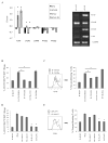
Figure 1
Figure 1
Compared with their parental metastatic 4T1 and 4T1.2 cells, non-metastatic cancer 4T1-PE and 4T1.2-PE cells preferentially expressed genes of the 5-LO/leukotriene pathway, such as 5-LO, FLAP, LTA4 hydrolase (LTA4H) and LTC4 synthase (LTC4S), as shown by mRNA microarray analysis (right panel, A) and RT-PCR (left panel, A) of 3–4 independent samples. Expression of 5-LO/FLAP in 4T1-PE cells was required for tBreg generation, as B cells incubated with 50% CM from 4T1-PE cells treated with zileuton (Zil, 50 μM) or CAY10416 (CAY, 5 μM) did not up regulate tBreg-associated surface markers (B, CD81High CD25+) and suppress proliferation of anti-CD3 antibody -stimulated CD4+ T cells (C). In contrast, CM from ibuprofen (Ibu, 10 μM) or mock (DMSO) -treated cells readily generated tBregs (B, C). Similarly, 4T1-PE cells with shRNA-induced FLAP knocked down (sh FLAP) also did generate tBregs from B cells, as shown by the inability to up regulate CD81High CD25+ (D) and suppress T cell responses (E). Shown in D and E are the results of two independent 4T1-PE cell clones transduced with sh-FLAP (sh FLAP 1 and 2) and control shRNA (sh control 1 and 2). In C and E, B cells and CFSE-labeled T cells (responder) cells were cultured at a 1:1 ratio for four days in the presence of anti-CD3 Ab and 50 U/ml IL-2. Y-axis (% ± SEM of at least three independent triplicate experiments) is for CD81High CD25+ cells (within CD19+ cells, B and D) and non-proliferating CD4+ T cells (CFSE retained, C and E). A representative histogram of CFSE dilution in CD4+ T cells is depicted in C and E (left panel). From here on, *P<0.05; and tBregs designate B cells treated with CM of 4T1-PE cells.
Figure 2
Figure 2
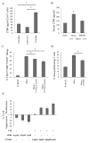
(A) LTB4 expression in 4T1-PE cells is reduced by inactivating 5-LO/FLAP with MK886 (MK, 10 μM) and CAY10416 (CAY, 5 μM ), but not ibuprofen (Ibu, 10 μM). Shown are ELISA results in CM of 2e6 cells cultured for 72 hours. (B) Serum levels of LTB4 is enhanced in 4T1 tumor-bearing mice, which was reduced back to the levels of naïve mice by treatment with MK886 (20 μg/mouse). Control tumor-bearing mice were treated with DMSO (mock). Y-axis levels of LTB4 pg/ml ± SEM in sera of three per group mice measured using ELISA. (C–D) 4T1-PE cells induced the generation of tBregs, at least in part, utilizing LTB4, as B cells treated with CM-PE together with inhibitors of BLT1 and BLT2 (U75302 and LY2552833, respectively) had reduced capability to up regulate CD81High CD25+ (C) and suppress proliferation of T cells (D, E). Conversely, B cells treated with free LTB4 in cRPMI slightly (but significantly) were rendered suppressive (E). In E, B cells were treated with indicated amounts of LTB4 in cRPMI or U75302 in CM of 4T1-PE cells (CM) and tested for suppression or proliferation of T cells, respectively. Y-axis shows % T cell suppression or proliferation ± SEM of T cells stimulated with indicated B cells compared with control B cells (100% tBregs or naïve B cells, respectively). Shown are results of triplicate experiments reproduced twice (A, B, E) and three times (C and D).
Figure 3
Figure 3
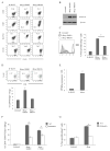
B cells treated with CM-PE in the presence of MK886 (10 μM) cannot generate tBregs, as shown by decreased expression of CD81, CD25, BAFF-R, and B7-H1 (A) and down regulation of pStat3 (B, western blot hybridization of a total cell lysate); and by the inability to suppress T cell proliferation (C) and induce conversion of FoxP3+ Tregs from CD25-CD4+ T cells (D). A representative histogram and dot plot of CFSE diluted (C, left panel) and FoxP3 –expressing (D, upper panel) CD4+ T cells are shown. (E–G) The generation of tBregs requires PPARα, as it is expressed in tBregs (E, qRT-PCR data); and B cells from PPARα KO mice cannot efficiently generate tBregs, i.e. up regulate CD81 and CD25 (F) and suppress proliferation of T cells (G). Control B cells were treated with BAFF. Importantly, BLT1 inhibitor U75302 only inhibited tBreg generation from WT, but not PPARα KO, B cells (F). Data shown in F are repeated twice, while the rest ones are reproduced at least three times in triplicate experiments.
Figure 4
Figure 4
A poor progression of B16 melanoma in μMT mice (23) is reversed by adoptive transfer of WT, but not PPARα deficient, tBregs. μMT mice with s.c. challenged B16F10 melanoma cells (105 cells) were i.v. injected with tBregs (5×10⁶) from PPARα KO or WT C57BL/6 mice one day before and 5 days after tumor challenge. Y-axis shows mean ± SEM of 4–5 mice per group experiment, such as tumor size (mm2, A) and weight (g, B), numbers of FoxP3+CD4+ T cells (C) and IFNγ+CD8+ T cells (D) per 106 splenocytes or IFNγ+CD8+ T cells per mm2 blood (E). IFNγ (intracellular staining) was assessed in CD8+ T cells after one week ex vivo expansion with melanoma-specific gp10025–32 peptide. The results were reproduced at least twice.
Figure 5
Figure 5
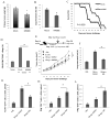
4T1 cancer progression and metastasis are reduced by in vivo inactivation of 5-LO/PPARα using MK886. BALB/C mice were i.p. treated with MK886 (20 μg/mouse) or DMSO every other day for 7 times starting from day 3 post s.c. challenge with 5×10⁴ 4T1 cells in mammary gland. Mice were sacrificed at day 28 to assess the weight of the primary tumor in the mammary gland (A) and count metastatic foci in the lungs (B), as we previously described (22). The survival benefit of MK886 treatment was tested in 8 mice per group experiment (C). Control mice were treated with DMSO (mock). The MK886 treatment reduces the proportion of tBregs in tumor-bearing mice to the levels of naïve mice (D). Antitumor activity of MK886 involves inactivation of tBregs (E–L). Tumor bearing BALB/C mice were treated with MK886 as in (A) to reduce tBregs, but until day 12. At day 14 post tumor challenge (when free MK886 is presumably cleared) the mice were randomized and either injected with PBS, or adoptively transferred with naive B cells, or tBregs ex vivo pretreated with DMSO or MK886 (E). Progression of primary tumor in mammary gland (E, mm2) was measured every other day. Metastatic foci in the lungs (F) and numbers of FoxP3+ (G) and IFNγ+ (H) CD4+ T cells and INFγ+CD8+ T cells (L) per 106 splenocytes were counted at day 28. Shown are mean ± SEM of a representative 4–5 mice per group experiment reproduced at least three times.

"V体育官网" References

    1. Greene ER, Huang S, Serhan CN, Panigrahy D. Regulation of inflammation in cancer by eicosanoids. Prostaglandins Other Lipid Mediat. 2011;96:27–36. - PMC (VSports手机版) - PubMed
    1. Sano H, Kawahito Y, Wilder RL, Hashiramoto A, Mukai S, Asai K, Kimura S, Kato H, Kondo M, Hla T. Expression of cyclooxygenase-1 and -2 in human colorectal cancer. Cancer research. 1995;55:3785–3789. - VSports注册入口 - PubMed
    1. Dannenberg AJ, Subbaramaiah K. Targeting cyclooxygenase-2 in human neoplasia: rationale and promise. Cancer Cell. 2003;4:431–436. - "VSports app下载" PubMed
    1. Ding XZ, Iversen P, Cluck MW, Knezetic JA, Adrian TE. Lipoxygenase inhibitors abolish proliferation of human pancreatic cancer cells. Biochem Biophys Res Commun. 1999;261:218–223. - PubMed
    1. Ghosh J, Myers CE. Inhibition of arachidonate 5-lipoxygenase triggers massive apoptosis in human prostate cancer cells. Proceedings of the National Academy of Sciences of the United States of America. 1998;95:13182–13187. - PMC - PubMed

Publication types

"V体育2025版" MeSH terms

Associated data

V体育官网入口 - Grants and funding