Skip to main page content
U.S. flag

An official website of the United States government

Dot gov

The . gov means it’s official VSports app下载. Federal government websites often end in . gov or . mil. Before sharing sensitive information, make sure you’re on a federal government site. .

Https

The site is secure. The https:// ensures that you are connecting to the official website and that any information you provide is encrypted and transmitted securely. V体育官网.

. 2012;7(10):e47649.
doi: 10.1371/journal.pone.0047649. Epub 2012 Oct 19.

VSports注册入口 - WNT10A plays an oncogenic role in renal cell carcinoma by activating WNT/β-catenin pathway

Affiliations

WNT10A plays an oncogenic role in renal cell carcinoma by activating WNT/β-catenin pathway

Ren-Jun Hsu et al. PLoS One. 2012.

Abstract (VSports在线直播)

Renal cell carcinoma (RCC) is a malignancy with poor prognosis VSports手机版. WNT/β-catenin signaling dysregulation, especially β-catenin overactivation and WNT antagonist silencing, is associated with RCC carcinogenesis and progression. However, the role of WNT ligands in RCC has not yet been determined. We screened 19 WNT ligands from normal kidney and RCC cell lines and tissues and found that WNT10A was significantly increased in RCC cell lines and tissues as compared to that in normal controls. The clinical significance of increase in WNT10A was evaluated by performing an immunohistochemical association study in a 19-year follow-up cohort comprising 284 RCC and 267 benign renal disease (BRD) patients. The results of this study showed that WNT10A was dramatically upregulated in RCC tissues as compared to that in BRD tissues. This result suggests that WNT10A, nuclear β-catenin, and nuclear cyclin D1 act as independent risk factors for RCC carcinogenesis and progression, with accumulative risk effects. Molecular validation of cell line models with gain- or loss-of-function designs showed that forced WNT10A expression induced RCC cell proliferation and aggressiveness, including higher chemoresistance, cell migration, invasiveness, and cell transformation, due to the activation of β-catenin-dependent signaling. Conversely, WNT10A siRNA knockdown decreased cell proliferation and aggressiveness of RCC cells. In conclusion, we showed that WNT10A acts as an autocrine oncogene both in RCC carcinogenesis and progression by activating WNT/β-catenin signaling. .


Conflict of interest statement

Competing Interests: The authors have declared that no competing interests exist.

"V体育平台登录" Figures

Figure 1
Figure 1. Expression of 19 WNT genes in kidney cell lines and tissues.
(A) mRNA expression profiles of WNT genes from kidney cell lines obtained using RT-PCR. mRNA expression profiles of 19 WNT genes from 5 RCC cell lines (786-O, Caki-1, RCC-1, A498, and ACHN) and 1 immortalized proximal tubule epithelial cell line from a normal adult human kidney (HK-2) were obtained using RT-PCR. Higher WNT10A expression was observed in RCC cell lines Caki-1, RCC-1, and ACHN, but lower expression was observed in RCC cell lines 786-O and A498. WNT10A expression was undetectable in the normal kidney cell line HK-2. (B) mRNA expression profile of WNT genes from BRD and RCC specimens obtained using RT-PCR. mRNA expression profiles of 19 WNT genes from 6 paired RCC (T1–T6) and paratumoral (N1–N6) tissues. mRNA expression profiles of WNT genes from other 4 BRD tissues (N7–N10) were also examined. Higher expression of WNT10A was observed in most RCC tissues than in paratumoral and BRD tissues. (C) Quantitative real-time PCR for detecting the expression of WNT genes. Expression of WNT genes in each cell line was determined using SYBR Green real-time PCR. An independent data set comprising 10 RCC and 10 normal kidney tissues was used. The results obtained were similar to those obtained for RT-PCR, i.e., higher expression of WNT10A was observed in most RCC cell lines and tissues than in normal controls. All experiments were performed in triplicate. Relative expression of each gene from each cell line was normalized with that of the gene from HK-2 normal kidney cell line; relative expression of each gene of tissues was normalized with the mean of 10 normal kidney tissues.
Figure 2
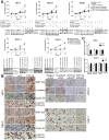
Figure 2. Immunohistochemical staining of WNT10A, β-catenin, and cyclin D1 in kidney tissues.
(A) WNT10A showed very low cytoplasmic expression in renal tubular cells of non-tumoral tissues from both the cortex and medulla. Moreover, WNT10A showed a dramatically higher cytoplasmic expression in RCC tissues. To confirm WNT10A immunohistochemical profiles, WNT10A antibody (sc69135) was used for a serial tissue microarray section, and antibody ablation was performed using its competitive peptide (sc69135P). WNT10A antibody (sc69135) showed similar profiles, and antibody ablation with WNT10A peptide abolished the staining pattern (sc69135P). (B) WNT10A showed higher cytoplasmic expression in RCC cells (A: cortex, B: CCRCC; 400×). β-catenin showed higher intracellular accumulation (cytoplasmic and nuclear; red arrows indicate some representative nuclear-stained cells) in RCC tissues (C: cortex, D: CCRCC; 400×). However, β-catenin showed higher membranous expression in renal tubular cells from non-tumoral normal kidney tissue. Moreover, c-myc (E: cortex, F: CCRCC; 400×) and cyclin D1 (G: cortex, H: CCRCC; 400×) showed higher nuclear expression in RCC tissues, but cyclin D1 showed weak cytoplasmic expression in renal tubular cells from non-tumoral normal kidney tissue. (C) Kaplan-Meier analysis of WNT10A, nuclear β-catenin, and nuclear cyclin D1 accumulative effects on overall survival (OS) and disease-free survival (DFS). There exists an accumulated dose effect, in that patients with higher WNT10A, nuclear β-catenin, and nuclear cyclin D1 levels have poor RCC prognosis (n  =  initial patient number, s  =  number of survivors at the end of the study). The 4 groups were defined as follows: 0, carriers with lower expression of all the 3 markers; 1, carriers with higher expression of any 1 of the 3 markers; 2, carriers with higher expression of any 2 of the 3 markers; 3, carriers with higher expression of all the 3 markers. P of log-rank test <0.001.
Figure 3
Figure 3. Forced WNT10A expression and WNT10A siRNA knockdown in kidney cell lines.
(A) Cell growth curve of pcDNA-WNT10A-transfected cells and WNT10A siRNA knockdown cells. WNT10A gain-of-function was achieved by transfecting pcDNA-WNT10A in normal kidney cell line HK-2 and RCC cell lines 786-O and A498, which had relatively lower endogenous WNT10A expression. Cotransfection of pcDNA-WNT10A and β-catenin siRNA was also performed. Conversely, WNT10A loss-of-function was achieved by WNT10A siRNA knockdown in RCC-1 and Caki-1, which have higher endogenous WNT10A. After transient transfection for 48 h, cell viability was determined using the MTT assay and was compared with that of cells transfected with Lipofectamine only (reagent control), pcDNA3.1 vector only (vector), and pcDNA-WNT10A and with that of cells cotransfected with pcDNA-WNT10A and β-catenin siRNA at 12, 24, 48, and 72 h. HK-2, 786-O, and A498 transfected with pcDNA-WNT10A showed significant increase in cell proliferation after 48 h; however, cotransfection of pcDNA-WNT10A and β-catenin siRNA reduced WNT10A-induced cell proliferation. Conversely, RCC-1 and Caki-1 transfected with WNT10A siRNA showed significant decrease in cell proliferation as compared to cells transfected with reagent and scrambled siRNA controls. (**P<0.001, *P<0.05; Student’s t-test). Western blot analysis of pcDNA-WNT10A- and WNT10A siRNA-transfected cells from each cell line was also performed. HK-2, 786-O, and A498 were transfected with 3 µg of pcDNA-WNT10A, pcDNA3.1 vector alone, and cotransfected with β-catenin siRNA for 48 h. Conversely, RCC-1 and Caki-1 were transfected with 1 µg WNT10A siRNA or scrambled siRNA controls for 72 h. Twenty micrograms of total protein extract from each cell line was loaded onto SDS-polyacrylamide gel and western blot analysis was performed. Forced WNT10A expression in HK-2, 786-O, and A498 remarkably increased the concentration of WNT10A than that of the vector control in these cells. Forced WNT10A expression also upregulated nuclear β-catenin, cyclin D1, and c-myc levels, and cotransfection with β-catenin siRNA reduced cyclin D1 and c-myc expression levels. Conversely, WNT10A siRNA knockdown in RCC-1 and Caki-1 markedly reduced endogenous WNT10A levels, thus reducing nuclear β-catenin, cyclin D1, and c-myc levels. (B) Immunocytochemical analysis of pcDNA-WNT10A-transfected cells and WNT10A siRNA knockdown cells. After pcDNA-WNT10A was transfected in HK-2 and 786-O, expression of WNT10A, β-catenin, and cyclin D1 was determined by immunocytochemistry. WNT10A levels were significantly increased in the transfected cells. β-catenin showed high intracellular accumulation in WNT10A-transfected cells and low membranous expression in vector- and reagent-transfected controls. Moreover, cyclin D1 was upregulated in the nucleus of pcDNA-WNT10A-transfected cells but showed low cytoplasmic expression in vector- and reagent-transfected controls. Upregulated nuclear β-catenin and cyclin D1 were also observed in the same pcDNA-WNT10A-transfected cells (red staining, β-catenin; brown staining, cyclin D1). Conversely, WNT10A siRNA-transfected RCC-1 and Caki-1 showed an obvious reduction in endogenous WNT10A expression and reduced intracellular β-catenin accumulation and cyclin D1 expression. (C) TCF/LEF reporter assay. Forced WNT10A expression in 786-O for 48 h significantly induced luciferase activity in these cells than that in vector- and reagent-transfected controls. Conversely, WNT10A siRNA-transfected Caki-1 showed significantly reduced luciferase activity after 72 h as compared to that in vector- and reagent-transfected controls.
Figure 4
Figure 4. Forced WNT10A expression induced higher chemoresistance in 786-O, and WNT10A siRNA knockdown reduced chemoresistance in Caki-1.
Effects of WNT10A on cell survival were analyzed using PI staining with flow cytometry. pcDNA-WNT10A-transfected 786-O showed slight increment in G1 phase as compared to that observed in vector-transfected controls after 48 h of solvent (DMSO) treatment. Moreover, cotransfection of pcDNA-WNT10A and β-catenin siRNA showed an obvious G1 arrest (left lane of 786-O). However, pcDNA-WNT10A-transfected 786-O showed lower sub-G1 cell population than vector-transfected controls after 48 h of treatment with 2 µM epirubicin (middle lane of 786-O) or 10 µM cisplatin (right lane of 786-O). Besides, cotransfection of pcDNA-WNT10A and β-catenin siRNA increased the chemosensitivity of 786-O. Conversely, WNT10A siRNA-transfected Caki-1 showed minor sub-G1 and G1 phase modifications as compared to those observed in scrambled siRNA-transfected controls. However, β-catenin siRNA induced both higher sub-G1 and G1 arrest (left lane of Caki-1). WNT10A siRNA- and β-catenin siRNA-transfected Caki-1 showed significantly increase sub-G1 cell population than scrambled siRNA-transfected controls after 48 h of treatment with 2 µM epirubicin (middle lane of Caki-1) or 10 µM cisplatin (right lane of Caki-1). The proportion of each cell cycle phase belonging to both cell lines are shown using bar charts.
Figure 5
Figure 5. Western blot analysis of apoptotic markers.
Effects of WNT10A on cell survival were evaluated by western blot analysis of apoptotic markers. pcDNA-WNT10A-transfected 786-O showed a slightly reduced basal level of cleaved caspase 3 and PARP as compared to that observed in vector-transfected controls after 48 h of solvent (DMSO) treatment. However, β-catenin siRNA-transfected 786-O showed higher levels of cleaved caspase 3 and PARP than those observed in other groups of cells. After 48 h of treatment with 2 µM epirubicin or 10 µM cisplatin, pcDNA-WNT10A-transfected 786-O showed lower levels of cleaved caspase 3 and PARP than those observed in vector-transfected controls or cells cotransfected with pcDNA-WNT10A and β-catenin siRNA. Conversely, both WNT10A siRNA- and β-catenin siRNA-transfected Caki-1 showed higher levels of cleaved caspase 3 and PARP than those observed in scrambled siRNA-transfected controls. β-catenin silencing seemed to induce the production of cleaved caspase 3 and PARP. Both WNT10A siRNA- and β-catenin siRNA-transfected Caki-1 showed significantly higher levels of cleaved caspase 3 and PARP than those observed in scrambled siRNA-transfected controls after 48 h of treatment with 2 µM epirubicin or 10 µM cisplatin.
Figure 6
Figure 6. WNT10A promotes wound healing ratio in RCC cells.
After 48 h of transfection with 3 µg pcDNA-WNT10A or WNT10A siRNA, cells were scraped with p200 tip (time 0) and photographed each day. Forced WNT10A expression in 786-O and A498 increased the migration ability of these cells in the wound healing assay, especially 48 h after scraping. Moreover, cotransfection of pcDNA-WNT10A and β-catenin siRNA reduced WNT10A promotive effect on cell migration (P<0.001, Student’s t-test between pcDNA-WNT10A-transfected cells and other cell groups). Conversely, each WNT10A siRNA knockdown in Caki-1 reduced the migration ability of these cells in the wound healing assay (P<0.001 at 48 h after scraping, Student’s t-test between WNT10A siRNA-transfected cells and scrambled siRNA-transfected controls or between WNT10A siRNA-transfected cells and reagent-transfected controls). Wound repair percentage of each cell line is shown using bar charts.
Figure 7
Figure 7. WNT10A promotes RCC cell migration and invasion in transwell assay.
Transwell assay was used to evaluate the effects of WNT10A on cell migration. After 48 h of transfection, 1×10⁴ cells were transferred into 8-µm inserts and incubated for another 48 h. Forced WNT10A expression in 786-O significantly increased the migration ability of these cells through the transwell; however, cotransfection with β-catenin siRNA reduced the WNT10A promotive effect on cell migration. Conversely, WNT10A siRNA knockdown in Caki-1 significantly reduced the migration ability of these cells. Bar charts show the cell migration ratio; significances were analyzed using the Student’s t-test. Invasion assay took advantage of the Matrigel-coated transwell assay. After 48 h of transfection, 2×10⁴ cells were transferred into 1 mg/mL Matrigel-coated insert and incubated for another 48 h. Forced WNT10A expression in 786-O significantly increased the invasive ability of these cells, but cotransfection with β-catenin siRNA reduced the WNT10A promotive effect on cell invasion. Conversely, WNT10A siRNA knockdown in Caki-1 significantly reduced the invasive ability of these cells. Bar charts show the cell invasion ratio; and significances were analyzed as mentioned above.
Figure 8
Figure 8. WNT10A increases colony formation ability.
Colony formation assay was used to evaluate the effect of WNT10A on cell transformation. After 48 h of transfection, 2×10⁴ cells were mixed with 0.3% agarose in 1× complete RPMI and transferred into a coated 6-cm dish, as described in Materials and Methods. To prolong the effects of transfection, pcDNA-WNT10A-transfected cells were maintained in 200 µg/mL G418, and WNT10A siRNA or β-catenin siRNA knockdown cells were maintained in 10 nM of WNT10A siRNA for 15 days. Forced WNT10A expression in 786-O significantly increased the number of colonies of these cells; cotransfection with β-catenin siRNA reduced the WNT10A promotive effect on colony formation ability. Moreover, each WNT10A siRNA knockdown in Caki-1 reduced the number of colonies of these cells. Bar charts show the number of colonies; significances were analyzed by using the Student’s t-test.

VSports手机版 - References

    1. Ljungberg B, Campbell SC, Choi HY, Jacqmin D, Lee JE, et al. (2011) The epidemiology of renal cell carcinoma. Eur Urol 60: 615–621. - PubMed
    1. Bureau of Health Promotion, Department of Health, ROC (Taiwan) website. Available: http://www.bhp.doh.gov.tw/BHPnet/English/Class.aspx?Sub=statistics&No=20.... Accessed 2012 Sep 25.
    1. Schedl A (2007) Renal abnormalities and their developmental origin. Nat Rev Genet 8: 791–802. - PubMed (V体育安卓版)
    1. Banumathy G, Cairns P (2010) Signaling pathways in renal cell carcinoma. Cancer Biol Ther 10: 658–664. - PMC - PubMed
    1. McCoy KE, Zhou X, Vize PD (2011) Non-canonical wnt signals antagonize and canonical wnt signals promote cell proliferation in early kidney development. Dev Dyn 240: 1558–1566. - PMC - PubMed

Publication types

MeSH terms