Skip to main page content
U.S. flag

An official website of the United States government

Dot gov

The VSports app下载. gov means it’s official. Federal government websites often end in . gov or . mil. Before sharing sensitive information, make sure you’re on a federal government site. .

Https

The site is secure. The https:// ensures that you are connecting to the official website and that any information you provide is encrypted and transmitted securely V体育官网. .

. 2012 Oct 2:12:445.
doi: 10.1186/1471-2407-12-445.

Correlation between Slug transcription factor and miR-221 in MDA-MB-231 breast cancer cells

Affiliations

Correlation between Slug transcription factor and miR-221 in MDA-MB-231 breast cancer cells

"V体育ios版" Elisabetta Lambertini et al. BMC Cancer. .

Abstract

Background: Breast cancer and its metastatic progression is mainly directed by epithelial to mesenchymal transition (EMT), a phenomenon supported by specific transcription factors and miRNAs VSports手机版. .

Methods: In order to investigate a possible correlation between Slug transcription factor and miR-221, we performed Slug gene silencing in MDA-MB-231 breast cancer cells and evaluated the expression of genes involved in supporting the breast cancer phenotype, using qRT-PCR and Western blot analysis. Chromatin immunoprecipitation and wound healing assays were employed to determine a functional link between these two molecules V体育安卓版. .

Results: We showed that Slug silencing significantly decreased the level of miR-221 and vimentin, reactivated Estrogen Receptor α and increased E-cadherin and TRPS1 expression V体育ios版. We demonstrated that miR-221 is a Slug target gene, and identified a specific region of miR-221 promoter that is transcriptionally active and binds the transcription factor Slug "in vivo". In addition, we showed that in Slug-silenced cells, wich retained residual miR-221 (about 38%), cell migration was strongly inhibited. Cell migration was inhibited, but to a less degree, following complete knockdown of miR-221 expression by transfection with antagomiR-221. .

Conclusions: We report for the first time evidence of a correlation between Slug transcription factor and miR-221 in breast cancer cells VSports最新版本. These studies suggest that miR-221 expression is, in part, dependent on Slug in breast cancer cells, and that Slug plays a more important role than miR-221 in cell migration and invasion. .


Figures
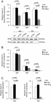
Figure 1
Figure 1
Effect of Slug knockdown on miR-221 expression in breast cancer cells. MDA-MB-231 (A, B) and MDA-MB-436 (C) breast cancer cells were transfected with 30nM si-Slug molecule or a non-relevant siRNA (si-Scr). Slug and miR-221 expression was determined at RNA level at three different times (24 h, 48 h, 72 h), and revealed by quantitative RT-PCR analysis. RT-PCR results were calculated using the ΔΔCt method and data are presented as fold change respect to control untreated cells (Ctr 24 h for MDA-MB-231, and Ctr 72 h for MDA-MB-436). Results represent means ± SEM of three independent experiments. p-values ≤ 0.05 were considered statistically significant. In panel A, Slug expression investigated at protein level and revealed by Western Blot, is reported. IP3K was used as loading control.
Figure 2
Figure 2
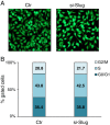
Effect of Slug knockdown on cell cycle and viability. MDA-MB-231 cells were transfected with si-Slug molecule and collected 72 hours after transfection. (A) Viability was determined by double staining assay with Calcein-AM and propidium iodide. Fluorescence photomicrographs (4X magnification) are representative merged images showing the presence of green fluorescence (calcein-AM)-labelled live cells and the absence of red fluorescence (PI)-labelled dead cells. (B) Cells were subjected to fluorescence-activated cell sorting analysis, and the relative G0/G1, S, and G2/M compartments calculated. Percentages of cells in each compartment are means of two independent experiments.
Figure 3
Figure 3
In vivo recruitment of Slug protein at the miR-221-222 locus. (A) The localization of predicted Slug consensus binding sites (5'-CAGGTG-3' or 5'-CACCTG-3') in the human miR-222/221 locus region is indicated with grey ovals. Protein-DNA complexes were in vivo formaldehyde-cross linked in MDA-MB-231. Chromatin fragments were subjected to immunoprecipitation with antibodies against endogenous Slug and Acetyl Histone H3 (Ac-H3). A negative control using nonspecific normal rabbit antibody against Ig λ chain was also included. After cross-link reversal, the coimmunoprecipitated DNA was amplified by PCR using the primers pairs spanning the reported regions of miR-221 promoter (PCR amplicons are indicated by horizontal bars). Aliquots of chromatin taken before immunoprecipitation were used as Input positive controls whereas chromatin eluted from immunoprecipitation lacking antibody was used as no antibody control (No Ab). All experiments were repeated at least three times and representative images shown. MDA-MB-231 cells were transfected with 30 nM si-Slug molecule or a non-relevant siRNA (si-Scr). Pri-miR-221 (B) and miR-222 (C) expression levels were determined at RNA level after 72 h of treatment, and revealed by quantitative RT-PCR analysis. RT-PCR results were calculated using the ΔΔCt method and data are presented as fold change respect to untreated cells (Ctr). Results represent means ± SEM of three independent experiments. p-values ≤ 0.05 were considered statistically significant.
Figure 4
Figure 4
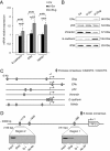
Effect of Slug knockdown on the expression of specific genes. MDA-MB-231 cells were transfected with si-Slug molecule or a non-relevant siRNA (si-Scr). (A) E-cadherin, ERα, TRPS1 expression was determined at mRNA level, and revealed by quantitative RT-PCR analysis. Data are represented as fold change respect to control sample (Ctr) for each gene analysed. Results represent means ± SEM of three independent experiments. p-values ≤ 0.05 were considered statistically significant. (B) ERα, p53, Vimentin, and E-cadherin expression was determined at protein level, and revealed by Western Blot. (C) Analysis of the 2 Kb in size promoter region of Slug, ERα, p53, Vimentin, E-cadherin and TRPS1 genes. Predicted E-boxes consensus-binding site are indicated with grey ovals. (D) Slug is recruited at TRPS1 promoter in vivo. The localization of predicted Slug consensus binding sites (5'-CAGGTG-3' or 5'-CACCTG-3') in the human TRPS1 promoter is reported. Protein-DNA complexes were in vivo formaldehyde-cross linked in MDA-MB-231 cells. Chromatin fragments were subjected to immunoprecipitation with antibody against endogenous Slug. A negative control using nonspecific normal rabbit antibody against Ig λ chain was also included. After cross-link reversal, the coimmunoprecipitated DNA was amplified by PCR using the primers pairs spanning the reported regions of TRPS1 promoter (PCR amplicons are indicated by horizontal bars). Aliquots of chromatin taken before immunoprecipitation were used as Input positive controls whereas chromatin eluted from immunoprecipitation lacking antibody was used as no antibody control (No Ab). All experiments were repeated at least three times and representative images shown.
Figure 5
Figure 5
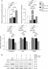
Effect of miR-221 overexpression on the expression of specific genes. MDA-MB-231 cells were transfected with si-Slug molecule alone, si-Slug in combination with miR-221 mimic or a non-relevant miR (miR-Scr) mimic, a non-relevant siRNA (si-Scr) in combination with miR-Scr mimic. (A) Slug, miR-221, E-cadherin, ERα, TRPS1 expression was determined at RNA level, and revealed by quantitative RT-PCR analysis. RT-PCR results were calculated using the ΔΔCt method. Data are represented as fold change respect to control sample (Ctr). Results represent means ± SEM of three independent experiments. p-values ≤ 0.05 were considered statistically significant. (B) Slug, ERα, Vimentin, and E-cadherin expression was determined at protein level, and revealed by Western Blot. Ten micrograms of whole cell lysates were assayed on a 12% SDS-PAGE, and the proteins were visualized using Supersignal Femto Substrate (Pierce). IP3K was used as loading control.
Figure 6
Figure 6
Effect of Slug and miR-221 knockdown on MDA-MB-231 cell migration ability. (A) Cells were transfected with 30nM si-Slug, a non-relevant siRNA (si-Scr), antagomiR-221 or a non-relevant antagomiR (antagomiR-Scr). Twenty-four hours after transfection, cells monolayer were scratch wounded with a 20-μl pipet tip (0 h), and observed over the indicated time periods, 0 and 24 hours (4x magnification). Scale bar =100 μm. (B) Proliferation curves of MDA-MB-231 cells exposed to si-Slug, si-Scr, antagomiR-221 and antagomiR-Scr up to six days. Statistical analysis was performed si-Slug or antagomiR-221 treated cells versus untreated cells (Ctr) (*), and si-Slug or antagomiR-221 treated cells versus si-Scr or antagomiR-Scr respectively (o); p ≤ 0.05. (C) Slug and miR-221 RNA levels were analysed by quantitative RT-PCR after antagomiR-221 or antagomiR-Scr treatment, and results were calculated using the ΔΔCt method. Data are presented as fold difference respect to control untreated cells (Ctr). Results represent means ± SEM of three independent experiments. p-values ≤ 0.05 were considered statistically significant. The expression of Slug, E-cadherin, ERα proteins was also analyzed by Western Blot. IP3K was used as loading control.

References

    1. Nieto MA. The ins and outs of the epithelial to mesenchymal transition in health and disease. Annu Rev Cell Dev Biol. 2011;27:347–376. doi: 10.1146/annurev-cellbio-092910-154036. - "V体育官网" DOI - PubMed
    1. Vincent-Salomon A, Thiery JP. Host microenvironment in breast cancer development: epithelial-mesenchymal transition in breast cancer development. Breast Cancer Res. 2003;5(2):101–106. doi: 10.1186/bcr578. - V体育官网 - DOI - PMC - PubMed
    1. De Herreros AG, Peiró S, Nassour M, Savagner P. Snail family regulation and epithelial mesenchymal transitions in breast cancer progression. J Mammary Gland Biol Neoplasia. 2010;15(2):135–147. doi: 10.1007/s10911-010-9179-8. - DOI - PMC - PubMed
    1. Barrallo-Gimeno A, Nieto MA. The Snail genes as inducers of cell movement and survival: implications in development and cancer. Development. 2005;132(14):3151–3161. doi: 10.1242/dev.01907. - V体育2025版 - DOI - PubMed
    1. Thiery JP, Acloque H, Huang RYJ, Nieto MA. Epithelial-mesenchymal transitions in development and disease. Cell. 2009;139(5):871–890. doi: 10.1016/j.cell.2009.11.007. - DOI - PubMed

Publication types (V体育官网入口)

MeSH terms

VSports最新版本 - Substances